Germline mutations in DNA repair genes are prevalent in myeloma patients and correlate with duration of response to a frontline genotoxic therapy.
Abstract
First-degree relatives of patients with multiple myeloma are at increased risk for the disease, but the contribution of pathogenic germline variants (PGV) in hereditary cancer genes to multiple myeloma risk and outcomes is not well characterized. To address this, we analyzed germline exomes in two independent cohorts of 895 and 786 patients with multiple myeloma. PGVs were identified in 8.6% of the Discovery cohort and 11.5% of the Replication cohort, with a notable presence of high- or moderate-penetrance PGVs (associated with autosomal dominant cancer predisposition) in DNA repair genes (3.6% and 4.1%, respectively). PGVs in BRCA1 (OR = 3.9, FDR < 0.01) and BRCA2 (OR = 7.0, FDR < 0.001) were significantly enriched in patients with multiple myeloma when compared with 134,187 healthy controls. Five of the eight BRCA2 PGV carriers exhibited tumor-specific copy number loss in BRCA2, suggesting somatic loss of heterozygosity. PGVs associated with autosomal dominant cancer predisposition were associated with younger age at diagnosis, personal or familial cancer history, and longer progression-free survival after upfront high-dose melphalan and autologous stem-cell transplantation (P < 0.01).
Significance: Our findings suggest up to 10% of patients with multiple myeloma may have an unsuspected cancer predisposition syndrome. Given familial implications and favorable outcomes with high-dose melphalan and autologous stem-cell transplantation in high-penetrance PGV carriers, genetic testing should be considered for young or newly diagnosed patients with a personal or family cancer history.
Introduction
There is considerable evidence to suggest that germline genetic variation contributes to multiple myeloma risk. First-degree relatives of patients with the disease have two- to fourfold higher risk of multiple myeloma or precursor conditions (1, 2) and a higher risk of developing other solid and hematologic cancers (3). Moreover, sequencing studies of families with multiple myeloma have detected a small number of rare, high-penetrance germline variants in candidate susceptibility genes (CDKN2A, KDM1A, USP45, ARID1A, DIS3, and EP300; refs. 4, 5). However, the contribution of pathogenic or likely pathogenic germline variants (PGV) in known genes associated with hereditary cancer (HC) syndromes to multiple myeloma risk remains to be systematically characterized.
It is estimated that approximately 3.9 million individuals in the United States harbor a PGV in a cancer susceptibility gene, with most being unaware of their cancer risk (6, 7). These PGVs are increasingly found across a broad spectrum of both solid tumors and hematologic malignancies, extending even to cancers without a recognized hereditary basis (8). Identifying PGVs holds significant value for healthcare providers and patients and their families, as it can inform the need for personalized screening and prevention strategies. Moreover, there may be therapeutic implications for patients with PGVs diagnosed with cancer (9).
Here, using a standard panel of HC genes and a clinically validated pipeline for PGV detection and annotation, we explore the prevalence of PGVs and their association with clinical outcomes in two independent cohorts of patients with multiple myeloma: a Discovery cohort comprising 895 patients and a Replication cohort of 786 patients.
Results
Study Population
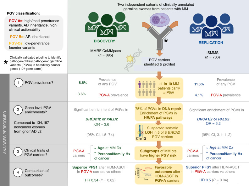
Our study encompassed a total of 1,681 individuals diagnosed with active multiple myeloma, explicitly excluding patients with precursor conditions (i.e., monoclonal gammopathy of undetermined significance or smoldering multiple myeloma) and those with immunoglobulin light chain (AL) amyloidosis. Two independent datasets were analyzed: 895 patients from the Multiple Myeloma Research Foundation (MMRF) CoMMpass dataset (Discovery cohort) and 786 patients from the Icahn School of Medicine at Mount Sinai (ISMMS; Replication cohort). Figure 1 shows the study schema, and Supplementary Table S1 provides an overview of the clinical characteristics for each cohort. The median age at diagnosis across both cohorts was 63 (range, 25–94) years, with 59% of patients being male. In terms of racial and ethnic background, 59% self-identified as White, 20% as Black/African-American, 6% as Hispanic, 4% as Asian, 3% as Other, and the remaining 8% as Unknown. Forty-three percent of patients with known cytogenetics had one or more high-risk abnormalities at diagnosis, including del17p, t(4;14), t(14;16), t(14;20), or +1q. Forty-four percent of patients received high-dose melphalan with autologous stem-cell transplantation (HDM-ASCT) as part of their first-line therapy. Comprehensive data on medical history and family history were available for 98% and 71% of the study population, respectively.
Figure 1.
Study schema. Study framework evaluating PGV prevalence in multiple myeloma through a two-stage analysis involving 895 patients from the MMRF CoMMpass study (Discovery cohort) and 786 from the ISMMS cohort (Replication cohort). PGVs are categorized as follows: PGV-As (high-/moderate-penetrance, AD inheritance), PGV-Bs (AR inheritance), and PGV-Cs (low-penetrance founder variants). AD, autosomal dominant; AR, autosomal recessive; Dx, diagnosis; HR/FA, homologous recombination and Fanconi anemia pathways; Hx, history; MM, multiple myeloma; mod, moderate; pts, patients.
PGVs Detected in ∼10% of Patients with Multiple Myeloma across Two Independent Cohorts
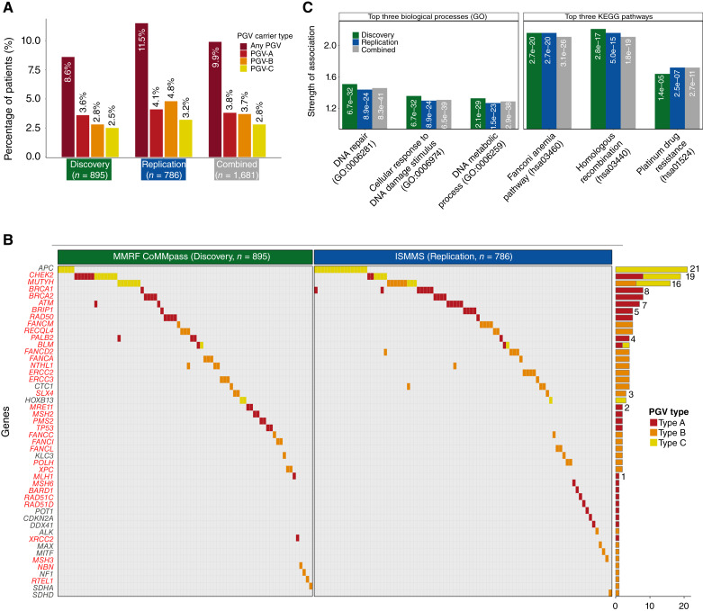
In the Discovery cohort, 77/895 patients with multiple myeloma (8.6%) were PGV carriers (Table 1; Fig. 2A). About 80 heterozygous PGVs (63 unique variants, Supplementary Table S2) were distributed across 32 HC genes, 81.3% of which were linked to DNA repair (Supplementary Table S3; Fig. 2B). The most significant pathway enrichments were in homologous recombination (strength = 2.21, FDR = 2.77e−17) and Fanconi anemia (strength = 2.16, FDR = 2.66e−20; Fig. 2C). About 32/895 patients (3.6%) carried a high-/moderate-penetrance PGV associated with autosomal dominant cancer predisposition (PGV-A, Table 2), whereas 25/895 patients (2.8%) carried a heterozygous PGV associated with autosomal recessive cancer predisposition (PGV-B) and 22/895 (2.5%) had a low-penetrance founder PGV (PGV-C; Supplementary Table S4). Two patients who were PGV-A carriers also harbored a PGV-C, and one patient carried two PGV-Bs. The remaining patients had a single PGV.
Table 1.
Clinical characteristics of patients with multiple myeloma by PGV status.
| Discovery (n = 895) | Replication (n = 786) | Combined (n = 1,681) | ||||||||||||||
|---|---|---|---|---|---|---|---|---|---|---|---|---|---|---|---|---|
| Clinical feature | Subgroup | Noncarrier | PGV-A carrier | P a | PGV-B/C carrier | P b | Noncarrier | PGV-A carrier | P a | PGV-B/C carrier | P b | Noncarrier | PGV-A carrier | P a | PGV-B/C carrier | P b |
| n (%) | 818 (91.4) | 32 (3.6) | 45 (5.0) | 696 (88.5) | 32 (4.1) | 58 (7.4) | 1,514 (90.1) | 64 (3.8) | 103 (6.1) | |||||||
| Gender (%) | Male | 498 (60.9) | 19 (59.4) | NS | 25 (55.6) | NS | 395 (56.8) | 16 (50.0) | NS | 38 (65.5) | NS | 893 (59.0) | 35 (54.7) | NS | 63 (61.2) | NS |
| Age [median (range)], years | 63 (27–93) | 62 (36–84) | NS | 65 (32–91) | NS | 61 (25–91) | 55 (39–82) | 0.07 | 66 (39–93) | NS | 62 (25–94) | 59 (36–84) | 0.04 | 66 (32–93) | 0.04 | |
| Self-reported race (%) | White | 546 (66.7) | 24 (75.0) | 0.05 | 30 (66.7) | NS | 337 (48.4) | 19 (59.4) | NS | 38 (65.5) | 0.03 | 883 (58.3) | 43 (67.2) | NS | 68 (66.0) | NS |
| Black | 127 (15.5) | 0 (0.0) | 8 (17.8) | 187 (26.9) | 6 (18.8) | 6 (10.3) | 314 (20.7) | 6 (9.4) | 14 (13.6) | |||||||
| Hispanic | 0 (0.0) | 0 (0.0) | 0 (0.0) | 87 (12.5) | 4 (12.5) | 10 (17.2) | 87 (5.7) | 4 (6.2) | 10 (9.7) | |||||||
| Asian | 14 (1.7) | 0 (0.0) | 0 (0.0) | 46 (6.6) | 2 (6.2) | 3 (5.2) | 60 (4.0) | 2 (3.1) | 3 (2.9) | |||||||
| Other | 36 (4.4) | 2 (6.2) | 3 (6.7) | 2 (0.3) | 0 (0.0) | 0 (0.0) | 38 (2.5) | 2 (3.1) | 3 (2.9) | |||||||
| Unknown | 95 (11.6) | 6 (18.8) | 4 (8.9) | 37 (5.3) | 1 (3.1) | 1 (1.7) | 132 (8.7) | 7 (10.9) | 5 (4.9) | |||||||
| International Staging System stage (%) | Stage 1 | 276 (34.8) | 12 (38.7) | NSc | 20 (45.5) | NSc | 191 (41.3) | 10 (58.8) | NSc | 24 (61.5) | NSc | 467 (37.2) | 22 (45.8) | NSc | 44 (53.0) | NSc |
| Stage 2 | 292 (36.9) | 10 (32.3) | 12 (27.3) | 141 (30.5) | 3 (17.6) | 7 (17.9) | 433 (34.5) | 13 (27.1) | 19 (22.9) | |||||||
| Stage 3 | 224 (28.3) | 9 (29.0) | 12 (27.3) | 130 (28.1) | 4 (23.5) | 8 (20.5) | 354 (28.2) | 13 (27.1) | 20 (24.1) | |||||||
| Cytogenetics (%) | Any high risk | 332 (40.6) | 9 (28.1) | NS | 18 (40.0) | NS | 281 (46.8) | 12 (44.4) | NSc | 23 (47.9) | NSc | 613 (43.2) | 21 (35.6) | NSc | 41 (44.1) | NSc |
| del17p (%) | 71 (8.7) | 3 (9.4) | NS | 3 (6.7) | NS | 74 (12.3) | 2 (7.4) | NSc | 6 (12.5) | NSc | 145 (10.2) | 5 (8.5) | NSc | 9 (9.7) | NSc | |
| 1q gain (%) | 234 (28.6) | 7 (21.9) | NS | 12 (26.7) | NS | 217 (36.2) | 10 (37.0) | NSc | 16 (33.3) | NSc | 451 (31.8) | 17 (28.8) | NSc | 28 (30.1) | NSc | |
| t(4;14) (%) | 82 (10.0) | 2 (6.2) | NS | 4 (8.9) | NS | 50 (8.3) | 2 (7.4) | NSc | 3 (6.2) | NSc | 132 (9.3) | 4 (6.8) | NSc | 7 (7.5) | NSc | |
| t(14;16) (%) | 27 (3.3) | 0 (0.0) | NS | 2 (4.4) | NS | 25 (4.2) | 2 (7.4) | NSc | 3 (6.2) | NSc | 52 (3.7) | 2 (3.4) | NSc | 5 (5.4) | NSc | |
| t(14;20) (%) | 10 (1.2) | 0 (0.0) | NS | 0 (0.0) | NS | 10 (1.7) | 0 (0.0) | NSc | 1 (2.1) | NSc | 20 (1.4) | 0 (0.0) | NSc | 1 (1.1) | NSc | |
| t(11;14) (%) | 144 (17.6) | 10 (31.2) | 0.07 | 12 (26.7) | NS | 123 (20.5) | 6 (22.2) | NSc | 13 (27.1) | NSc | 267 (18.8) | 16 (27.1) | NSc | 25 (26.9) | NSc | |
| Family history of cancer (%) | Yes | 319 (54.5) | 17 (81.0) | <0.01 c | 21 (65.6) | 0.02 c | 294 (61.6) | 23 (85.2) | 0.03 c | 25 (67.6) | NS | 613 (57.7) | 40 (83.3) | <0.01 c | 46 (66.7) | 0.02 c |
| Personal history of other cancerd (%) | Yes | 23 (2.8) | 3 (9.4) | 0.05 c e | 2 (4.4) | NSc | 60 (9.0) | 6 (19.4) | 0.05 c e | 15 (26.3) | <0.01 c | 83 (5.6) | 9 (14.3) | <0.01 c | 17 (16.7) | <0.01 c |
| First-line therapy (%) | Doublet | 130 (15.9) | 7 (21.9) | NS | 10 (22.2) | NS | 53 (8.4) | 3 (10.0) | NSc | 3 (5.5) | NSc | 183 (12.6) | 10 (16.1) | NSc | 13 (13.0) | NSc |
| Triplet | 665 (81.3) | 23 (71.9) | 35 (77.8) | 352 (55.8) | 16 (53.3) | 27 (49.1) | 1,017 (70.2) | 39 (62.9) | 62 (62.0) | |||||||
| Quadruplet | 0 (0.0) | 0 (0.0) | 0 (0.0) | 181 (28.7) | 6 (20.0) | 19 (34.5) | 181 (12.5) | 6 (9.7) | 19 (19.0) | |||||||
| Other | 23 (2.8) | 2 (6.2) | 0 (0.0) | 45 (7.1) | 5 (16.7) | 6 (10.9) | 68 (4.7) | 7 (11.3) | 6 (6.0) | |||||||
| HDM-ASCT with first-line therapy (%) | Yes | 396 (48.4) | 16 (50.0) | NS | 25 (55.6) | NS | 266 (38.2) | 19 (59.4) | 0.03 | 16 (27.6) | NS | 662 (43.7) | 35 (54.7) | NS | 41 (39.8) | NS |
| After ASCT lenalidomide maintenance (%) | Yes | 187 (47.2) | 4 (25.0) | NS | 12 (48.0) | NS | 184 (69.2) | 9 (47.4) | NS | 7 (43.8) | NS | 371 (56.0) | 13 (37.1) | 0.04 | 19 (46.3) | NS |
P value for the comparison of PGV-A carriers against noncarriers within each cohort.
P value for the comparison of PGV-B/C carriers against noncarriers within each cohort; NS, nonsignificant (P ≥ 0.05).
P values calculated based on imputed data using pooled logistic regression analysis across five imputed datasets, given >5% missing data.
Excluding cancer types strongly associated with environmental exposures (nonmelanoma skin cancer, lung cancer, and head and neck cancer).
Significant P values (<0.05) were rounded up to 0.05 for consistency with the displayed number of decimal places.
Figure 2.
Distribution and frequency of PGVs in patients with multiple myeloma. A, Prevalence of PGVs across the cohorts with multiple myeloma. B, Oncoplot detailing the distribution among the patients (columns) of PGVs in specific genes (rows). DNA repair–related gene names are denoted in red font. Adjacent is a bar plot summarizing the total PGVs detected per gene, differentiated by PGV type. C, Enriched biological pathways are shown for the gene sets carrying PGVs across the Discovery, Replication, and Combined cohorts. Left shows the top three biological processes from Gene Ontology (GO), and right displays the top three pathways from the Kyoto Encyclopedia of Genes and Genomes (KEGG). “Strength of Association” on the y-axis is obtained from the STRING protein interaction analysis and is defined as log10(observed/expected), reflecting the magnitude of enrichment for each pathway. The white text within each bar shows the FDR values for each association.
Table 2.
Comprehensive characterization of PGV-A carriers.
| ID | Gene | Variant | Set | Age | Sex | Race | ISS | High risk | FISHa | Prior CA | Fam Hx CA | Rx 1 | ASCT first-line |
|---|---|---|---|---|---|---|---|---|---|---|---|---|---|
| 1 | ATM | c.710delC (p.T237IfsX18) | D | 67 | M | W | 1 | No | None | No | Yes | Trip | Yes |
| 31b | ATM | c.1945G>T (p.E649X) | D | 59 | F | 3 | No | None | No | Trip | Yes | ||
| 896 | ATM | c.5762+1G>T (-) | R | 55 | M | W | Yes | 1q gain, t(4;14) | No | No | Other | Yes | |
| 897 | ATM | c.8266A>T (p.K2756X) | R | 39 | M | H | No | None | No | Yes | Quad | Yes | |
| 915c | ATM | c.2502dupA (p.V835SfsX7) | R | 54 | M | W | No | t(11;14) | No | Yes | Other | Yes | |
| 918 | ATM | c.5712dupA (p.S1905Ifs*25) | R | 72 | F | W | 2 | No | t(11;14) | Yesd | Trip | No | |
| 922 | ATM | c.8122G>A (p.D2708N) | R | 61 | M | A | 3 | No | None | No | Quad | No | |
| 927 | BARD1 | c.463A>T (p.R155*) | R | 70 | M | B | 3 | Yes | 1q gain | No | Yes | No | |
| 2 | BLM | c.1642C>T (p.Q548X) | D | 64 | M | W | 1 | No | None | No | Yes | Doubl | No |
| 898 | BLM | c.3875-2A>G (-) | R | 59 | M | W | 1 | Yes | del17p | No | Yes | Trip | Yes |
| 3 | BRCA1 | c.5177_5180delGAAA (p.R1726KfsX3) | D | 58 | M | W | 2 | No | None | No | Yes | Trip | Yes |
| 899 | BRCA1 | c.1687C>T (p.Q563X) | R | 64 | F | W | No | Yes | Doubl | No | |||
| 900 | BRCA1 | c.5123C>A (p.A1708E) | R | 57 | M | W | 3 | Yes | t(14;16) | No | Yes | Trip | No |
| 901 | BRCA1 | c.671-1G>T (-) | R | 50 | F | A | 1 | No | None | No | Yes | Trip | Yes |
| 902 | BRCA1 | c.68_69delAG (p.E23VfsX17) | R | 53 | F | W | 3 | No | Trip | Yes | |||
| 916b | BRCA1 | c.68_69delAG (p.E23VfsX17) | R | 66 | M | W | 1 | No | None | Yese | Yes | Trip | Yes |
| 917b | BRCA1 | c.68_69delAG (p.E23VfsX17) | R | 53 | M | W | 1 | No | t(11;14) | No | Yes | Other | Yes |
| 926 | BRCA1 | c.302-1G>A (-) | R | 78 | F | B | No | None | Yesd | Yes | Trip | No | |
| 4 | BRCA2 | c.2808_2811delACAA (p.A938PfsX21) | D | 67 | M | O | 1 | No | None | No | Doubl | No | |
| 5 | BRCA2 | c.4936_4939delGAAA (p.E1646QfsX23) | D | 84 | M | W | 3 | No | None | No | Yes | Doubl | No |
| 6 | BRCA2 | c.658_659delGT (p.V220IfsX4) | D | 61 | M | 2 | No | None | No | Trip | Yes | ||
| 7 | BRCA2 | c.6641dupC (p.Y2215LfsX10) | D | 63 | F | 1 | No | None | No | Trip | No | ||
| 903 | BRCA2 | c.3778_3779delTT (p.L1260IfsX4) | R | 47 | F | H | No | Yes | Quad | Yes | |||
| 904 | BRCA2 | c.5946delT (p.S1982RfsX22) | R | 55 | M | W | 1 | Yes | 1q gain | No | Yes | Trip | Yes |
| 905 | BRCA2 | c.7977-1G>C (-) | R | 41 | F | W | 1 | Yes | t(11;14), 1q gain | No | Yes | Other | No |
| 924 | BRCA2 | c.5946delT (p.S1982Rfs*22) | R | 71 | F | W | 2 | Yes | 1q gain, t(14;16) | Yesd | Yes | Trip | No |
| 8 | BRIP1 | c.440delA (p.Y147SfsX10) | D | 67 | F | W | 1 | No | None | No | Yes | Trip | No |
| 906 | BRIP1 | c.1A>G (p.M1?) | R | 43 | F | W | No | None | No | Trip | Yes | ||
| 907 | BRIP1 | c.2392C>T (p.R798X) | R | 53 | M | H | Yes | del17p, 1q gain | No | Yes | Other | Yes | |
| 908 | BRIP1 | c.3390_3393delCTAT (p.Y1131LfsX18) | R | 50 | F | B | 1 | No | None | No | No | Trip | No |
| 909 | BRIP1 | c.751C>T (p.R251C) | R | 48 | M | W | No | t(11;14) | No | No | Quad | Yes | |
| 923 | CDKN2A | c.176T>G (p.V59G) | R | 82 | M | W | Yesf | Yes | Trip | No | |||
| 9 | CHEK2 | c.1100delC (p.T367MfsX15) | D | 66 | F | W | 1 | Yes | t(11;14), del17p | No | Yes | Trip | No |
| 10 | CHEK2 | c.1100delC (p.T367MfsX15) | D | 52 | F | W | 1 | No | t(11;14) | Yesd | Yes | Doubl | No |
| 11 | CHEK2 | c.1100delC (p.T367MfsX15) | D | 51 | F | W | 2 | No | t(11;14) | No | Yes | Trip | Yes |
| 12 | CHEK2 | c.1232G>A (p.W411X) | D | 69 | M | W | 1 | Yes | del17p | No | Yes | Other | No |
| 13 | CHEK2 | c.1368dupA (p.E457RfsX33) | D | 60 | M | W | 3 | No | t(11;14) | No | Yes | Trip | Yes |
| 14 | CHEK2 | c.3G>A (p.M1?) | D | 58 | F | W | 3 | Yes | 1q gain | No | Yes | Trip | Yes |
| 910 | CHEK2 | c.349A>G (p.R117G) | R | 64 | F | H | Yes | 1q gain | Yesd | Yes | Quad | Yes | |
| 920 | CHEK2 | c.1100delC (p.T367Mfs*15) | R | 67 | F | B | No | Yes | Trip | Yes | |||
| 919 | DDX41 | c.388A>T (p.K130*) | R | 53 | M | 2 | No | None | No | Yes | Trip | No | |
| 15 | MLH1 | c.1489dupC (p.R497PfsX6) | D | 68 | M | W | 3 | No | None | Yesg | Trip | No | |
| 16 | MRE11 | c.1714C>T (p.R572X) | D | 83 | M | W | 3 | Yes | 1q gain | No | No | Trip | No |
| 17 | MRE11 | c.1852dupA (p.M618NfsX8) | D | 52 | F | 1 | No | t(11;14) | No | Trip | Yes | ||
| 18 | MSH2 | c.1862G>T (p.R621L) | D | 36 | F | W | 2 | No | None | No | Trip | Yes | |
| 19 | MSH2 | c.1906G>C (p.A636P) | D | 63 | M | W | 1 | Yes | del17p, 1q gain, t(4;14) | No | Yes | Trip | No |
| 911 | MSH6 | c.3439-1G>T (-) | R | 67 | M | W | Yes | 1q gain, t(4;14) | No | Yes | Trip | No | |
| 20 | PALB2 | c.1467_1468insTTAA (p.P490LfsX7) | D | 72 | M | W | 2 | No | t(11;14) | No | Doubl | No | |
| 21 | PALB2 | c.2727_2728delTT (p.T911LfsX16) | D | 40 | M | W | 1 | Yes | 1q gain | No | No | Trip | Yes |
| 32b | PALB2 | c.1142_1143delTT (p.L381QfsX19) | D | 50 | M | W | 2 | No | t(11;14) | No | Other | No | |
| 912 | PALB2 | c.120delA (p.A41LfsX12) | R | 67 | M | W | Yes | 1q gain | No | Yes | Yes | ||
| 22 | PMS2 | c.2444C>T (p.S815L) | D | 60 | F | W | 3 | No | t(11;14) | No | Yes | Trip | Yes |
| 23 | PMS2 | c.631C>T (p.R211X) | D | 56 | M | 2 | No | None | No | Trip | No | ||
| 921 | POT1 | c.809G>A (p.S270N) | R | 41 | F | B | 1 | No | None | No | No | Trip | No |
| 24 | RAD50 | c.2165dupA (p.E723GfsX5) | D | 63 | M | 2 | No | None | No | Trip | Yes | ||
| 25 | RAD50 | c.326_329delCAGA (p.T109NfsX20) | D | 58 | F | O | 3 | No | t(11;14) | No | Yes | Trip | Yes |
| 26 | RAD50 | c.3612_3618+5del (p.Q1205fs) | D | 55 | F | W | 3 | Yes | 1q gain | No | No | Trip | No |
| 27 | RAD50 | c.3G>A (p.M1?) | D | 58 | M | W | 2 | Yes | 1q gain, t(4;14) | No | No | Trip | Yes |
| 913 | RAD50 | c.1958C>A (p.S653X) | R | 57 | F | W | 1 | No | t(11;14) | No | Yes | Doubl | Yes |
| 925 | RAD51C | c.267dupA (p.L90Tfs*3) | R | 46 | F | B | Yes | 1q gain | No | Yes | Doubl | Yes | |
| 914 | RAD51D | c.1A>T (p.M1?) | R | 53 | F | W | 1 | No | None | No | Quad | Yes | |
| 28 | TP53 | c.438_439insT (p.V147CfsX2) | D | 69 | F | W | 2 | No | None | No | Yes | Trip | Yes |
| 29 | TP53 | c.473G>A (p.R158H) | D | 63 | M | W | Yes | t(11;14), 1q gain | Yesf | Yes | Doubl | Yes | |
| 30 | XRCC2 | c.651_652delTG (p.C217X) | D | 68 | M | W | 1 | No | None | No | Yes | Doubl | No |
Genomic information and clinical traits are provided for the 64 PGV-A carriers identified.
“None” indicates none of these abnormalities were detected, whereas a missing value indicates no FISH findings were available for this patient.
Abbreviations: A, Asian; B, Black/African-American; D, Discovery; Doubl, doublet (two-drug combination); F, female, Fam Hx CA, family history of cancer; H, Hispanic/Latino; ISS, International Staging System stage; M, male; Other, other drug combination; Prior CA, personal history of antecedent cancer; Quad, quadruplet (four-drug combination); R, Replication; Rx 1, first-line therapy; Trip, triplet (three-drug combination); W, White/European.
Reported FISH findings are: del17p, +1q, t(4;14), t(14;16), t(14;20), t(11;14).
Patient is also a PGV-C carrier.
Patient is also a PGV-B carrier.
Breast.
Melanoma.
Prostate.
Bladder.
In the Replication cohort, 90/786 patients (11.5%) were PGV carriers (Table 1; Fig. 2A). About 95 heterozygous PGVs (73 unique variants) were found across 36 HC genes, with 69.4% related to DNA repair (Fig. 2B), and the strongest pathway enrichments again in homologous recombination (strength = 2.11, FDR = 1.65e−14) and Fanconi anemia (strength = 2.14, FDR = 8.76e−22; Fig. 2C). About 32/895 patients (4.1%) had a PGV-A (Table 2), 25/895 (4.8%) had a PGV-B, and 22/895 (3.2%) had a PGV-C (Supplementary Table S4). One PGV-A carrier had a concurrent PGV-B, two PGV-A carriers had a concurrent PGV-C, and two PGV-B carriers had a concurrent PGV-C, with the remaining patients having a single PGV.
PGVs in 20 genes were detected in both the Discovery and Replication cohorts. The most common genes harboring PGV-As were BRCA1 (n = 8; one in Discovery, seven in Replication), BRCA2 (n = 8; four in Discovery, four in Replication), CHEK2 (n = 8; six in Discovery, two in Replication), and ATM (n = 7; two in Discovery, five in Replication). Of note, 95.3% of all PGV-As were in DNA repair genes (100% in Discovery, 91% in Replication). As for PGV-Bs, the most commonly affected genes represented in both cohorts were FANCM (n = 5; one in Discovery, four in Replication), RECQL4 (n = 5; three in Discovery, two in Replication), and FANCA (n = 4; three in Discovery, one in Replication). The most common PGV-Cs were the well-recognized Ashkenazi Jewish low-penetrance founder variants APC c.3920T>A (n = 21; five in Discovery, 16 in Replication), CHEK2 c.470T>C (n = 6, four in Discovery, two in Replication), CHEK2 c.1283C>T (n = 5; three in Discovery, two in Replication), MUTYH c.536A>G (n = 6, five in Discovery, one in Replication), and MUTYH c.1187G>A (n = 4; two in Discovery, two in Replication). Of note, three other well-described Ashkenazi Jewish founder variants were found in these datasets but were categorized as PGV-As rather than PGV-Cs due to their protein-truncating nature and higher penetrance: CHEK2 c.1100delC (n = 4; three in Discovery, one in Replication), BRCA1 c.68_69delAG (n = 3; all in Replication), and BRCA2 c.5946delT (n = 2; both in Replication).
PGV Enrichment in Multiple Myeloma Compared with Healthy Controls
To assess the enrichment of PGVs in patients with multiple myeloma relative to the general population, we conducted gene-specific burden analyses comparing each cohort against data from 134,187 healthy controls in the gnomAD database. Recognizing the differences in population composition between our cohorts and the gnomAD dataset, we opted for a conservative comparison approach. This involved using the gnomAD subpopulation that exhibited the highest frequency of pathogenic or likely pathogenic alleles for each gene as the benchmark control.
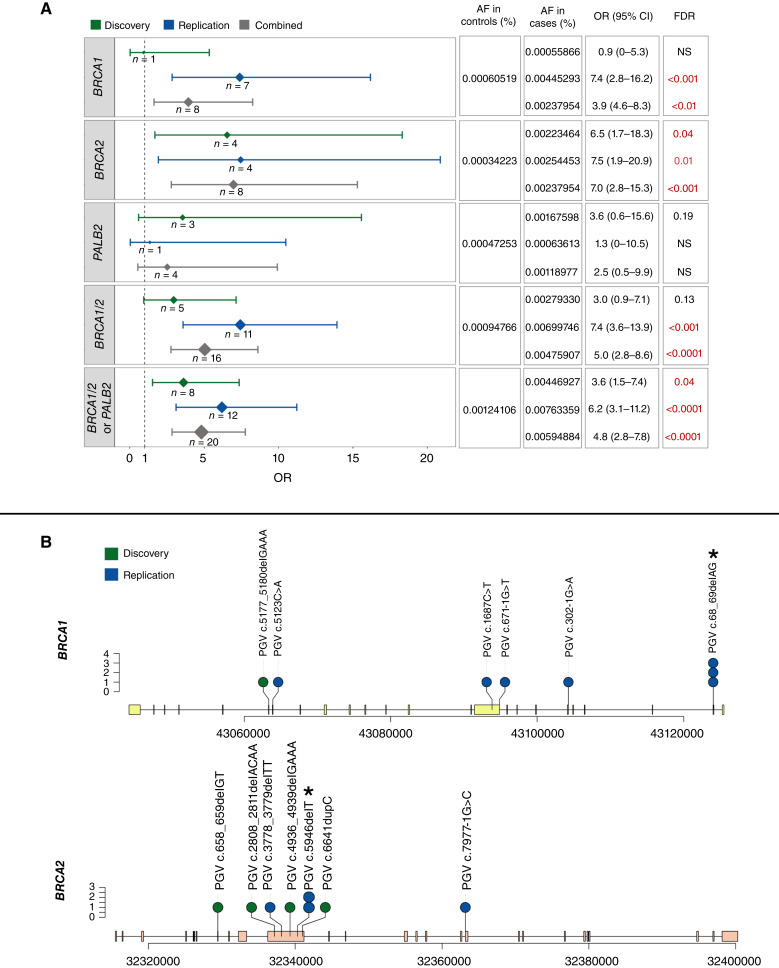
In the Discovery cohort, we found a significant enrichment of PGV-As in BRCA2 [OR = 6.5, 95% confidence interval (CI), 1.7–18.3, FDR = 0.04]. Similarly, in the Replication cohort, significant enrichment of PGV-As was seen in both BRCA1 (OR = 7.4, 95% CI, 2.8–16.2, FDR < 0.001) and BRCA2 (OR = 7.5, 95% CI, 1.9–20.9, FDR = 0.01). In a combined analysis of both cohorts, a marked enrichment of PGV-As in BRCA1/2 was apparent, with an OR of 5.0 (95% CI, 2.8–8.6, FDR < 0.0001; Fig. 3A). Individual PGVs in BRCA1 and BRCA2 across both cohorts are shown in Fig. 3B. Although the analysis did not reveal significant enrichment of PGV-As in other genes within these cohorts with multiple myeloma under our conservative criteria, it is worth noting that four additional genes exhibited a two- to fourfold PGV-A enrichment as compared with controls, suggesting a potential association that might achieve statistical significance with increased study power: PALB2 (OR = 2.5, 95% CI, 0.5–9.9), TP53 (OR = 3.4, 95% CI, 0.4–14.2), MSH2 (OR = 3.5, 95% CI, 0.3–30.2), and CDKN2A (OR = 2.5, 95% CI, 0.1–16.9; Supplementary Table S5). PALB2, in particular, stands out due to its strong biological links and shared cancer risk profile with BRCA1/2, and in fact, the enrichment of PGV-As across PALB2, BRCA1, and BRCA2 in the combined cohort was observed to be highly significant (OR 4.8, 95% CI, 2.8–7.8, FDR < 0.0001). In contrast to the significant findings for PGV-As, genes linked to PGV-Bs did not exhibit significant enrichment in our cohorts with multiple myeloma (Supplementary Table S6) nor did any PGV-Cs analyzed at the variant level (Supplementary Table S7).
Figure 3.
Association of BRCA1, BRCA2, and PALB2 PGV-As with disease across independent cohorts with multiple myeloma. A, Forest plot displaying OR with 95% CI of PGVs in BRCA1, BRCA2, and PALB2 occurrence in Discovery, Replication, and Combined cohorts with multiple myeloma. The size of the diamond for each OR represents the case count, with larger diamonds indicating a higher number of PGVs. FDR was considered significant when <0.05. B, Lollipop plots detailing the landscape of pathogenic variants along the BRCA1 (top) and BRCA2 (bottom) genes. Each “lollipop” represents a distinct variant, with its position on the gene denoted along the x-axis and the frequency or number of observations indicated by the lollipop’s height. Asterisks (*) denotes Ashkenazi Jewish founder PGVs in these genes. AF, allele frequency; NS, nonsignificant.
Genomic Findings Suggestive of LOH in a Subset of BRCA2 PGV Carriers
Given that BRCA2-encompassing deletions of chromosome 13q are common in multiple myeloma, we investigated the LOH in tumor DNA from the 16 BRCA1/2 PGV carriers in our dataset using whole-exome sequencing (WES; Supplementary Table S8). FISH analysis showed large or complete deletions of chromosome 13 spanning BRCA2 (monosomy 13; as indicated by the loss of both 13q14 and 13q34 loci) in five out of eight BRCA2 PGV cases. Consistently, the five tumors had lost one copy of the BRCA2 locus. The variant allele frequency (VAF) of the PGVs in tumor samples versus germline was increased in two and decreased in three of these five monosomy 13 BRCA2-PGV cases. Given the significant effects that sample purity and intratumoral clonal heterogeneity can have on VAF measurements in bulk sequencing methods, further confirmatory testing would be necessary for more definitive insights.
By contrast, in the eight BRCA1 PGV carriers, single-nucleotide variants (SNV) matching the germline PGVs were present in the tumor but without additional pathogenic SNVs or copy number losses in BRCA1, suggesting that LOH did not occur.
PGV-As Linked to Younger Age at Diagnosis and Personal/Familial Cancer History
Next, we examined the clinical phenotypes of PGV carrier subgroups (Table 1). As compared with noncarriers, PGV-A carriers in the Discovery cohort were significantly more likely to report a family history of cancer in a first- or second-degree relative [81% in carriers vs. 55% in noncarriers, OR = 3.8 (95% CI, 1.3–10.7); P = 0.02 by Fisher exact test (FET)]. The same pattern was observed in the Replication cohort: 85% of PGV-A carriers had family history of cancer versus 62% of noncarriers [OR = 3.4 (95% CI, 1.1–10.1); P = 0.03 by FET]. By contrast, although PGV-B/C carriers in both cohorts were more likely to report a family history of cancer than noncarriers, the difference only reached statistical significance in the Discovery cohort [66% vs. 55%, OR = 2.2 (95% CI, 1.1–4.4); P = 0.02 by FET].
A personal history of other cancers at the time of multiple myeloma diagnosis was also significantly more common in PGV-A carriers than in noncarriers, both in the Discovery cohort [9.4% vs. 2.8%, OR = 3.6 (95% CI, 1.0–12.6); P = 0.048 by FET] and the Replication cohort [19.4% vs. 9%, OR = 2.7 (95% CI, 1.1–6.7); P = 0.047 by FET]. The combined analysis of both datasets further solidified the statistical significance of this association: 14.3% versus 5.6%, OR = 2.7 (95% CI, 1.3–5.7); P < 0.01 by FET. PGV-B/C carriers had higher rates of prior cancers than noncarriers in both cohorts, but the difference was only significant in the Replication cohort [26% vs. 9%, OR = 3.7 (95% CI, 1.9–7.1); P < 0.01 by FET].
On age at multiple myeloma diagnosis, PGV-A carriers were consistently younger than noncarriers across both datasets. This trend approached statistical significance within each cohort, and achieved it when the datasets were analyzed together (median age, 59 vs. 62 years, P = 0.04 by Mann–Whitney U test). Multivariate logistic regression revealed that, within the combined dataset, a younger age at diagnosis independently increased the odds of being a PGV-A carrier after controlling for both personal and family cancer histories. Specifically, the analysis showed that each additional year of age was associated with a 3% decrease in the odds of harboring a PGV-A (OR = 0.97, 95% CI, 0.95–0.99). Notably, in this analysis, personal and family histories of cancer were independently linked to PGV-A carrier status, suggesting distinct contributions to the risk profile. Interestingly, PGV-B/C carriers were diagnosed at an older age than noncarriers, although this was only significant in the Replication cohort (median age, 66 vs. 61 years; P = 0.02 by Mann–Whitney U test).
PGV-A Carriers Show Superior Progression-Free Survival after First-Line Therapy with HDM-ASCT than Other Patients with Multiple Myeloma
Previous studies have indicated that cancer cells with high-/moderate-penetrance PGVs in DNA repair genes are more susceptible to DNA-damaging treatments (10). In multiple myeloma, the use of HDM-ASCT introduces extensive DNA damage in cancer cells, primarily through covalent DNA interstrand cross-links (11). Based on this, we hypothesized that patients with multiple myeloma carrying PGV-As in DNA repair genes might respond more favorably to HDM-ASCT than those without such variants, due to weakened DNA repair mechanisms in their cancer cells.
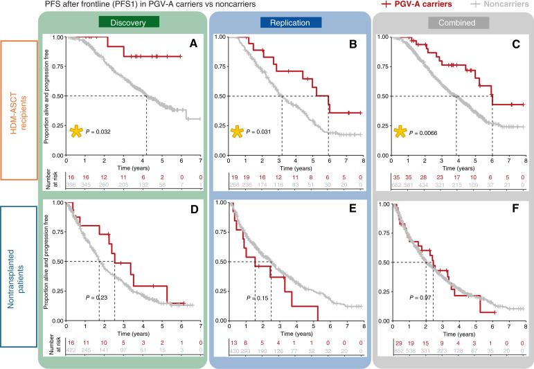
To test this, we first examined the effect of PGV-A on first progression-free survival after first-line therapy (PFS1) in the Discovery cohort, focusing on the 437/895 patients with multiple myeloma in this dataset who received HDM-ASCT as part of their first-line therapy. Transplanted PGV-A carriers demonstrated a notable improvement in PFS1 compared with transplanted noncarriers (median PFS not reached vs. 4.2 years, log-rank P = 0.03; Fig. 4A). This benefit persisted after adjusting for confounders like age, stage as per the International Staging System, cytogenetics, and treatment regimen used for induction and maintenance, showing an adjusted HR of 0.34 [95% CI, 0.14–0.83; P = 0.02 by Cox proportional hazards model (CPH)]. In the Replication cohort, in which 301/786 patients with multiple myeloma underwent upfront HDM-ASCT, transplanted PGV-A carriers again experienced significantly extended PFS1 over transplanted noncarriers (median PFS 5.9 vs. 3.2 years, log-rank P = 0.03; Fig. 4B), with multivariate analysis yielding an adjusted HR of 0.5 (95% CI, 0.25–0.97; P = 0.04 by CPH). Combining data from both cohorts reinforced the strong association between PGV-A status and prolonged PFS1 after HDM-ASCT in both univariate (median PFS 6.0 vs. 3.7 years, log-rank P < 0.01, Fig. 4C) and multivariate analyses (adjusted HR 0.47, 95% CI, 0.26–0.84; P = 0.01 by CPH; Supplementary Fig. S1). Conversely, PGV-A carriers who did not receive HDM-ASCT as part of their first-line therapy (458/895 in Discovery, 485/786 in Replication) had no significant difference in PFS1 as compared with noncarriers (Fig. 4D–F). As for the overall survival (OS), within the harmonized follow-up duration of 7.83 years (adjusted to ensure comparability across both cohorts), we found no significant differences between PGV-A carriers and noncarriers (Supplementary Fig. S2A–S2F). Likewise, PGV-B/C carriers, regardless of HDM-ASCT in their first-line treatment regimen, did not exhibit PFS1 or OS benefits over noncarriers in either cohort (Supplementary Fig. S3).
Figure 4.
PFS1 Outcomes in patients with multiple myeloma with and without PGV-As, stratified by HDM-ASCT. Kaplan–Meier plots of PFS1 in patients with multiple myeloma after first-line therapy including HDM-ASCT (A–C) or alternative therapy (D–F), comparing those with and without high-/moderate-penetrance PGVs (PGV-As) across the Discovery (A and D), Replication (B and E), and Combined (C and F) cohorts. Yellow stars denote statistical significance (P < 0.05) as measured by the log-rank test, used to compare survival distributions between groups. PFS, progression-free survival.
Discussion
As genetic testing becomes more widely available, the range of malignancies linked to inherited genetic predispositions is expanding, revealing previously unrecognized associations. Our comprehensive germline exome analysis of 1,681 patients with multiple myeloma reveals ∼10% prevalence of PGVs in cancer predisposition genes across two independent cohorts, with a significant fraction found in DNA repair genes. Particularly, our study highlights the nearly 4% prevalence of high-/moderate-penetrance PGVs (PGV-As) in multiple myeloma, and the statistically significant enrichment of PGV-As in BRCA1/2 genes as compared with healthy controls. It also identifies subsets of patients with multiple myeloma at increased risk of carrying PGV-As, including those diagnosed at a young age and those with a personal or family history of cancer, and demonstrates a robust association between PGV-As and PFS1. Taken together, our novel findings position multiple myeloma within the spectrum of malignancies influenced by hereditary genetic predispositions and support the integration of germline genetic testing into evaluations of patients with the disease.
The integrity of DNA repair processes, especially those addressing interstrand cross-links and double-strand breaks, is crucial for B-cell development, given the role these mechanisms play in V(D)J recombination, class-switch recombination and somatic hypermutation. We found that most PGVs in multiple myeloma are in genes associated with the homologous recombination and Fanconi anemia pathways, crucial for interstrand cross-links and double-strand break repair. Dysregulation of these pathways, in which BRCA proteins play a central role, has been linked to genomic instability and progression in multiple myeloma (12). Notably, our study demonstrates a fivefold increased occurrence of BRCA1/2 PGV-As among patients with multiple myeloma compared with healthy individuals. This enrichment may indicate a possible mechanistic link between BRCA pathway dysfunction and multiple myeloma predisposition. Although others have suggested an association between BRCA1/2 and plasma cell disorders (13–17), this study is, to our knowledge, the first to demonstrate statistically that multiple myeloma may be a component of hereditary cancer syndromes due to PGVs in these genes.
Bulk tumor sequencing performed in BRCA1/2 PGV carriers demonstrated findings suggestive of somatic LOH in a subset of BRCA2 PGV carriers. Although confirmatory techniques like single-cell sequencing, digital droplet PCR, or SNP phasing were not within this project’s scope, they could provide more precise validation in future research. In all of these cases, the loss of BRCA2 occurred in the context of large or complete deletions in chromosome 13, which is very commonly observed in multiple myeloma and can arise from a range of genomic instabilities and selective pressures independent of BRCA2 mutations. However, even if PGVs are not directly driving somatic loss of the wild-type allele, the resulting bi-allelic inactivation of BRCA2 may still hold significant implications for tumor biology and impact susceptibility to DNA repair–targeting therapies in these patients. Also importantly, the absence of LOH does not negate the oncogenic potential of a PGV. Studies across various cancers such as prostate, pancreatic, breast, and ovarian cancers have demonstrated that BRCA1/2 PGVs can drive tumorigenesis without LOH, potentially through mechanisms like haploinsufficiency or dominant-negative effects, and through alternative pathways affecting gene expression or DNA repair fidelity (18, 19). The absence of LOH in some BRCA1/2 PGV carriers in our study might similarly suggest alternative oncogenic mechanisms warranting further investigation.
Although our conservative analysis only identify significant PGV-A enrichment in BRCA1/2, suggestive trends in PALB2, TP53, and MSH2 indicate potential associations with multiple myeloma warranting further investigation with increased sample sizes. PALB2 is of particular interest due to its intimate biological association with BRCA1/2, especially in light of the highly significant PGV-A enrichment observed when analyzing these three closely related genes as a group. As for TP53, the detection of PGV-As within our cohort with multiple myeloma, although limited to two cases, gains significance in the context of emerging literature: two recent abstracts (20, 21) and a peer-reviewed study reporting multiple myeloma in a Li-Fraumeni syndrome family (22) suggest that the cancer spectrum of Li-Fraumeni syndrome could be broader than previously thought, potentially encompassing multiple myeloma.
Detecting PGVs allows for personalized health management plans, including specific surveillance and prevention strategies for individuals at risk, while also advising families on genetic testing and proactive measures for potentially at-risk members. We observed that patients with multiple myeloma with personal and/or family history of cancer, and those with an earlier onset of the disease, had significantly higher rates of PGVs than others. As an illustrative example, among patients with multiple myeloma in the combined dataset with an antecedent cancer diagnosis, we found one in four patients to be a PGV carrier. These findings suggest that subsets of patients with multiple myeloma with these characteristics could particularly benefit from targeted germline genetic screening.
Our findings reveal that PGV-As in DNA repair genes not only contribute to multiple myeloma risk but also significantly influence treatment outcomes. Specifically, across two independent cohorts, patients with multiple myeloma carrying PGV-As who received upfront HDM-ASCT exhibited significantly longer PFS1 than those without these variants. This observation aligns with existing knowledge that PGVs in DNA repair genes can enhance sensitivity to DNA-damaging therapies, a phenomenon observed in various other cancers. In multiple myeloma, polymorphisms in DNA repair genes including BRCA1 have been correlated with improved responses to HDM (23). In melphalan-resistant multiple myeloma cell lines, knockdown of key components of the FA/BRCA pathway results in heightened susceptibility to the drug, whereas overactivation of the pathway in sensitive cells induces melphalan resistance (24). In the evolving therapeutic landscape of multiple myeloma, in which the role of HDM-ASCT is being reevaluated amidst a shift toward novel immunotherapies, our findings suggest PGV-As might become useful biomarkers when assessing transplant candidacy. Furthermore, most of the PGV-As that we identified fall within DNA repair genes that are therapeutically targetable in cancers outside of multiple myeloma (9), using drugs such as PARP inhibitors, which have demonstrated in vitro activity against multiple myeloma (25, 26). Our results advocate for expanded research into targeted treatment strategies for patients with multiple myeloma harboring DNA repair mutations, whether germline or acquired.
Despite the compelling associations between PGVs, multiple myeloma predisposition, and treatment outcomes, our study acknowledges inherent limitations stemming from its retrospective design. Additionally, the study was designed to investigate associations between multiple myeloma and known HC genes rather than discovering novel predisposition genes, which may have limited our ability to identify other genetic factors contributing to multiple myeloma predisposition beyond the scope of the selected HC genes. Finally, we were unable to individually match controls to cases but instead used aggregated healthy population controls from gnomAD, which could introduce bias due to population structure or other factors. We argue, however, that our conservative analytical approach and the robust and statistically significant association between BRCA1/2 and multiple myeloma mitigate against this possibility, as does the independent association of PGV-As with disease-related outcomes. Notably, our results were successfully replicated across two independent cohorts, further bolstering their validity. However, to firmly establish the role of PGVs in DNA repair genes in multiple myeloma risk and treatment response, prospective studies and validation in diverse cohorts are warranted.
In conclusion, our study positions multiple myeloma as a potential component of cancer-predisposing conditions associated with known HC genes, notably BRCA1/2. The discovery of PGV-As in patients with multiple myeloma not only highlights the importance of genetic testing for early detection and familial risk assessment but also opens up avenues for targeted therapeutic strategies that could significantly impact patient management and outcomes. Future research should aim to confirm and expand these findings, fostering a deeper understanding of the genetic underpinnings and therapeutic vulnerabilities of multiple myeloma.
Methods
Patient Data Collection
We analyzed genomic and clinical data from two well-annotated datasets of patients with multiple myeloma, designated as Discovery and Replication cohorts. The Discovery cohort includes 895 patients from the CoMMpass clinical trial [NCT014542922 (10)], a longitudinal observational study sponsored by the MMRF. These 895 patients comprise all individuals in the IA16 data release for whom germline WES raw data were available. The Replication cohort comprises 786 patients treated at the Multiple Myeloma Center of Excellence at the ISMMS, excluding those who participated in the CoMMpass trial. Diagnoses for the patients from the CoMMpass dataset occurred between 2011 and 2015, in which the majority of the ISMMS patients were diagnosed between 2015 and 2022. Self-reported race was used as a proxy for genetically determined ancestry, supported by a high concordance rate observed in a subset analysis (see Supplementary Fig. S4). In our analysis of personal cancer history, nonmelanoma skin cancer, lung, and head and neck cancers were excluded due to their strong links with environmental exposures. ISMMS data were collected under an institutional review board–approved study (Precision Medicine for Multiple Myeloma, STUDY-18-00458). Data from the CoMMpass trial were obtained through the MMRF Researcher Gateway (release IA16). For reference, new case identifiers in our study are linked to the original MMRF CoMMpass IDs in Supplementary Table S9.
Sample Collection and Sequencing Protocols
In patients from the CoMMpass dataset, germline DNA was extracted from the peripheral blood and tumor DNA from CD138-positive bone marrow aspirate cells as described previously (27). In the ISMMS cohort, germline DNA was sourced from granulocytes isolated from the peripheral blood, while tumor DNA was derived from CD138-positive cells in bone marrow aspirates. In the ISMMS dataset, WES libraries were prepared using two distinct approaches. Some samples were processed using the Agilent SureSelect Human All Exon kit (version 7) and sequenced on various Illumina platforms at Mount Sinai, including HiSeq 3000/4000, NextSeq 500/550, and HiSeq 2500. Other samples were prepared using the KAPA HyperPlus kits with TWIST Exome capture and sequenced on Illumina NovaSeq 6000 at Sema4.
WES Data Processing and Analysis
FASTQ WES files were aligned to the reference human genome (GRCh38) using Burrows–Wheeler Aligner (BWA version 0.7.15). After alignment, variant calling was conducted using the Genome Analysis Toolkit HaplotypeCaller version 4.2.2.0. The Sema4 pipeline, a clinically validated workflow for variant interpretation from whole exome/genome samples, was used for extracting and analyzing genotype data. This analysis focused on 107 genes from the Sema4 HC Universal panel (Supplementary Table S10).
A rigorous quality check was performed on all samples. Variants were filtered and recalibrated using the Genome Analysis Toolkit’s Best Practices Workflow for germline variant discovery, ensuring the identification of high-confidence variants. Specifically, we used HaplotypeCaller for calling variants across the cohort, using the reference genome GRCh38. Variants were scored using convolutional neural network approaches with the CNNScoreVariants tool, which annotated the haplotype Variant Call Format (VCF) files based on a trained neural network model to predict variant effects. Subsequently, we applied FilterVariantTranches to filter the scored variants by using resource files, including a hapmap dataset and gold-standard INDELs dataset, both referenced against the hg38 human genome assembly. The filtering criteria were set to retain variants falling within the top 99.95% tranche for SNPs and the top 99.4% tranche for INDELs based on the CNN_1D score, resulting in a final, filtered VCF file.
Variant annotation used the Sema4 HC bioinformatics pipeline, integrating snpEff-based predictions (v4_3t_core; ref. 28) with variant-specific metadata from ClinVar, HGMD (data downloads as of 01/31/2021), and gnomAD (version 2.1; ref. 29). Sema4’s internal database, reviewed by American Board of Medical Genetics and Genomics lab directors, further supplemented this annotation, aligning with the American College of Medical Genetics guidelines. Variants predicted to be pathogenic (P), likely pathogenic (LP), or of unknown significance had this prediction confirmed (including through inspection of the primary literature where needed) by two independent analysts trained in variant interpretation. To ensure accurate identification of germline variants and mitigate the capture of clonal hematopoiesis of indeterminate potential or myeloma clones from the peripheral blood, a VAF cutoff of 20% was applied. All remaining variants classified as P/LP were considered PGVs.
Estimating the Frequency of Putative Multiple Myeloma PGVs in Controls
To estimate the burden of PGVs per gene in the general population, we analyzed 134,187 exomes from healthy individuals in the gnomAD database (version 2.1.1, “Noncancer” dataset). This analysis focused on genes in which PGVs were identified in our cohort with multiple myeloma. Initially, variants with error flags identified during the gnomAD dataset quality control process were excluded. PGVs in each gene were then defined using two criteria: (i) variants classified as pathogenic (P), likely pathogenic (LP), or P/LP in ClinVar and (ii) variants with “conflicting interpretations of pathogenicity” in ClinVar, but predicted by Variant Effect Predictor to result in loss of function.
Next, to ensure a precise evaluation of low-penetrance founder variants, we set these aside for separate analysis. Unlike the broader set of PGVs, founder variants were examined at the variant level. For the remaining PGVs, we undertook a gene-level analysis, calculating the combined allele count and frequency of all PGVs within each respective gene in gnomAD. This comprehensive analysis included all identified PGVs within each gene, not limited to those discovered in our cohorts with multiple myeloma. In some cases, we also conducted gene group analyses, supported by a biological rationale, to enhance statistical power and provide a more nuanced understanding of genetic predisposition to multiple myeloma.
To account for differences in population structure, the frequency of pathogenic alleles in each gene in the cohort with multiple myeloma was compared with the maximum allele frequency in the same gene across five non–bottlenecked gnomAD populations: European Non-Finnish, African/African-American, Latino/Admixed American, East Asian, and South Asian. In the variant-level analysis of low-penetrance founder variants, we extended our comparison to include the three bottlenecked gnomAD populations (i.e., Ashkenazi Jewish, European Finnish, and “Other”). We used FET to compare PGV frequencies between cases and controls, calculating ORs, CIs, and P values, with adjustments for multiple comparisons using the FDR method. We considered PGVs to be significantly enriched in multiple myeloma when the FDR was <0.05.
PGV Classification and Pathway Enrichment
Based on the clinical implications of PGVs, we categorized them into three types: (i) PGV-As: high-/moderate-penetrance variants associated with autosomal dominant cancer predisposition, characterized by their clear clinical actionability; (ii) PGV-Bs: heterozygous variants linked to autosomal recessive cancer predisposition, highlighting the importance of partner testing for family planning; and (iii) PGV-Cs: low-penetrance founder variants analyzed separately due to their association with a modestly increased lifetime cancer risk. This classification provides a nuanced understanding of PGVs, tailoring follow-up actions and interventions according to their clinical significance.
To elucidate the biological context of the identified PGVs in our cohorts with multiple myeloma, we performed pathway enrichment analyses using the STRING database (version 11.0). This analysis identified significantly enriched Gene Ontology terms and Kyoto Encyclopedia of Genes and Genomes pathways associated with the genes harboring these PGVs, using a significance threshold of FDR <0.05. The enrichment strength was calculated as log10(observed/expected), assessing the overrepresentation of genes in specific biological processes and pathways.
Evaluation of Somatic Alterations in Tumor Cells in Selected PGV Carriers
Although our study’s initial scope did not encompass a comprehensive analysis of tumor DNA for somatic alterations, we performed targeted analyses in selected cases of interest among PGV carriers to assess for somatic second hits or LOH. Somatic alterations were evaluated using WES on CD138-selected cells obtained from tumor samples. These samples were derived from bone marrow aspirates of patients with multiple myeloma at the time of diagnosis. We assessed both SNV and copy number alterations as relevant somatic alterations. FISH findings were also examined when available as part of this analysis. We considered genomic findings to be indicative of somatic LOH in PGV carriers when two specific conditions were met: (i) the tumor sample contained an SNV matching the PGV, indicating the presence of the inherited mutation in the tumor and (ii) there was a concurrent detection of either a somatic copy number loss or another pathogenic somatic mutation in the same gene, suggesting additional alterations in the tumor cells. We also measured and reported the VAFs in both tumor and germline samples to provide additional context.
Statistical Considerations for Clinical Analyses
In our clinical datasets, missing data were limited and carefully managed, with no variable having more than 30% missing values. We assumed that variables were missing at random and used multiple imputation using predictive mean matching via the “mice” package in R. This approach generated multiple complete datasets, which were each analyzed independently. Results were then pooled to provide consolidated estimates and CIs. The imputation process was standardized with the following parameters: m = 5 imputations, maxit = 50 iterations, method = “pmm” for predictive mean matching, and a seed of 500 to ensure reproducibility of our analyses.
The statistical significance of comparisons involving categorical variables was determined using χ2 test or FET, as appropriate, with a two-sided P value threshold set at <0.05. For survival analyses, the Kaplan–Meier method was used to assess OS and PFS1, using the “survival” package in R. To address differences in follow-up durations between the Discovery and Replication cohorts (maximum follow-up of 7.83 and 27.1 years, respectively), we implemented a harmonization approach in our outcomes analysis, restricting the analysis to a uniform maximum follow-up period of 7.83 years for both cohorts to ensure comparability and reduce biases arising from varying observation times. The CPH was applied for multivariate survival analysis. To ensure Cox models were not overfitted, we adhered to the events per variable criterion, commonly recommended to be at least 10 events per variable included in the model. This ratio was calculated for all Cox models to assess model stability and the reliability of the estimated coefficients. Analyses were performed in R version 4.0.2.
Data Availability
Germline WES raw data for the 895 patients in the Discovery cohort (MMRF CoMMpass dataset) is publicly available for download (dbGaP Study Accession, phs000748). Access to genomic and clinical data for CoMMpass patients can also be obtained by request at https://mmrf.formstack.com/forms/research_gateway_registration. Germline WES raw data for the 786 patients in the Replication cohort (ISMMS dataset) has been deposited in NCBI Sequence Read Archive, with submission ID 1099633 and accession number PRJNA1099633. Further data can be made available upon reasonable request.
Supplementary Material
SUPPLEMENTARY TABLE 1: Clinical Characteristics by Cohort
SUPPLEMENTARY TABLE 2: PGV Classification and Allele Count by Cohort
SUPPLEMENTARY TABLE 3: Per-Gene PGV Counts and Gene Pathway Enrichments
SUPPLEMENTARY TABLE 4: Comprehensive characterization of PGV-B/C carriers with multiple myeloma
SUPPLEMENTARY TABLE 5: Comparative Per-gene PGV-A Burden Analysis in Multiple Myeloma Cohorts versus Non-Cancer gnomAD Control Populations
SUPPLEMENTARY TABLE 6: Comparative Per-gene PGV-B Burden Analysis in Multiple Myeloma Cohorts versus Non-Cancer gnomAD Control Populations
SUPPLEMENTARY TABLE 7: Comparative Per-variant PGV-C Burden Analysis in Multiple Myeloma Cohorts versus Non-Cancer gnomAD Control Populations
SUPPLEMENTARY TABLE 8: Assessment of Loss of Heterozygosity (LOH) in Carriers of PGVs in BRCA1 and BRCA2
SUPPLEMENTARY TABLE 9: Matching Study Identifiers for MM patients included in the Discovery cohort to Publicly Available MMRF CoMMpass Ids
SUPPLEMENTARY TABLE 10: Sema4 Hereditary Cancer (HC) Gene Panel Composition
SUPPLEMENTARY FIGURE 1: Output of Cox Proportional Hazards Model for Multivariate PFS1 Analysis in HDM-ASCT Recipients with MM (Combined Cohort)
SUPPLEMENTARY FIGURE 2: Overall Survival (OS) in PGV-A carriers vs non-carriers
SUPPLEMENTARY FIGURE 3: PFS1 and OS in PGV-B/C carriers vs non-carriers (combined cohort)
SUPPLEMENTARY FIGURE 4: Concordance of Genetically-Determined Ancestry and Self-Reported Race in a subset of patients from the Discovery Cohort (MMRF CoMMpass dataset)
Acknowledgments
Germline whole exome sequencing data and the corresponding clinical annotation for the Discovery cohort in this study were generated as part of the Multiple Myeloma Research Foundation's personalized medicine initiatives (https://research.themmrf.org and www.themmrf.org). This work is supported by the MMRF Research Scholar Award, the Winn Diversity in Clinical Trials: Career Development Award sponsored by the Bristol Myers Squibb Foundation, and NIH KL2 Scholars Award 1KL2TR004421-01 (to S. Thibaud); NCI R01 CA244899 and CA252222 and the Tisch Cancer Institute P30 CA196521 (to S. Parekh); and the American Society of Hematology Bridge Grant (to A. Lagana); as well as the Multiple Myeloma Center of Excellence Philanthropic fund.
Footnotes
Note Supplementary data for this article are available at Blood Cancer Discovery Online (https://bloodcancerdiscov.aacrjournals.org/).
Authors’ Disclosures
T.H. Mouhieddine reports personal fees from Sanofi outside the submitted work. J. Houldsworth reports nonfinancial support from the Cancer Genomics Consortium outside the submitted work. H.J. Cho reports other support from the Multiple Myeloma Research Foundation, Genentech/Roche, Bristol Myers Squibb, and Takeda outside the submitted work. J. Richter reports personal fees from Janssen, Bristol Myers Squibb, Karyopharm, Genentech, AbbVie, Pfizer, Sanofi, Adaptive Biotechnologies, and Regeneron outside the submitted work. C. Rodriguez reports personal fees from Janssen (consultant), Bristol Myers Squibb (consultant), AbbVie (consultant), Takeda (consultant), Sanofi (consultant), and Artiva (consultant). A. Rossi reports other support from Bristol Myers Squibb, Johnson & Johnson, Adaptive Biotechnologies, and Sanofi outside the submitted work. S. Jagannath reports personal fees from Bristol Myers Squibb, Caribou Biosciences, Janssen, Legend Biotech, Regeneron, Sanofi, Takeda, GRAIL, and Poseida Therapeutics outside the submitted work. No disclosures were reported by the other authors.
Authors’ Contributions
S. Thibaud: Conceptualization, data curation, formal analysis, methodology, writing–original draft, project administration, writing–review and editing. R.L. Subaran: Data curation, formal analysis, methodology, writing–review and editing. S. Newman: Data curation, formal analysis, methodology, writing–review and editing. A. Lagana: Data curation, formal analysis, methodology, writing–review and editing. D.T. Melnekoff: Data curation, formal analysis, writing–review and editing. S. Bodnar: Data curation, formal analysis, writing–review and editing. M. Ram: Data curation, formal analysis, writing–review and editing. Z. Soens: Data curation, formal analysis, writing–review and editing. W. Genthe: Writing–review and editing. T. Brander: Writing–review and editing. T.H. Mouhieddine: Writing–review and editing. O. Van Oekelen: Writing–review and editing. J. Houldsworth: Writing–review and editing. H.J. Cho: Writing–review and editing. S. Richard: Writing–review and editing. J. Richter: Writing–review and editing. C. Rodriguez: Writing–review and editing. A. Rossi: Writing–review and editing. L. Sanchez: Writing–review and editing. A. Chari: Writing–review and editing. E. Moshier: Data curation, formal analysis, writing–review and editing. S. Jagannath: Supervision, writing–review and editing. S. Parekh: Conceptualization, supervision, project administration, writing–review and editing. K. Onel: Conceptualization, supervision, methodology, project administration, writing–review and editing.
References
- 1. Alexander DD, Mink PJ, Adami H-O, Cole P, Mandel JS, Oken MM, et al. Multiple myeloma: a review of the epidemiologic literature. Int J Cancer 2007;120(Suppl 12):40–61. [DOI] [PubMed] [Google Scholar]
- 2. Landgren O, Kristinsson SY, Goldin LR, Caporaso NE, Blimark C, Mellqvist UH, et al. Risk of plasma cell and lymphoproliferative disorders among 14621 first-degree relatives of 4458 patients with monoclonal gammopathy of undetermined significance in Sweden. Blood 2009;114:791–5. [DOI] [PMC free article] [PubMed] [Google Scholar]
- 3. Kristinsson SY, Björkholm M, Goldin LR, Blimark C, Mellqvist UH, Wahlin A, et al. Patterns of hematologic malignancies and solid tumors among 37,838 first-degree relatives of 13,896 patients with multiple myeloma in Sweden. Int J Cancer 2009;125:2147–50. [DOI] [PMC free article] [PubMed] [Google Scholar]
- 4. Catalano C, Paramasivam N, Blocka J, Giangiobbe S, Huhn S, Schlesner M, et al. Characterization of rare germline variants in familial multiple myeloma. Blood Cancer J 2021;11:33. [DOI] [PMC free article] [PubMed] [Google Scholar]
- 5. Pertesi M, Went M, Hansson M, Hemminki K, Houlston RS, Nilsson B. Genetic predisposition for multiple myeloma. Leukemia 2020;34:697–708. [DOI] [PubMed] [Google Scholar]
- 6. Frey MK, Kahn RM, Chapman-Davis E, Tubito F, Pires M, Christos P, et al. Prospective feasibility trial of a novel strategy of facilitated cascade genetic testing using telephone counseling. J Clin Oncol 2020;38:1389–97. [DOI] [PMC free article] [PubMed] [Google Scholar]
- 7. Offit K, Tkachuk KA, Stadler ZK, Walsh MF, Diaz-Zabala H, Levin JD, et al. Cascading after peridiagnostic cancer genetic testing: an alternative to population-based screening. J Clin Oncol 2020;38:1398–408. [DOI] [PMC free article] [PubMed] [Google Scholar]
- 8. Huang K-L, Mashl RJ, Wu Y, Ritter DI, Wang J, Oh C, et al. Pathogenic germline variants in 10,389 adult cancers. Cell 2018;173:355–70.e14. [DOI] [PMC free article] [PubMed] [Google Scholar]
- 9. Stadler ZK, Maio A, Chakravarty D, Kemel Y, Sheehan M, Salo-Mullen E, et al. Therapeutic implications of germline testing in patients with advanced cancers. J Clin Oncol 2021;39:2698–709. [DOI] [PMC free article] [PubMed] [Google Scholar]
- 10. Torgovnick A, Schumacher B. DNA repair mechanisms in cancer development and therapy. Front Genet 2015;6:157. [DOI] [PMC free article] [PubMed] [Google Scholar]
- 11. Tonon G. Myeloma and DNA damage. Blood 2024;143:488–95. [DOI] [PubMed] [Google Scholar]
- 12. Shammas MA, Shmookler Reis RJ, Koley H, Batchu RB, Li C, Munshi NC. Dysfunctional homologous recombination mediates genomic instability and progression in myeloma. Blood 2009;113:2290–7. [DOI] [PMC free article] [PubMed] [Google Scholar]
- 13. Sobol H, Vey N, Sauvan R, Philip N, Noguchi T, Eisinger F. Re: familial multiple myeloma: a family study and review of the literature. J Natl Cancer Inst 2002;94:461–2. [DOI] [PubMed] [Google Scholar]
- 14. Li S, Silvestri V, Leslie G, Rebbeck TR, Neuhausen SL, Hopper JL, et al. Cancer risks associated with BRCA1 and BRCA2 pathogenic variants. J Clin Oncol 2022;40:1529–1541. [DOI] [PMC free article] [PubMed] [Google Scholar]
- 15. Lynch HT, Watson P, Tarantolo S, Wiernik PH, Quinn-Laquer B, Isgur Bergsagel K, et al. Phenotypic heterogeneity in multiple myeloma families. J Clin Oncol 2005;23:685–93. [DOI] [PubMed] [Google Scholar]
- 16. Akkus E, Tuncalı T, Akın HY, Aydın Y, Beşışık SK, Gürkan E, et al. Germline genetic variants in Turkish familial multiple myeloma/monoclonal gammopathy of undetermined significance cases. Br J Haematol 2024;204:931–8. [DOI] [PubMed] [Google Scholar]
- 17. Went M, Duran-Lozano L, Halldorsson GH, Gunnell A, Ugidos-Damboriena N, Law P, et al. Deciphering the genetics and mechanisms of predisposition to multiple myeloma. Nat Commun 2024;15:6644. [DOI] [PMC free article] [PubMed] [Google Scholar]
- 18. Minello A, Carreira A. BRCA1/2 haploinsufficiency: exploring the impact of losing one allele. J Mol Biol 2024;436:168277. [DOI] [PubMed] [Google Scholar]
- 19. Maxwell KN, Wubbenhorst B, Wenz BM, De Sloover D, Pluta J, Emery L, et al. BRCA locus-specific loss of heterozygosity in germline BRCA1 and BRCA2 carriers. Nat Commun 2017;8:319. [DOI] [PMC free article] [PubMed] [Google Scholar]
- 20. Wiedmeier JE, Golafshar MA, Kunze KL, Esplin ED, Nussbaum RL, Stewart AK, et al. Pathogenic germline variants in a prospective multi-center cohort of patients with multiple myeloma. Blood 2022;140(supplement 1):7162–3. [Google Scholar]
- 21. Conry MT, Camp N, Vachon C, Hildebrandt M, Brown EE, Lipkin SM, et al. Abstract 5229: pathogenic germline mutations and risk of multiple myeloma. Cancer Res 2023;83(7 supplement):5229. [Google Scholar]
- 22. Caron O, Frebourg T, Benusiglio PR, Foulon S, Brugières L. Lung adenocarcinoma as part of the Li-fraumeni syndrome spectrum: preliminary data of the LIFSCREEN randomized clinical trial. JAMA Oncol 2017;3:1736–7. [DOI] [PMC free article] [PubMed] [Google Scholar]
- 23. Dumontet C, Landi S, Reiman T, Perry T, Plesa A, Bellini I, et al. Genetic polymorphisms associated with outcome in multiple myeloma patients receiving high-dose melphalan. Bone Marrow Transplant 2010;45:1316–24. [DOI] [PubMed] [Google Scholar]
- 24. Chen Q, Van der Sluis PC, Boulware D, Hazlehurst LA, Dalton WS. The FA/BRCA pathway is involved in melphalan-induced DNA interstrand cross-link repair and accounts for melphalan resistance in multiple myeloma cells. Blood 2005;106:698–705. [DOI] [PMC free article] [PubMed] [Google Scholar]
- 25. Caracciolo D, Scionti F, Juli G, Altomare E, Golino G, Todoerti K, et al. Exploiting MYC-induced PARPness to target genomic instability in multiple myeloma. Haematologica 2021;106:185–95. [DOI] [PMC free article] [PubMed] [Google Scholar]
- 26. Neri P, Ren L, Gratton K, Stebner E, Johnson J, Klimowicz A, et al. Bortezomib-induced “BRCAness” sensitizes multiple myeloma cells to PARP inhibitors. Blood 2011;118:6368–79. [DOI] [PMC free article] [PubMed] [Google Scholar]
- 27. ClinicalTrials.gov . Relating clinical outcomes in multiple myeloma to personal assessment of genetic profile - full text view - ClinicalTrials.gov [Internet]. National Library of Medicine. [cited 2022 Nov 20]. Available from:https://clinicaltrials.gov/ct2/show/NCT01454297. [Google Scholar]
- 28. SourceForge . SnpEff - browse files at SourceForge.Net [Internet]. [cited 2024 Jan 9]. Available from:https://sourceforge.net/projects/snpeff/files/.
- 29. Releases [Internet]. [cited 2024 Jan 9]. Available from:https://gnomad.broadinstitute.org/news/category/releases/.
Associated Data
This section collects any data citations, data availability statements, or supplementary materials included in this article.
Supplementary Materials
SUPPLEMENTARY TABLE 1: Clinical Characteristics by Cohort
SUPPLEMENTARY TABLE 2: PGV Classification and Allele Count by Cohort
SUPPLEMENTARY TABLE 3: Per-Gene PGV Counts and Gene Pathway Enrichments
SUPPLEMENTARY TABLE 4: Comprehensive characterization of PGV-B/C carriers with multiple myeloma
SUPPLEMENTARY TABLE 5: Comparative Per-gene PGV-A Burden Analysis in Multiple Myeloma Cohorts versus Non-Cancer gnomAD Control Populations
SUPPLEMENTARY TABLE 6: Comparative Per-gene PGV-B Burden Analysis in Multiple Myeloma Cohorts versus Non-Cancer gnomAD Control Populations
SUPPLEMENTARY TABLE 7: Comparative Per-variant PGV-C Burden Analysis in Multiple Myeloma Cohorts versus Non-Cancer gnomAD Control Populations
SUPPLEMENTARY TABLE 8: Assessment of Loss of Heterozygosity (LOH) in Carriers of PGVs in BRCA1 and BRCA2
SUPPLEMENTARY TABLE 9: Matching Study Identifiers for MM patients included in the Discovery cohort to Publicly Available MMRF CoMMpass Ids
SUPPLEMENTARY TABLE 10: Sema4 Hereditary Cancer (HC) Gene Panel Composition
SUPPLEMENTARY FIGURE 1: Output of Cox Proportional Hazards Model for Multivariate PFS1 Analysis in HDM-ASCT Recipients with MM (Combined Cohort)
SUPPLEMENTARY FIGURE 2: Overall Survival (OS) in PGV-A carriers vs non-carriers
SUPPLEMENTARY FIGURE 3: PFS1 and OS in PGV-B/C carriers vs non-carriers (combined cohort)
SUPPLEMENTARY FIGURE 4: Concordance of Genetically-Determined Ancestry and Self-Reported Race in a subset of patients from the Discovery Cohort (MMRF CoMMpass dataset)
Data Availability Statement
Germline WES raw data for the 895 patients in the Discovery cohort (MMRF CoMMpass dataset) is publicly available for download (dbGaP Study Accession, phs000748). Access to genomic and clinical data for CoMMpass patients can also be obtained by request at https://mmrf.formstack.com/forms/research_gateway_registration. Germline WES raw data for the 786 patients in the Replication cohort (ISMMS dataset) has been deposited in NCBI Sequence Read Archive, with submission ID 1099633 and accession number PRJNA1099633. Further data can be made available upon reasonable request.


