



Abstract
Background:
RERE is a highly conserved transcriptional co-regulator that is associated with a human neurodevelopmental disorder with or without anomalies of the brain, eye, or heart (NEDBEH, OMIM: 616975).
Results:
We show that the zebrafish rerea mutant (babyface) robustly recapitulates optic fissure closure defects resulting from loss of RERE function, as observed in humans. These defects result from expansion of proximal retinal optic stalk (OS) and reduced expression of some of the ventral retinal fate genes due to deregulated shh signaling. Using zebrafish and cell-based assays, we determined that NEDBEH-associated human RERE variants function as hypomorphs in their ability to repress shh signaling and some exhibit abnormal nuclear localization. Inhibiting shh signaling by the shh inhibitor HPI-1 rescues coloboma, confirming our observation that coloboma in rerea mutants is indeed due to deregulation of shh signaling.
Conclusions:
Zebrafish rerea mutants exhibit OS and optic fissure closure defects. The optic fissure closure defect was rescued by an shh signaling inhibitor, suggesting that this defect could arise due to deregulated shh signaling.
Keywords: coloboma, optic fissure, optic stalk, RERE and SHH
1 |. INTRODUCTION
Although numerous transcription factors have been implicated in human genetic conditions, relatively few transcriptional regulators (co-repressors and co-activators) have been identified that are associated with human developmental disorders. This is particularly important since precise spatio-temporal regulation of gene expression during development is not only achieved by transcriptional activation but also via repression. De novo mutations in the human RERE gene (ATROPHIN 2, OMIM *605226) are associated with a neurodevelopmental disorder that includes ophthalmic, cardiac, genitourinary, and hearing defects.1 There is a strong phenotypic overlap between individuals with RERE syndrome and rerea mutant zebrafish such as sensorineural hearing loss,2 craniofacial jaw defects and dysphagia,3 and defects in the central nervous system (CNS).3,4 Hearing and CNS defects have been well characterized in the rerea mutant zebrafish.3,4
The Rere (arginine-glutamic acid dipeptide repeats) protein is a form of Atrophin-2 and belongs to the highly conserved atrophin family of proteins known to act as transcriptional corepressors in various nuclear receptor signaling pathways. Rere is thought to utilize its domains (e.g., ELM2 and SANT) as scaffolds for transcriptional co-factors and protein complexes to bind and regulate DNA expression. For instance, Rere’s SANT domain, a 50-amino acid motif conserved among atrophins, consists of three alpha helices that bind and recruit enzymes such as HDAC1/2 and G9a to regulate and repress transcription via alterations in methylation and acetylation.5 Although Rere has been found to display punctate localization patterns in the nucleus, it has also been identified throughout the cytoplasm, where it may interact with cytoplasmic protein such as Fat1 cadherin.6 Previous studies have demonstrated that Rere and other atrophins bind to Fat1 via C-terminal domains to modulate vascular smooth muscle cell migration and orientation, evidencing its essential role in various aspects of cellular migration and embryologic development.6
Previous studies have suggested a role for Rere in developmentally important signaling pathways like fibroblast growth factor (FGF),3 retinoic acid (RA),7 and Sonic hedgehog (SHH),8 through which it carries out essential functions such as maintaining symmetrical somite formation and regulating cellular differentiation. Recently, Rere was reported to be involved in regulating Notch signaling target genes also.9 Fgf, RA, and shh are known to play an important role in early optic cup patterning and are associated with coloboma and microphthalmia. It is currently unknown as to which of these pathways contribute toward the ophthalmic phenotype in rerea mutant zebrafish. Previously, we have also shown that Rere binding partner Fat1 plays an important role during optic fissure closure in human, mouse, and zebrafish.10 Ophthalmic defects observed in RERE mutant zebrafish, mouse, and humans1,11 further suggest a highly conserved role in eye development.
In this manuscript, we demonstrate that rerea mutant zebrafish exhibit several developmental eye defects, including an enlarged optic stalk (OS), coloboma, disruption of the OS/neuro-retina boundary, and the presence of ectopic retinal tissue in the ventral diencephalon region. The coloboma and blowout-like phenotype observed in rerea mutant embryos is morphologically similar to that observed in patched1,12 aussicht,13 six3a: six3b mutants,14 and zebrafish morphants for sox11,15 sox4,16 and vax1:vax2.17 These observations suggest that deregulation of shh signaling could be involved in ophthalmic defects associated with rerea mutants. Inhibiting shh signaling using a shh inhibitor HPI-1, but not shh inhibitor cyclopamine, rescued the ophthalmic defects associated with rerea mutation, confirming our observation that these defects could be due to de-regulated shh signaling.
2 |. RESULTS
2.1 |. Rerea mutant zebrafish exhibits multiple developmental eye defects
To investigate the role of rerea in eye development, we performed a detailed histological time-course analysis of zebrafish wild-type (WT) and rerea mutant embryos. We selected the rereatb210/tb210 (babyface) mutant caused by a nonsense mutation (Y361X) in the SANT domain of rerea, which is well characterized in the literature.3,4 The most apparent eye phenotypes observed between 21s to prim5 stage in rereatb210/tb210 (from now on referred to as mutant) embryos are the enlarged OS (Figure 1A–C,F–H, A and F imaged from ventral side, and D and I imaged from sagittal side) and failure of the optic fissure margins to appose, resulting in a coloboma (Figure 1D,E,I,J). Sagittal sections (plane of section provided in zebrafish schematic, K) of 24 hours post fertilization (hpf) embryos through the proximal optic cup confirmed a significantly enlarged OS area relative to the eye surface area (Figure 1B,C,G,H; quantified in L and M) and optic fissure margins that are not in apposition (Figure 1E,J; quantified in N and O). At this time point, enlarged OS phenotype was observed to be completely penetrant, whereas the delayed apposition of optic fissure margins displayed variable expressivity. Phalloidin staining of whole embryos followed by z-optical sectioning using confocal microscopy from the ventral side of the embryo revealed a thickened, undifferentiated OS in rerea mutant embryos as compared to WT embryos of the same clutch and time point (Figure 2A–D, Prim16–30 hpf). This thickened OS phenotype was very similar to the ptc2 mutant phenotype,12 which was originally identified as the “blowout” mutant. The OS phenotype was observed to be variable between clutches (Figure 2 table). The overall morphology of the optic cup and organization of fissure margins around the lens were similar in the WT and rerea mutant embryos (Figure 1E,J) at approximately 24 hpf.
FIGURE 1.

Zebrafish rerea homozygous mutants exhibit enlarged optic stalk and optic fissure margins that are not apposed. Brightfield images of live fish embryos (A, F), ventro-lateral angle focused on the optic stalk [OS] and histology sections of wild-type (B, C) and rerea mutant (G, H) zebrafish embryos (24 hpf) where OS (black arrow) is present on the ventral side of the eye (lateral view). Brightfield images (D, I) and histology sections of wild-type (E) and rerea mutant (J) zebrafish embryos (24 hpf) depicting optic fissure margins that are far apart (square bracket) on the ventral side of the eye (lateral view). Schematic of plane of histology sections (K). Quantification of area of the eye (L) and optic stalk (M). Distance between optic fissure margins of brightfield images (N) and histology images (O) displayed as a box and whisker plot with dots indicating individual measurements and the horizontal bar the median value. Student’s t-test was applied for P values. Scale bar is 20 μm.
FIGURE 2.
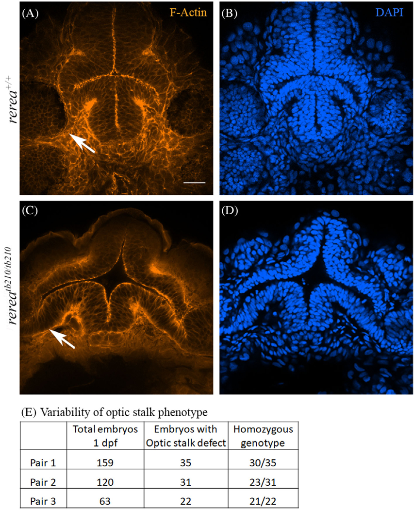
Phalloidin and DAPI stained WT (A, B) and rerea (C, D) mutant zebrafish embryos (~30 hpf) imaged from the ventral side showing optic stalk (white arrows).
Table showing variability in optic stalk phenotype (E). Scale bar is 20 μm.
The overall growth of 3 days post fertilization (dpf) WT and mutant rerea larvae was comparable (Figure 3A,E), whereas the optic fissure margins remained unfused in the mutant (Figure 3A–D,E–H). Histology of 3 dpf larval eyes demonstrated presence of periocular mesenchymal tissue within the optic fissure margins, suggesting this tissue may be obstructing the edges of the fissure from meeting and fusing (Figure 3D,H). Overall organization of the optic cup (Figure 3C,G) and optic fissure margins (Figure 3D,H) was observed to be normal, suggesting that the optic fissure closure defect could be due to a combinatorial effect of delayed morphogenetic movements (as margins were apart at 24 hpf, as shown in Figure 1E,J) and physical obstruction of the optic fissure margins. By 3 dpf, retinal pigment epithelium (RPE) differentiation was morphologically complete in both WT and rerea mutant larvae (Figure 3C,G). We observed variability in the penetrance of the coloboma phenotype and the table 3I provides the data from three heterozygous breeding pairs for percentage of embryos with coloboma. Plaster et al previously noted microphthalmia in homozygous rerea mutant fish embryos at 4 dpf.3 To quantify this defect, we measured the area of the zebrafish eye at 3 and 5 dpf; although rerea mutant eyes were smaller than WT at 3 dpf (P < .05), this difference was not observed at 5 dpf (Figure 3J). The coloboma phenotype was partially penetrant with only 70% of the homozygous mutants displaying the phenotype, as observed on 5 dpf. The original rereawt/tb210 line obtained from ZIRC was out-crossed to WT ABTL for three generations including different transgenic lines and coloboma was consistently observed regardless of the background (data not shown). For this manuscript, we focused more on defects observed during optic fissure closure and defects observed in the retinal ganglion cell (RGC) layer are being further investigated.
FIGURE 3.
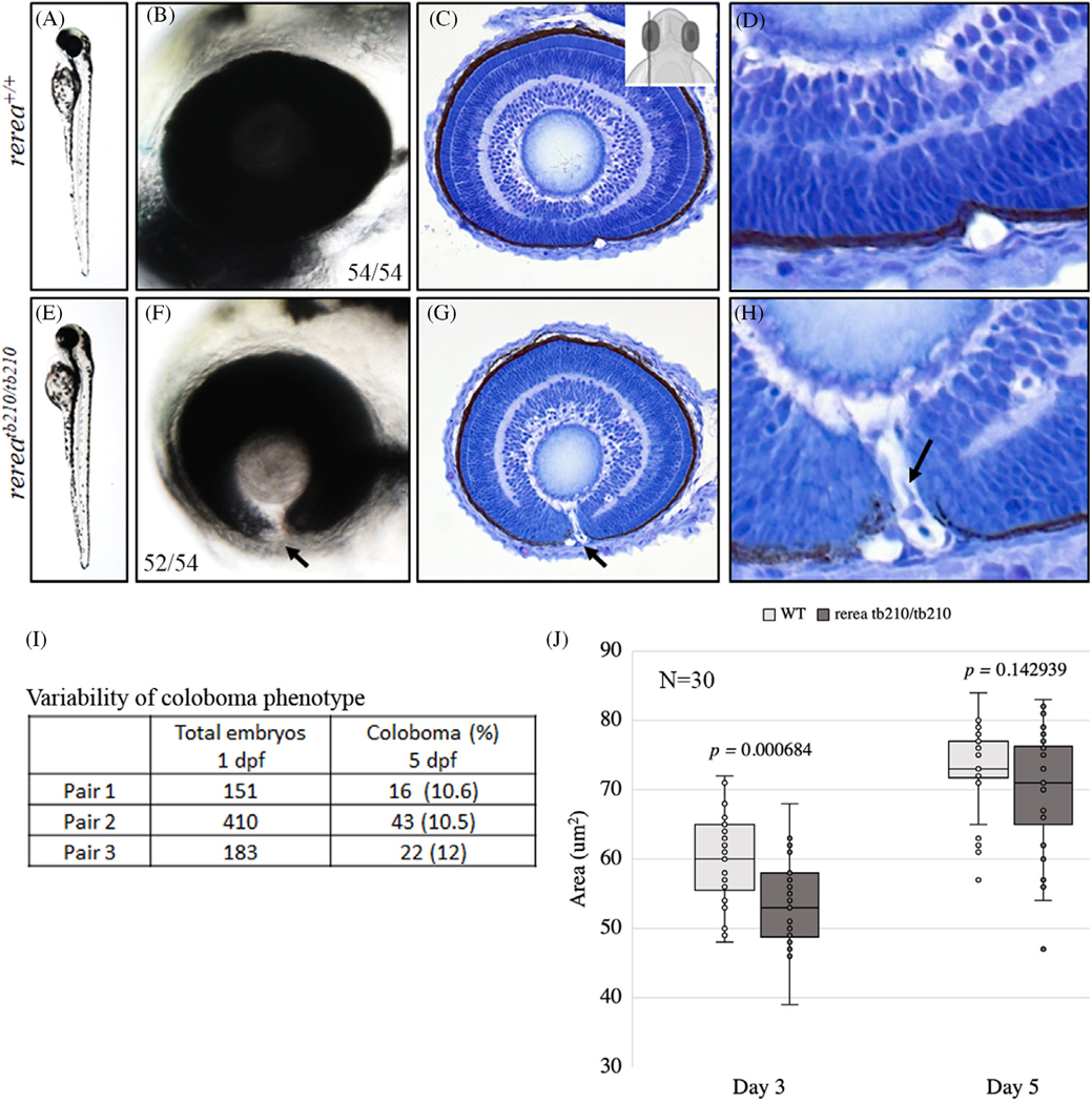
Zebrafish rerea homozygous mutants exhibit coloboma. Three-day post fertilization WT zebrafish embryos (A, B) exhibit fused optic fissure margins (54 genotyped embryos), whereas rerea homozygous mutants exhibit coloboma (E, F, 52/54 genotyped embryos). Sagittal histological sectioning of WT (C, D, N = 10 genotyped embryos) and rerea mutant (G, H, N = 10 genotyped embryos) zebrafish eye (3 dpf). The schematic in C gives the approximate plane of sectioning for the histologic sections. Coloboma showed variable penetrance in embryos at 5 dpf (I). Measurement of eye area (lateral view, Figure 2K) revealed significantly smaller eye at 3 dpf (P < .05) of rerea mutant larvae compared with WT. No statistical difference in eye size was observed at 5 dpf (J). Student’s t test was used to determine level of significance and P value is provided (N = 30 genotyped embryos/group).
2.2 |. Rerea mutant zebrafish exhibits disruption of retinal/OS boundary
The RPE and neural retina laminae are limited to the optic cup/OS boundary, with axons from RGCs passing along the path of the OS to synapse in the diencephalon (Figure 4A–C). In 5 dpf rerea mutant larvae, however, retinal laminae and, to a lesser extent, RPE, extend well into the region of OS and into the ventral diencephalon, in approximately 70% (~17% of total embryos derived from three heterozygous crosses, 92/522) of the mutant embryos. This abnormal extension of retinal tissue was pronounced and led to a large opening at the back of the eye (Figure 4A–E, two red arrow in panel D), similar to the previously reported “blowout” phenotype12 observed in ptc2 mutant. A similar observation has been made for the ptc2 mutants where only 3% to 22% of total embryos display the blowout phenotype from heterozygous mating; variability was noted between clutches.12 Unfused margins of the optic fissure (Figure 4A,D, single red arrow) could be observed as late as 6.5 dpf. The retinal tissue invading the ventral diencephalon appeared fully differentiated and immunostaining for ZPR-1 (photoreceptor) and HuC/D (amacrine and ganglion cell layer) was observed in the ectopic retinal tissue (Figure 4C,F). Other retinal cell layers including the choroid, retinal pigment epithelium, photoreceptors, inner nuclear layer, and other aspects of eye development including lens formation appeared morphologically normal, as observed using histology (Figure 4B,E). Interestingly, the disrupted retinal/OS boundary phenotype is not completely penetrant in rerea mutants, which is also the case for blowout phenotype associated with the homozygous ptc2 mutations. These observations suggest the possibility of shared mechanisms between rerea and ptc2 mutants causing the blowout-like phenotype.
FIGURE 4.
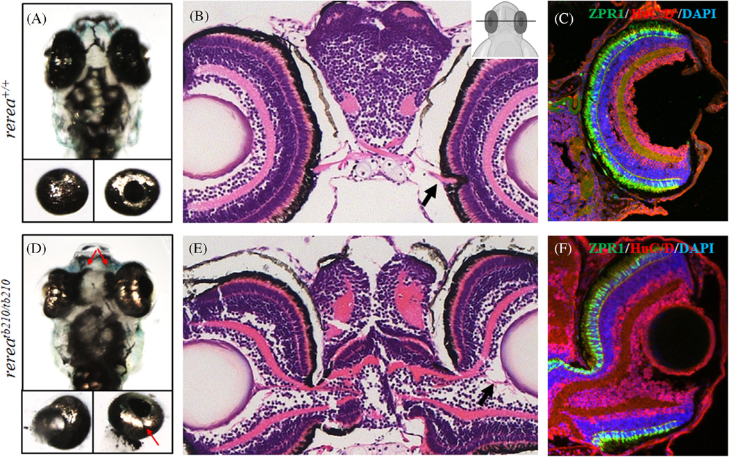
Zebrafish rerea homozygous mutants exhibit disruption of retinal optic stalk boundary. Zebrafish larvae homozygous for rerea mutation exhibit disruption of retinal optic stalk boundary (A–C, D–F), which was further confirmed by isolation of eye cups (A, D, bottom panels) and detailed coronal histological sectioning (B, E) of 6 dpf larvae. Unfused margins of optic fissure can be observed (D bottom panel, red arrow). The presence of ectopic differentiated retinal layers (C, F) was confirmed by immunostaining with ZPR-1 (photoreceptors) and HuC/D (amacrine cells and ganglion cells).
2.3 |. Rerea mutation affects OS patterning
The ocular phenotype displayed by rerea mutants is highly reminiscent of the zebrafish blowout and aus mutant,12,13 where upregulation of pax2a and fgf8 is associated with optic cup, stalk, and nerve defects. A phenotype similar to blowout was reported for the aus mutant, for which the underlying genetic cause has not been identified; according to authors, the mutation was hypothesized to be in a gene regulating fgf8 expression.13 Interestingly, Fgf8 is a known target of Rere-mediated repression during mouse somitogenesis18 and rerea mutant zebrafish embryos exhibit deregulated fgf8 signaling.3 Also, Fgf8 is known to pattern the OS and neural retina as well as the differentiation of RGCs in zebrafish and chick embryos.19 Because of the enlargement of the OS and presence of extraocular retinal tissue, we hypothesized that rerea mutations might affect fgf8 expression. We performed whole mount in situ hybridization (WISH) of zebrafish embryos at 24 hpf and observed expanded fgf8 expression domains in the OS region of rerea mutant embryos (Figure 5A–D, black arrows). No change was observed in the expression pattern of fgf3 (data not shown). Similarly, pax2a, one of the most well-studied markers of OS, was expanded toward the anterior midline (Figure 5E,G, black arrows) and ventral retina (Figure 5F,H, black arrow) in rerea mutant embryos compared with WT at 24 hpf. Loss of pax2 expression is strongly associated with coloboma in humans, mice, and zebrafish (23, 24). Previous studies on zebrafish and chick embryos have shown that upregulation of fgf8 and/or pax2 can also result in coloboma.13,20 The exact mechanism how pax2 over expression leads to coloboma is unknown, but persistence of a Pax2+ glial scar in the optic fissure has been suggested.20
FIGURE 5.

Whole mount in situ hybridization staining of zebrafish rerea homozygous mutants exhibit expanded expression domains of optic stalk markers fgf8 and pax2a. Dorsal and lateral views of 24 hpf zebrafish embryos stained with fgf8 (A–D) and pax2a (E–H) exhibited expanded expression domains (arrows) in rerea mutants (C, D, G, H) as compared with WT (A, B, E, F) embryos
2.4 |. Proximal-distal and dorsoventral symmetry of eye is disrupted in rere mutant embryos
The disruption of the retinal/OS boundary and the presence of the blowout-like phenotype in the rerea mutant larvae suggested that rerea might be involved in regulating the retinal/OS boundary directly or indirectly. Vax1 and vax2, genes under the control of shh and/or fgf signaling, have been implicated in the maintenance of retinal/OS boundary by regulating proximal-distal patterning of the eye.17 A previous study in Drosophila and zebrafish morphants suggested the role of rerea in regulating shh signaling target genes.8 In WT zebrafish at ~24 hpf, after the initial eye patterning is complete, vax1 was observed to be expressed strongly in the OS area (black arrow, Figure 6A,B), vax2 was prominently expressed in the pre optic area (POA), ventral retina, and weakly in the OS (data not shown). In rerea mutant embryos, vax1 expression was reduced in the OS (Figure 6A,C), but the expression domain expanded from the OS region into the ventral retina (Figure 6B,D, white arrow). Vax2 expression was not changed between the WT and rerea mutant embryos (data not shown). These observations suggest that vax genes expression is differentially affected in rerea mutant embryos.
FIGURE 6.

Zebrafish rerea mutants exhibit disruption of proximo-distal patterning. Whole mount in situ hybridization staining of 24 hpf zebrafish embryos with vax1, aldh1a2, aldh1a3, and nlz1 probes. Numbers of embryos with patterns of staining similar to that pictured are given in each panel
Zebrafish rerea mutants display patterning defects in dorsoventral symmetry of telencephalon, leading to dorsalization of telecephalon.3 Ventral optic cup defects like coloboma can be caused by dorsalization of the optic cup.20,21 As such, we studied the expression pattern of dorsal (aldh1a2) and ventral (aldh1a3 and nlz1) optic cup markers (Figure 6E–H) during optic cup morphogenesis at 24 hpf. We observed expression of aldh1a2 in the dorsal optic cup, where the expression domain of aldh1a2 was expanded in mutant embryos compared with WT (Figure 6E,G). Ventral optic cup marker nlz1 was significantly down-regulated in rerea mutant embryos compared with WT embryos (Figure 6F,H, arrow). On the contrary, aldh1a3, another important marker for ventral optic cup expressed at the apposing edges of the optic fissure margin, remained unaffected (Figure 6E,G). The optic fissure margins stained with aldh1a3 were observed to be separated in the rerea mutants in contrast to WT embryos, further providing evidence of lack of optic fissure closure. Since we observed consistent expansion of aldh1a2 and downregulation of nlz1, we concluded that the optic cup could be dorsalized in rerea mutants. Thus, ventral optic cup defects observed in rerea mutant could also be due to dorsalization of the optic cup and expansion of proximal retinal structures like OS, and reduced expression of some of the ventral fate genes.
2.5 |. Human WT RERE mRNA rescue coloboma in rerea morphant zebrafish
Fregeau et al had reported a cohort of 10 patients with de novo mutations in RERE exhibiting partially penetrant ophthalmic features including cerebral-visual defects, coloboma, and microphthalmia.1 We decided to characterize the variant of RERE (c.3466 G > A, p.Gly1156Arg) that was associated with coloboma. First, we performed a dose response assay with rerea morpholino and observed an increased percentage of coloboma with increasing morpholino concentration (Figure 7A). The control morpholino injected at the highest tested rerea morpholino concentration did not display coloboma (Figure 7A,B). To ensure that coloboma phenotype displayed by the morphants was mechanistically similar to the mutants, we performed in situ staining for pax2a on control and morphant rerea embryos and observed its upregulation in the OS region (Figure 7C) of the rerea morphants. Downregulation of pax6 in the presumptive neural retina and nlz1 in the ventral optic cup was also observed in the in rerea morphants at 24 hpf (data not shown). Expression of aldh1a2 was observed in the dorsal region of the optic cup with expansion in the rerea morphants. Aldh1a3 expression domain, which marks the optic fissure margins, was not affected in rerea morphants as compared with WT embryos. These observations are consistent with what was observed in rerea mutant embryos and suggests a shared mechanism for coloboma in mutants and morphants.
FIGURE 7.

Morpholino-mediated knockdown of rerea causes coloboma. Increasing concentrations of rerea morpholino was used for knocking down rerea expression (A). Severity of coloboma is denoted by: CI-mild, CII-moderate, CIII-severe. Zebrafish embryos injected with rerea morpholino consistently exhibited coloboma at 48 hpf compared with embryos injected with control morpholino (B). Representative whole mount in situ hybridization staining with pax2a in control and rerea morpholino injected embryos 24 hpf (n ~20 embryos each) (C). WT human RERE mRNA rescues coloboma in rerea morphant fish embryos (D), this rescue was observed to be statistically significant (P < .05) for 20, 40, and 80 pg of mRNA injected, but not for 10 pg. Equal amounts of the human RERE WT and mutant (c.3466 G > A, p.Gly1156Arg) mRNA (40 pg) can rescue coloboma in rerea morphants (P < .05), but the mutant mRNA is not as efficient as WT mRNA (P < .05, E). Student’s t test was used to determine level of significance and “*” denotes significant difference. NS, not significant
Next, we injected one-cell stage zebrafish embryos with rerea morpholino (5 ng) and increasing dose (10–80 pg) of WT human RERE mRNA and observed a dose-dependent significant rescue of the coloboma phenotype at 20, 40, and 80 pg of the mRNA injected but not at 10 pg (Figure 7D; P ≤ .05). Injection of human RERE mutant (c.3466 G > A, p.Gly1156Arg) mRNA (40 pg) also resulted in a slight (~20%) rescue of the coloboma phenotype but was not as efficient as WT mRNA (~40%, Figure 7E), suggesting that the mutant mRNA is a hypomorphic allele. Interestingly, injection of up to 80 pg of WT human RERE mRNA along with rerea morpholino consistently induced cyclopia in the zebrafish embryos, albeit at a lower frequency (~10%). Cylcopia is a phenotype associated with midline sonic hedgehog (shh) signaling,22 suggesting that RERE might play a role in this pathway, either through changes to the ligand or its downstream components. This observation is also in agreement with previous observations suggesting Atro (homologous to vertebrate rere) and Cubitus interruptus (Ci) (homologous to vertebrate gli1/2/3) mediated regulation of Shh signaling in Drosophila.8
2.6 |. Suppression of shh/gli signaling rescues coloboma in rerea mutants
To further probe the relationship between rerea and hedgehog signaling, we injected WT human RERE mRNA in one-cell stage zebrafish embryos and observed a dose-dependent increase in the cyclopia phenotype, suggesting that RERE indeed suppress shh signaling (Figure 8A–C). Unilateral and/or bilateral anophthalmia and microphthalmia was also observed with human RERE mRNA injection at all doses (Figure 8A). Patched, the receptor for and a target of shh signaling, demonstrated increased expression in rerea mutant embryos at 24 hpf compared with WT embryos (Figure 8D,F), further confirming upregulation of shh signaling. In situ hybridization, however, demonstrated no upregulation of shha transcripts in rerea mutants and morphants, at 24 hpf (Figure 8E,G and data not shown), pointing to a downstream effect. Cyclopamine, a smoothened inhibitor, has been previously used to rescue the coloboma phenotype associated with upregulated shh signaling.12,15,16 We used cyclopamine (5 μM treatment from 5.5 to 13.5 hpf) to rescue the coloboma phenotype in rerea mutant embryos but did not observe any rescue (data not shown). Higher doses of cyclopamine (100 μM) did cause cyclopia in embryos obtained from the same breeding (data not shown). This observation was in contrast to what has been observed previously with other alterations of the pathway,12,15,16 where the coloboma phenotype due to upregulated shh signaling was significantly rescued by cyclopamine treatment from 5.5 to 13.5 hpf.12 Also, treatment of rerea heterozygous and homozygous mutant embryos with smoothened agonist purmorphamine (75 μM, 5.5–24 hpf) did not increase the percentage of embryos displaying coloboma or the severity of phenotype (data not shown), as has been reported previously.15 These observations suggested that deregulation of shh signaling is occurring downstream of patched and smoothened. Consistent with this hypothesis, Zhou et al had reported that Drosophila Atro represses shh signaling by interacting with Ci,8 a point downstream of the molecules we addressed above. We used shh pathway inhibitors HPI-1 and HPI-4 that act at the level of gli proteins (gli1, gli2, and gli3), to repress upregulated shh signaling.23 HPI-4 treatment resulted in significant embryo death and could not be used for rescue experiments.
FIGURE 8.

Injection of WT human RERE mRNA results in cyclopia. Injecting increasing concentration of human RERE mRNA results in dose-dependent increase in cyclopia phenotype (A–C). Whole mount in situ hybridization staining of 24 hpf WT and rerea mutant zebrafish embryos with patched2 (D, F) and shha (E, G) probes. Representative images with numbers of embryos showing this pattern are given in each panel.
Heterozygous rerea mutant zebrafish were crossed, and embryos were randomly allocated to DMSO and HPI-1 (5 μM) group. Drug treatment was started 5.5 hpf and continued till 3 dpf, unlike previous reports from other groups where cyclopamine treatment was stopped at 13.5 hpf.12 Embryo media was changed every 24 hours and fresh drug was added. Homozygous mutant embryos were scored starting from 3 to 5 dpf (since the mutant phenotype was observed to get severe with time and is easily observable) based on the presence of fin defects, absence of swim bladder, curved body, and presence or absence of coloboma. We observed significant rescue of the coloboma phenotype with gli inhibitor HPI-1 (P = .014, 17.2% vs 7.2%; Figure 9, arrow). Of note, HPI-1 only rescued the coloboma phenotype but not the fin or swim bladder defects (Figure 9, arrowhead) that were also observed in the rerea mutant embryos. Moreover, a similar treatment regime with cyclopamine was unable to rescue the coloboma defect (data not shown). Taken together, these experiments suggest that deregulated shh signaling in rerea mutant embryos could be occurring at the level of gli proteins as the HPI-1 drug specifically inhibits Gli-induced Hh pathway activation.23 Given the role of rerea in fgf, shh, RA, and notch signaling, we cannot exclude the possibility of the contribution of these signaling pathways toward optic fissure closure defects observed with rerea mutations.
FIGURE 9.

Shh signaling inhibitor HPI-1 rescues the blowout coloboma phenotype in rerea mutant zebrafish embryos. Treatment of rerea mutant zebrafish embryos with HPI-1 (5 μM) reduces the occurrence of coloboma phenotype from 71.3% to 26.3% (Student’s t test, P < .05) coloboma in homozygous mutant embryos.
2.7 |. In vivo and in vitro characterization of human RERE syndrome associated variants
As shown above, overexpression of human RERE in the zebrafish embryos can cause cyclopia phenotype, suggesting suppression of shh signaling. A similar observation was also made upon injection of sox11 and sox4 mRNA, where overexpression resulted in cyclopia.15,16 We used this assay to determine the functionality of human RERE variants in vivo. Equimolar amounts of synthesized mRNA of human WT and mutant RERE isoforms were injected in one-cell stage zebrafish embryos to test their efficiency to cause cyclopia and unilateral or bilateral anophthalmia. All of the variants except p.Gln760* were not as efficient as the human WT rerea mRNA in causing cyclopia, and this difference was observed to be significant (P ≤ .05) prior to Bonferroni correction, whereas post correction only three variants (p.Val471Ile, p.Pro1041Lysfs*40, and p.Leu369Cysfs*16) exhibited significant difference (Figure 10A), suggesting their reduced potential in suppressing shh/gli signaling.
FIGURE 10.

Human RERE variants exhibit hypomorphic activity in vivo and mis-localized expression in vitro. In vivo assay to determine the efficiency of human RERE variants (80 pg) to cause cyclopia in zebrafish model (A, χ2 test was used to determine level of significance and “*” denotes significant difference). In vitro assay to determine the nuclear localization pattern of human RERE wild type (WT, B) and mutant proteins (C) in HEK293 cells. Scale bar is 10 μm.
To determine how the mutations associated with RERE syndrome affected the RERE protein localization, we performed in vitro characterization of the mutant proteins. RERE protein has been reported to localize in the nucleus in a characteristic punctate pattern.5 Upon overexpression of human WT and mutant RERE protein isoforms in HEK293 cells, it translocated to the nuclei (Figure 10B, blue) and displayed a punctate staining pattern (Figure 10B, red) in the nucleus. Interestingly, all the missense RERE variants did localize to the nucleus and displayed characteristic punctate staining, whereas all the truncating variants with frame-shift mutations did not exhibit the characteristic punctate staining in the nuclei (Figure 10C), suggesting that the targeting of RERE to specific regions of the genome was disrupted and/or was unable to form a co-repressor/activator complex with partner proteins. The nuclear targeting of truncating mutations was not affected as all of them did exhibit strong nuclear localization.
3 |. DISCUSSION
The developmental origins of structural ophthalmic defects like coloboma are poorly understood, both in isolated and syndromic forms of congenital conditions. Mouse and zebrafish models of human genetic disorders can be generated but may not faithfully replicate all aspects of the human disease phenotype. This is especially true for syndromic forms of human genetic disorders where multiple organ systems are affected. Thus, identifying appropriate model systems for studying phenotypes associated with syndromic genetic disorders is essential to understand the underlying mechanisms and find potential targets for rescuing those defects. Fregeau et al reported a syndromic form of neurodevelopmental condition associated with de novo mutations in transcriptional co-repressor RERE, also presenting with partially penetrant anatomical visual pathway defects like microphthalmia, coloboma, optic nerve defects, associated anterior commissure and CNS anomalies, and cerebral visual defects.1 Here, we describe in detail the developmental ophthalmic defects displayed by rerea mutant zebrafish, study the possible mechanisms involved, and identified a small molecule inhibitor that can be used to rescue coloboma, one of the major developmental ophthalmic defects associated with rerea mutation.
During initial stages of development in rerea mutants, the OS is enlarged, but the optic cup appears morphologically normal (Figure 1) and subsequent differentiation of retinal layers is also normal (Figure 3). However, the choroid fissure fails to fuse causing coloboma, due, at least in part, to the presence of periocular mesenchymal tissue in the optic fissure (Figure 3H). Although we did observe developmental delay in differentiation of inner and outer nuclear layers (Figure 3C,G) in rerea mutant embryos, the coloboma can be observed as late as 7 dpf, making delay less likely as a cause of this phenotype. We also show that this defect is due to the expanded ectopic OS in the form of pax2a and fgf8 expression domains (Figure 5) and reduced expression of some of the ventral retinal fate genes like nlz1 (Figure 6). A similar phenomenon has been observed in chick embryos where ectopic expression of Pax2 results in coloboma.20 The loss of function mutations of PAX2 is known to cause coloboma in human, mouse, and zebrafish.24 However, coloboma caused by ectopic expression of Pax2 as observed in chick follows a completely different mechanism compared to what is caused by loss of function.20 Ectopic expression of Pax2 results in a glial scar at the optic fissure which acts as a physical barrier inhibiting the fusion of optic fissure margins.20 Reduction of retinal markers like nlz1 within the ventral region of the retina in rerea mutants may also underlie the defects in optic fissure closure. The ventral optic cup defects could also be due to over-dorsalization of the eye field, and we observed expansion of aldh1a2 expression domains in the dorsal optic cup, and downregulation of ventral optic cup marker (nlz1, Figure 6).
Previous studies in mouse, zebrafish, and chick embryos have suggested that shh promotes pax2 expression via fgf8.25 Additionally, rerea also repress shh signaling,8 suggesting multiple mechanisms that may result in fgf8 and pax2 upregulation. An important role of shh signaling in the OV is to promote proximal cell fates like OS and repress distal cell fates (i.e., RPE, retina, and lens). Expression of patched2 in the OS and disruption of retinal/OS boundary in patched2 mutants further confirms the important role of shh signaling in determining patterning of retinal/OS boundary.12 The transcription factor Pax2 is an shh target gene in the OS26–29 and overexpression of shh is sufficient to induce the expression of pax2 in more distal optic vesicle territories where it is normally absent22,27,28,30 and suppression of pax6.22,30 Another set of genes that are influenced by shh signaling, directly or via fgf8 are, vax1 and vax2. Both vax1 and vax2 are expressed in the OS and morpholino mediated knockdown of both results in the disruption of neural retina/OS boundary.17 Given the importance of shh signaling in patterning of OS and neural retina, we tried to rescue the coloboma phenotype by suppressing shh signaling by cyclopamine as has been previously reported.12,15,16 The coloboma associated with rerea mutants was not rescued by cyclopamine nor increased in severity by treatment with smoothened agonist purmorphamine (data not shown), suggesting that rerea acts downstream of shh, patched, and smoothened. It has been reported previously that Drosophila Atro (homologous to vertebrate Rere) interacts with Ci (homologous to vertebrate Gli) to inhibit shh signaling; therefore, we used a gli inhibitor HPI-1 to rescue coloboma in rerea mutants. Since, HPI-1 can inhibit both gli1 and gli2, the specific Gli protein(s) involved in this process are currently under investigation. Of note, WT zebrafish embryos can tolerate high concentration of HPI-1 (50 μM) treatment without exhibiting any obvious structural defects. Recently, Gordon et al reported that coloboma due to ptch2 mutation can be rescued by introducing heterozygous or homozygous gli1 mutation, suggesting the role of shh/gli1-mediated signaling in the coloboma phenotype.31 Transcriptional co-repression activity of RERE is mediated by its interaction with HDAC1, and HDAC1 mutant zebrafish exhibit similar ophthalmic defects including an enlarged OS, coloboma, and blowout-like phenotype.32 Suggesting that transcriptional co-repression mediated by RERE and HDAC1 can also be an underlying mechanism for optic fissure closure defects.
In conclusion, we show that expanded fgf8 and pax2a domains in rerea mutant zebrafish embryos is associated with a persistent OS and disruption of retinal/OS boundary, affecting specification of ventral retinal fates that leads to coloboma. This upregulation of fgf8 and pax2a is mediated by de-repression of shh signaling, but not necessarily shh itself and could be mediated via shh/gli pathway. This is further substantiated by rescues of coloboma phenotype in rerea mutants by repressing shh/gli signaling using a small molecule inhibitor HPI-1.
4 |. EXPERIMENTAL PROCEDURES
The study was carried out in accordance with the recommendations of Guide for the Care and Use of Laboratory Animals of the National Institutes of Health. All animal experiments were conducted under protocols approved by the National Eye Institute’s Animal Care and Use Committee (NEI ACUC) at the National Institutes of Health (ASP#NEI-648).
4.1 |. Zebrafish maintenance and breeding
Adult zebrafish were maintained in an automated fish housing system following a standard protocol.33 Embryos were maintained at 28.5°C in fish embryo medium as previously described.34 The reretb210 heterozygous adults (ZIRC, Eugene, OR) were outcrossed to WT ABTL strain for three generations.
4.2 |. Zebrafish histology and immunostaining of cryo-sections
Plastic, paraffin sectioning and cryo-sectioning followed by immunostaining was performed as described previously.35,36 Antibodies used for immunostaining: ZPR-1 (Abcam: ab174435) and HuC/D (Abcam: ab210554). For immuno-fluorescence staining zebrafish larvae were fixed in 4% paraformaldehyde and stained with phalloidin (Abcam: ab176756) to stain for cell boundaries. Stained embryos were embedded in cover-glass bottom four-well chamber slides using 1.5% low melting agarose and imaged from the ventral side (ZEISS LSM 800; ×40 lens) of the embryos by collecting z-stacks with a step size of 0.5 μm. The images were analyzed using ZEN Software (Carl Zeiss Microscopy LLC, Thornwood, NY).
4.3 |. Morpholino and RNA injection
Splice-blocking morpholino oligonucleotides (MO, Gene Tools LLC., Philomath, OR) that have been reported previously2 diluted in 0.1 M KCl, nuclease free water and phenol red, were injected in freshly fertilized zebrafish embryos at one-cell stage. WT and mutant human RERE (Genewiz LLC., NJ) open reading frame were used to in vitro synthesize mRNA for injections (mMACHINE kit, Ambion, Grand Island, NY). In morpholino rescue experiments, the embryos were co-injected with rerea MO and in vitro synthesized WT and mutant human RERE mRNA (0–80 pg of mRNA). For testing the ability of human variants to induce cyclopia, 80 pg of each of the variant and WT mRNA was injected.
4.4 |. Whole mount in situ hybridization and brightfield imaging
Antisense RNA probes were synthesized from cDNA IMAGE clones (Open Biosystems) using digoxigenin RNA-labeling kit (Roche, Indianapolis, IN). Whole mount in situ hybridization was carried out as described earlier34 at 65°C using probes for fgf8, pax2a, vax1, pax6, nlz1, aldh1a2, aldh1a3, shha and patched. In situ embryos were washed and mounted in glycerol on a glass slide after removal of the yolk sac and imaged using an upright brightfield microscope (Zeiss Imager 1). Twenty embryos per group (WT and rerea mutant) were stained for each probe. DNA was isolated from individual embryos by incubating them in 10 μL NaOH (50 mM) at 95°C for 10 minutes and then at 4°C for 5 minutes. Subsequently, 1 μL of Tris-HCl (1 M, pH 8.0) was added to lysate and vortexed thoroughly. The resulting mix was diluted 1:10 before being used for PCR amplification (1–2 μL/25 μL reaction mix). Individual embryos were genotyped by Sanger sequencing (Psomagen, Rockville, MD) using primers described previously.3 For measurement of eye area, lateral view brightfield images of 30 genotyped zebrafish embryos were acquired at same magnification, and free hand tool of image J was used to mark the boundary of the eye and the area was measured. Zebrafish schematics were created with BioRender.com.
4.5 |. Cell culture and transfection experiments
HEK293T cells (~1 × 10⁴/well) maintained in DMEM with 10% FBS and 1% penicillin to streptomycin were seeded onto four-well chamber slides, maintained for 24 hours, and transiently transfected with RERE plasmid (WT or mutant) constructs using X-treme Gene HP (Roche, Indianapolis, IN, USA) following manufacturer’s instructions. Twenty-four hours post transfection, transfected cells were fixed for 15 minutes in 4% paraformaldehyde in PBST. After washing with 1× PBST and permeabilization and blocking in ICC buffer (0.5% BSA 0.5% Tween and 0.1% triton X100 1× PBS). Cells were then incubated overnight at 4°C with the primary antibody in ICC buffer. After multiple washes in PBST, the cells were incubated for 1 hour at room temperature in Alexa488 or 555 conjugated goat anti-rabbit and/or anti-mouse antibody and Hoechst33342 (1:1000 dilution in ICC buffer). Cells were then washed in PBST before mounting with Fluoromount-G (SouthernBiotech, Birmingham, AL, USA) imaging. Primary antibodies used were RERE (Novus: H00000473-M06).
4.6 |. Statistical analysis
All experiments where a statistical test was performed, represent three replicates with sample number (N) provided, except for Figure 10A where samples were pooled. Microsoft Excel was used to perform appropriate analyses (Student’s t and χ2 test) and level of significance is indicated in respective figure legends and text.
ACKNOWLEDGMENTS
Dr. Robert Farris (NEI, Biological Imaging Core) for expert assistance with confocal laser scanning microscopy. Andrew Wegerski for zebrafish breeding and maintenance. This work was supported by the intramural program of the National Eye Institute.
Funding information
National Eye Institute, Grant/Award Number: Intramural program
Footnotes
CONFLICT OF INTEREST
The authors declare no competing financial interests.
REFERENCES
- 1.Fregeau B, Kim BJ, Hernández-García A, et al. De novo mutations of RERE cause a genetic syndrome with features that overlap those associated with proximal 1p36 deletions. Am J Hum Genet. 2016;98(5):963–970. doi: 10.1016/j.ajhg.2016.03.002 [DOI] [PMC free article] [PubMed] [Google Scholar]
- 2.Asai Y, Chan DK, Starr CJ, Kappler JA, Kollmar R, Hudspeth AJ. Mutation of the atrophin2 gene in the zebrafish disrupts signaling by fibroblast growth factor during development of the inner ear. Proc Natl Acad Sci. 2006;103(24):9069–9074. doi: 10.1073/pnas.0603453103 [DOI] [PMC free article] [PubMed] [Google Scholar]
- 3.Plaster N, Sonntag C, Schilling TF, Hammerschmidt M. REREa/Atrophin-2 interacts with histone deacetylase and Fgf8 signaling to regulate multiple processes of zebrafish development. Dev Dyn. 2007;236(7):1891–1904. doi: 10.1002/dvdy.21196 [DOI] [PubMed] [Google Scholar]
- 4.Neuhauss SC, Biehlmaier O, Seeliger MW, et al. Genetic disorders of vision revealed by a behavioral screen of 400 essential loci in zebrafish. J Neurosci. 1999;19(19):8603–8615. [DOI] [PMC free article] [PubMed] [Google Scholar]
- 5.Wang L, Charroux B, Kerridge S, Tsai CC. Atrophin recruits HDAC1/2 and G9a to modify histone H3K9 and to determine cell fates. EMBO Rep. 2008;9(6):555–562. doi: 10.1038/embor.2008.67 [DOI] [PMC free article] [PubMed] [Google Scholar]
- 6.Hou R, Sibinga NES. Atrophin proteins interact with the Fat1 cadherin and regulate migration and orientation in vascular smooth muscle cells. J Biol Chem. 2009;284(11):6955–6965. doi: 10.1074/jbc.M809333200 [DOI] [PMC free article] [PubMed] [Google Scholar]
- 7.Vilhais-Neto GC, Maruhashi M, Smith KT, et al. Rere controls retinoic acid signalling and somite bilateral symmetry. Nature. 2010;463(7283):953–957. doi: 10.1038/nature08763 [DOI] [PubMed] [Google Scholar]
- 8.Zhang Z, Feng J, Pan C, et al. Atrophin-Rpd3 complex represses hedgehog signaling by acting as a corepressor of CiR. J Cell Biol. 2013;203(4):575–583. doi: 10.1083/jcb.201306012 [DOI] [PMC free article] [PubMed] [Google Scholar]
- 9.Wang H, Gui H, Rallo MS, Xu Z, Matise MP. Atrophin protein RERE positively regulates notch targets in the developing vertebrate spinal cord. J Neurochem. 2017;141(3):347–357. doi: 10.1111/jnc.13969 [DOI] [PubMed] [Google Scholar]
- 10.Lahrouchi N, George A, Ratbi I, et al. Homozygous frameshift mutations in FAT1 cause a syndrome characterized by colobomatous-microphthalmia, ptosis, nephropathy and syndactyly. Nat Commun. 2019;10(1):1180. doi: 10.1038/s41467-019-08547-w [DOI] [PMC free article] [PubMed] [Google Scholar]
- 11.Kim BJ, Zaveri HP, Shchelochkov OA, et al. An allelic series of mice reveals a role for RERE in the development of multiple organs affected in chromosome 1p36 deletions. PLoS One. 2013;8(2):e57460. doi: 10.1371/journal.pone.0057460 [DOI] [PMC free article] [PubMed] [Google Scholar]
- 12.Lee J, Willer JR, Willer GB, Smith K, Gregg RG, Gross JM. Zebrafish blowout provides genetic evidence for Patched1-mediated negative regulation of hedgehog signaling within the proximal optic vesicle of the vertebrate eye. Dev Biol. 2008;319(1):10–22. doi: 10.1016/j.ydbio.2008.03.035 [DOI] [PMC free article] [PubMed] [Google Scholar]
- 13.Heisenberg CP, Brennan C, Wilson SW. Zebrafish aussicht mutant embryos exhibit widespread overexpression of ace (fgf8) and coincident defects in CNS development. Development. 1999;126(10):2129–2140. doi: 10.1242/dev.126.10.2129 [DOI] [PubMed] [Google Scholar]
- 14.Samuel A, Rubinstein AM, Azar TT, Ben-Moshe Livne Z, Kim SH, Inbal A. Six3 regulates optic nerve development via multiple mechanisms. Sci Rep. 2016;6:20267. doi: 10.1038/srep20267 [DOI] [PMC free article] [PubMed] [Google Scholar]
- 15.Pillai-Kastoori L, Wen W, Wilson SG, et al. Sox11 is required to maintain proper levels of hedgehog signaling during vertebrate ocular morphogenesis. PLoS Genet. 2014;10(7):e1004491. doi: 10.1371/journal.pgen.1004491 [DOI] [PMC free article] [PubMed] [Google Scholar]
- 16.Wen W, Pillai-Kastoori L, Wilson SG, Morris AC. Sox4 regulates choroid fissure closure by limiting hedgehog signaling during ocular morphogenesis. Dev Biol. 2015;399(1):139–153. doi: 10.1016/j.ydbio.2014.12.026 [DOI] [PMC free article] [PubMed] [Google Scholar]
- 17.Take-uchi M, Clarke JDW, Wilson SW. Hedgehog signalling maintains the optic stalk-retinal interface through the regulation of vax gene activity. Development. 2003;130(5):955–968. doi: 10.1242/dev.00305 [DOI] [PubMed] [Google Scholar]
- 18.Kumar S, Duester G. Retinoic acid controls body axis extension by directly repressing Fgf8 transcription. Development. 2014; 141(15):2972–2977. doi: 10.1242/dev.112367 [DOI] [PMC free article] [PubMed] [Google Scholar]
- 19.Martinez-Morales JR, Del Bene F, Nica G, Hammerschmidt M, Bovolenta P, Wittbrodt J. Differentiation of the vertebrate retina is coordinated by an FGF signaling center. Dev Cell. 2005; 8(4):565–574. doi: 10.1016/j.devcel.2005.01.022 [DOI] [PubMed] [Google Scholar]
- 20.Sehgal R, Karcavich R, Carlson S, Belecky-Adams TL. Ectopic Pax2 expression in chick ventral optic cup phenocopies loss of Pax2 expression. Dev Biol. 2008;319(1):23–33. doi: 10.1016/j.ydbio.2008.03.041 [DOI] [PMC free article] [PubMed] [Google Scholar]
- 21.Wang X, Lupo G, He R, Barsacchi G, Harris WA, Liu Y. Dorsoventral patterning of the Xenopus eye involves differential temporal changes in the response of optic stalk and retinal progenitors to Hh signalling. Neural Dev. 2015;10(1):7. doi: 10.1186/s13064-015-0035-9 [DOI] [PMC free article] [PubMed] [Google Scholar]
- 22.Ekker SC, Ungar AR, Greenstein P, et al. Patterning activities of vertebrate hedgehog proteins in the developing eye and brain. Curr Biol. 1995;5(8):944–955. doi: 10.1016/s0960-9822(95)00185-0 [DOI] [PubMed] [Google Scholar]
- 23.Hyman JM, Firestone AJ, Heine VM, et al. Small-molecule inhibitors reveal multiple strategies for Hedgehog pathway blockade. Proc Natl Acad Sci U S A. 2009;106(33):14132–14137. doi: 10.1073/pnas.0907134106 [DOI] [PMC free article] [PubMed] [Google Scholar]
- 24.Schimmenti LA. Renal coloboma syndrome. Eur J Hum Genet. 2011;19(12):1207–1212. doi: 10.1038/ejhg.2011.102 [DOI] [PMC free article] [PubMed] [Google Scholar]
- 25.Kobayashi T, Yasuda K, Araki M. Coordinated regulation of dorsal bone morphogenetic protein 4 and ventral Sonic hedgehog signaling specifies the dorso-ventral polarity in the optic vesicle and governs ocular morphogenesis through fibroblast growth factor 8 upregulation. Dev Growth Differ. 2010;52(4):351–363. doi: 10.1111/j.1440-169X.2010.01170.x [DOI] [PubMed] [Google Scholar]
- 26.Chiang C, Litingtung Y, Lee E, et al. Cyclopia and defective axial patterning in mice lacking Sonic hedgehog gene function. Nature. 1996;383(6599):407–413. doi: 10.1038/383407a0 [DOI] [PubMed] [Google Scholar]
- 27.Perron M, Boy S, Amato MA, et al. A novel function for Hedgehog signalling in retinal pigment epithelium differentiation. Development. 2003;130(8):1565–1577. doi: 10.1242/dev.00391 [DOI] [PubMed] [Google Scholar]
- 28.Varga ZM, Amores A, Lewis KE, et al. Zebrafish smoothened functions in ventral neural tube specification and axon tract formation. Development. 2001;128(18):3497–3509. doi: 10.1242/dev.128.18.3497 [DOI] [PubMed] [Google Scholar]
- 29.Zhang XM, Yang XJ. Regulation of retinal ganglion cell production by Sonic hedgehog. Development. 2001;128(6):943–957. doi: 10.1242/dev.128.6.943 [DOI] [PMC free article] [PubMed] [Google Scholar]
- 30.Macdonald R, Barth KA, Xu Q, Holder N, Mikkola I, Wilson SW. Midline signalling is required for Pax gene regulation and patterning of the eyes. Development. 1995;121(10): 3267–3278. doi: 10.1242/dev.121.10.3267 [DOI] [PubMed] [Google Scholar]
- 31.Gordon HB, Lusk S, Carney KR, Wirick EO, Murray BF, Kwan KM. Hedgehog signaling regulates cell motility and optic fissure and stalk formation during vertebrate eye morphogenesis. Development. 2018;145(22):dev165068. doi: 10.1242/dev.165068 [DOI] [PMC free article] [PubMed] [Google Scholar]
- 32.Stadler JA, Shkumatava A, Norton WHJ, et al. Histone deacetylase 1 is required for cell cycle exit and differentiation in the zebrafish retina. Dev Dyn. 2005;233(3):883–889. doi: 10.1002/dvdy.20427 [DOI] [PubMed] [Google Scholar]
- 33.Westerfield M. The Zebrafish Book: A Guide for the Laboratory Use of Zebrafish (Danio Rerio). 4th ed. Eugene: University of Oregon Press; 2000. [Google Scholar]
- 34.Babcock HE, Dutta S, Alur RP, et al. aldh7a1 regulates eye and limb development in zebrafish. PLOS One. 2014;9(7):e101782. doi: 10.1371/journal.pone.0101782 [DOI] [PMC free article] [PubMed] [Google Scholar]
- 35.Sullivan-Brown J, Bisher ME, Burdine RD. Embedding, serial sectioning and staining of zebrafish embryos using JB-4 resin. Nat Protoc. 2011;6(1):46–55. doi: 10.1038/nprot.2010.165 [DOI] [PMC free article] [PubMed] [Google Scholar]
- 36.Uribe RA, Gross JM. Immunohistochemistry on cryosections from embryonic and adult zebrafish eyes. CSH Protoc. 2007; 2007:pdb.prot4779. doi: 10.1101/pdb.prot4779 [DOI] [PubMed] [Google Scholar]