Abstract
Objectives
Deep Brain Stimulation (DBS) is an effective, yet underused therapy for people living with Parkinson’s disease (PD) in whom tremor, motor fluctuations and/or dyskinesia are not satisfactorily controlled by oral medical therapy. Fear of vascular complications related to the operative procedure remains a strong reason for both the referrer and patient reluctance. We review the incidence of vascular complications in the first 600 patients with Parkinson’s disease treated at our centre by a single neurologist/neurosurgical team.
Methods
Surgical data routinely collected for patients who underwent DBS implantation for the management of PD between the years 2001–2023 was retrospectively reviewed. Incidences of vascular complication were analysed in detail, examining causal factors.
Results
Including reimplantations, 600 consecutive DBS patients underwent implantation with 1222 DBS electrodes. Three patients (0.50%) experienced vascular complications.
Conclusion
This vascular complication rate is at the low end of that reported in the literature. Risk mitigation strategies discussed include a consistent neurosurgical team, dual methodology target and trajectory planning, control of cerebrospinal fluid egress during the procedure, use of a specialised microelectrode recording (MER)/macrostimulation electrode without an introducing brain cannula and low number of MER passes. A reduced vascular complication rate may improve the acceptability of DBS therapy for both patients and referrers.
Keywords: PARKINSON'S DISEASE, NEUROSURGERY, STROKE, MOVEMENT DISORDERS
WHAT IS ALREADY KNOWN ON THIS TOPIC.
WHAT THIS STUDY ADDS
We report a novel operative technique whereby DBS electrode implantation is undertaken without the use of a brain cannula. Operative stroke risk across a large sequential cohort of PD cases undergoing DBS was 0.5%.
HOW THIS STUDY MIGHT AFFECT RESEARCH, PRACTICE OR POLICY
This raises questions as to the optimal electrode implantation technique. A consistently low stroke rate alters perception regarding the risk/benefit of DBS and increases accessibility of the therapy.
Introduction
Deep Brain Stimulation (DBS) for the therapeutic management of Parkinson’s disease (PD) and other movement disorders is a widely practiced and accepted treatment option,1 with an estimated 12 000 DBS procedures performed each year worldwide.2 In PD, DBS has been shown to improve motor control, activities of daily living, sleep, urinary dysfunction, quality of life and longevity, as well as reduce levodopa-induced motor and non-motor complications.3 4 It is at least as effective as infusional therapies5 in patients experiencing motor fluctuations and dyskinesia, and the only effective treatment option in patients with L-dopa refractory tremor or intolerance to dopaminergic therapies.
Despite 30 years of clinical experience and widespread availability in developed countries, uptake remains relatively modest, with only an estimated 10–15%6 7 of eligible patients electing to pursue the therapy. Therapeutic reluctance has been shown to relate to both referrer and patient factors, noting that some patients report having to ‘convince’ or ‘demand’ their physician refer them for surgical consideration.8 Referral reluctance is proposed to relate to an overestimation of adverse events experienced post DBS surgery,7 as well as a limited understanding of DBS indications in PD.7
Patient hesitancy has been shown to relate to uncertainty regarding the benefits, concerns about the implantation procedure itself and the possibility of adverse events.9 Procedural concerns include the potential economic burden6 10 11 and fear of being awake during the surgery.6 Prospective patients worry about medium and long-term alterations in mood and emotional well-being,6 10 and the possibility of surgical complications.6 11
Studies indicate that fear of surgical complications is a significant contributor to patient hesitancy, with one cohort reporting 46.3% of patients held fear around the risk of intracranial bleeding or permanent neurological deficits.7 Patient concern is also reflected by the findings Kim M.R et al, who reported that 74% of patients identified that a fear of adverse events related to surgery or DBS outcomes contributed to their initial reluctance to proceed.11
The reported rates for intracranial haemorrhage (ICH) in DBS for PD are highly variable (ranging from 0.44% to 8%412,26 with an average of 2.4% per patient).24 Concerns about ICH weigh heavily in a risk/benefit evaluation of surgery for patients, particularly in those earlier in the course of PD who may be managing, although suboptimally, with their Parkinsonian symptoms.
Together, these physician and patient considerations contribute to a prevailing view, despite strong scientific evidence to the contrary,27 that DBS is a ‘last resort’ therapy. This perspective assumes DBS is not to be considered in the earlier stages of disease process6 8 when the response is, in fact, most durable and there is the most effective delay on disability that progressively impacts relationships, employment and social participation.3
Reducing vascular complications of DBS is critical in improving the acceptance and accessibility of the procedure. Here, we report on the incidence of cerebrovascular complications from a single surgeon/neurologist implanting team in a large consecutive series of 600 bilateral DBS implantations for PD from 2001 to 2023. Operative risk reduction strategies are explored, including those facilitated by advances in imaging quality, image fusion software and electrode implantation technique.
Methods
Study population and data collection
We retrospectively reviewed the surgical data of 600 patients who received DBS for PD at North Shore Private Hospital, Sydney, Australia from 2001 to 2023. Indications for surgery included motor fluctuations, dyskinesia or tremor that are refractory to alterations in medical therapy and/or medication intolerance and the absence of moderate or severe cognitive impairment. All but the first~100 patients underwent formal preoperative psychiatric and cognitive assessment by a neuropsychiatrist.
All patients included were treated by a single neurosurgeon/intraoperative neurologist with a consistent intraoperative nursing and radiography team at our single centre site. All bilateral implantations were performed in the same procedure (table 1). 1222 leads were implanted in total in this cohort, initially Medtronic (3387 or 3389) (n=980) and subsequently Boston Cartesia electrodes (n=242). Lead re-implantations were undertaken for lead fracture (6), short circuit (1), suboptimal positioning (5), infection (2) or placement of additional globus pallidus internus (GPi) electrodes after the development of refractory dyskinesia post subthalamic nucleus DBS (8) as rescue therapy.28
Table 1. Distribution of procedures.
| Procedure | Number (patients) |
| Bilateral STN DBS | 568 |
| Bilateral GPi DBS | 22 |
| Bilateral PPN DBS | 3 |
| Bilateral VIM DBS | 2 |
| Unilateral VIM DBS | 3 |
| Bilateral STN DBS+unilateral VIM DBS | 1 |
| Unilateral VIM+unilateral STN DBS | 1 |
DBSdeep brain stimulationGPiglobus pallidus internus PPNpedunculopontine nucleusSTNsubthalamic nucleusVIMventralis intermedius nucleus
Patient demographics including age, clinical diagnosis, surgical date, surgical target, number of microelectrode recording (MER) passes on both right and left sides of the brain and vascular complications routinely collected throughout the series were collated.
All surgeries were performed stereotactically using a Radionics CRW (Cosman-Roberts-Wells) stereotactive system (Integra NeuroSciences) and CT/MRI fusion and stereotactic mapping software, the latter evolving through the surgical series (Radionics, replaced by Brainlab iPlan software and more recently Brainlab Elements software). Target localisation was confirmed by MER, intraoperative macrostimulation and stereotactic X-ray. All operations were performed using a MicroDrive (Integra CRWPMDD Digital Probe MicroDrive). Both the microelectrode/macrostimulator electrode and the definitive DBS electrode implant were introduced to the brain without the use of an inserted brain cannula. Lead implantation was undertaken with a combination of light sedation, local anaesthesia and systemic analgesia. Cervical lead insertion and implantation of the implanted programmable device was performed under general anaesthesia. A postoperative CT scan was performed within 24 hours of surgery on all patients treated.
Cerebral haemorrhages were identified due to the development of focal neurological deficits 2 days postoperatively (Case 1), on routine postoperative CT brain (Case 2) or the development of intraoperative changes in neurological status (Case 3).
Surgical methodology
The operative planning MRI is undertaken 1–3 days preoperatively. Initially, MRI imaging was at 1.5T with only region of interest scans in the axial anterior commissure–posterior commissure (AC–PC) plane and orthogonal coronal and sagittal planes. Currently, imaging is undertaken at 3T using a Siemens 3T MAGNETOM Vida MRI machine. Sequences used are volumetric T1 and T2 FLAIR (Fluid-Attenuated Inversion Recovery); axial, coronal and sagittal T2 images in the AC–PC plane and orthogonally, with the addition of FGATIR (Fast Grey Matter Acquisition T1 Inversion Recovery) sequencing for GPi.
Frame application
The patient is placed in the Radionics CRW head frame sitting in a chair under local anaesthesia with sedation, the base plate aligned with the AC–PC plane by placement in line with orbitomeatal line. A three-dimensional (3D) CT brain scan is performed through the frame and the fiducials.
Target and trajectory planning
Initial targeting is performed by the neurosurgeon based on proportional measurements with respect to the AC–PC plane. This is achieved with standard anteroposterior measurements based on this AC–PC length and laterality determined from the preoperative MRI with reference to atlas coordinates29 (detailed in table 2).
Table 2. Micro/macro stimulation targeting guidelines.
| Target | Distance from midpoint: anterior to posterior commissure (STN/GPi only) | Laterality of target | MER and macrostimulation |
| STN | <2 mm | Assessed along the Bejjani line (anterior aspect of the red nucleus relating to the posterior lateral part of the subthalamic region). Between 9 and 13 mm. The subthalamic plane is 4–6 mm below the AC–PC plane. | Map nucleus (microelectrode recording) between 4 and 6 mm length. Macrostimulation performed to observe threshold of the capsule—minimum of 5 mA optimally. Between 5 and 7 mA confirming laterality within subthalamic nucleus with respect to capsule stimulation. |
| GPi | +2 mm | Varies according to width of the third ventricle. Usually on the lateral side of the optic tract between 4 and 6 mm below the AC–PC plane and 18–22 mm lateral to the brain midpoint. | Identify the floor (caudal aspect) of the pallidum with microelectrode recording. The macro recorder is then used in a darkened operating room, provided pulsed flashes of electrical stimulus to identify a threshold (<3 mA at caudal aspect) on the lateral side of the optic tract. Capsule threshold is measured throughout the pass~5–7 mA. |
| VIM | Quarter of the distance of the AC–PC measurement anterior to the posterior commissure | Level of the AC–PC plane and 0–4 mm superior to the plane with the laterality of 11–15 mm dependent on width of the third ventricle and how the target relates to the medial aspect of the posterior limb of internal capsule. | Identify the threshold on the stimulation of the main sensory nucleus of the thalamus identifying paraesthesia. Usually in the hand or wrist contralateral to the stimulation transient on low mA (<5 mA). |
AC–PCanterior commissure–posterior commissureGPiglobus pallidus internusSTNsubthalamic nucleus
Preoperative MRI and CT imaging are fused using Brainlab Elements software. The target and the trajectory are then planned independently by the neurologist using Brainlab Elements software. The optimum target is arrived based on the anatomical variances. Gyrus entry point is determined, aiming in close apposition to the dura. A trajectory is then planned to avoid visualised cortical and dural veins, pial interfaces, vascular anomalies and the lateral ventricles. The burr hole is then made 1 degree wider than the planned cerebral entry point to account for skull width.
Targets and trajectories are then reviewed by both the neurosurgeon and neurologist for cross-checking purposes and agreed on prior to skin preparation.
A non-sterile CRW frame is then used to mark the optimal entry point on the scalp and then curvilinear incision is designed as a flap, based posteriorly. The scalp flap is injected with local anaesthetic and draped in a sterile, translucent bag. The frame is placed over the bag to optimise the sterile field. The incision is opened, and a pocket is made posterior to the incision for direct placement of the wires once implanted in the appropriate surgical target.
The team will operate on the most severely afflicted side (contralateral brain), or in cases where it has been identified during the target mapping process, the more surgically challenging side, to minimise operative complexity due to potential brain shift. While some patients may have predominantly unilateral symptoms, the vast majority (99.1%) of our DBS implantations were completed bilaterally in a single operation.
MER/macrostimulation and electrode implantation
A 14 mm burr hole is then cut using the automatic perforator. A matchstick cutting burr head is then used to breach the inner table.
The dura is opened minimally to allow for the insertion of a custom-made Fred Haer (FHC, Bowdoin, Maine, USA) electrode. This FHC electrode consists of an external macroelectrode that encases an internal microelectrode. The contact used for test stimulation is sited at the distal end of the macroelectrode. The internal microelectrode can be advanced independently of the external macroelectrode casing.
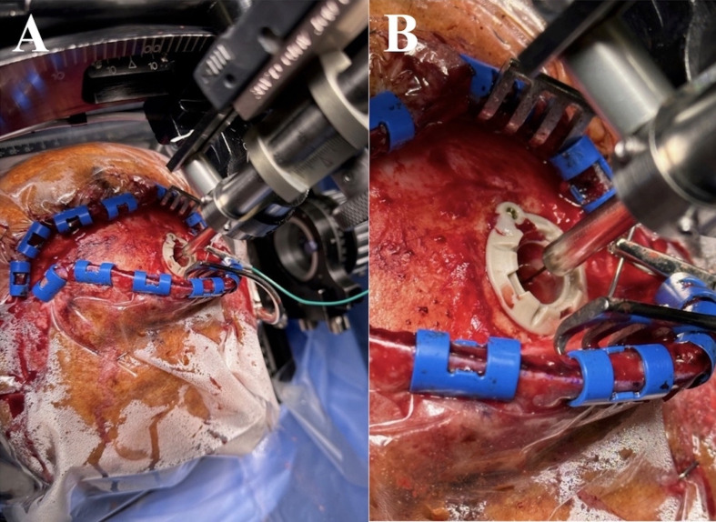
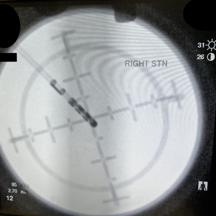
The FHC electrode is inserted into the brain along the planned trajectory without an introducing brain cannula (figure 1)—a technique we have termed a ‘naked’ MER. Once the FHC electrode is inserted 10–12 mm superior to the chosen target, fibrin sealant glue (Tisseel-Baxter) is then placed around the burr hole to prevent cerebrospinal fluid (CSF) loss and subsequent brain shift. The microelectrode is then advanced in 1 mm steps to determine the entry and exit level from the target structure (and associated trajectory length). The imaging intensifier is used to ensure the microelectrode is coursing along the planned tract to the centre of the stereotactic frame using the ‘bomb sites’ on the CRW frame (figure 2).
Figure 1. FHC microtargeting ‘Naked’ microelectrode recording diagram. Used with permission from FHC.
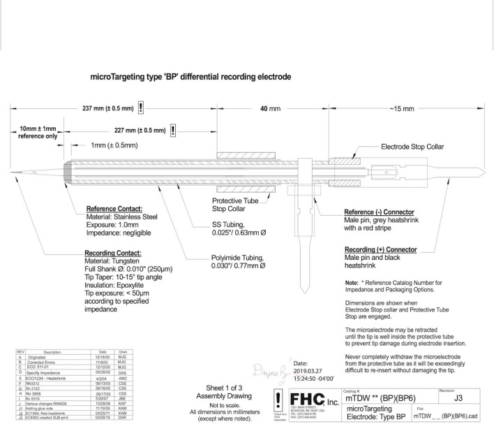
Figure 2. Cosman-Roberts-Wells ‘Bomb site’ on imaging intensifier.
The internal microelectrode is then withdrawn within the macroelectrode sheath and macrostimulation is achieved by progressing the macrostimulation electrode along the same trajectory (figure 3). Macrostimulation is undertaken at the planned levels of electrode placement to determine efficacy and side effect thresholds with respect to surrounding white matter tracts. This augments localisation information provided by MER recording (refer to table 2).
Figure 3. Panel A: macroelectrode insertion using MicroDrive. Panel B: zoomed in view of macroelectrode entering the brain. Note the absence of a cannula.
Once the image intensifier confirms the passage of the macroelectrode through the bomb site centre, the FHC electrode is withdrawn from the brain. A thermistor rod 1.6 mm in diameter (COSMAN (2) (250)) is placed along the length of the proposed DBS electrode implant, creating a path of least resistance for the definitive DBS electrode. This position is confirmed with X-ray with respect of the centre of the frame and the depth established from the earlier MER and the macro-stimulation. The thermistor rod is then removed, and the DBS electrode is placed and secured with the Medtronic (Stimlock), or Boston cap (Suretek), as per the implanted electrode. It is important to note, no insertional brain cannula is used at any stage. The only probe inserted into the brain is the naked microelectrode, guided to the brain cortex by the insertional cannula which never enters the brain parenchyma. The surgical sequence is then repeated on the other side. The electrodes are then positioned in situ under the scalp, the wound closed temporally with scalp clips.
The patient is then placed under general anaesthetic for the second stage of the procedure. The head is rotated contralaterally to the planned side of the passage of the cervical wire. A pocket is made either in the anterior chest wall superficial to the pectoralis fascia or the anterior abdominal wall to the anterior abdominal muscles. Two extension electrodes are then placed and connected to the burrowed DBS electrodes. There is a parietal incision above the ear allowing passage of the introducing cannula for the extension of electrodes. The circuit is then connected, impedances tested and all wounds are closed. Antibiotic coverage is either vancomycin or cephalin at a dose of 1 g three times a day for two doses throughout the course of the operation. Antibiotic dosing continues postoperatively for 72 hours with cephalin 1 g three times a day. The wound is closed with galeal subcuticular Vicryl and with clips to the skin on the scalp. There is a subcuticular Vicryl made to either the chest or the abdominal incision. The clips are removed on day 7 postoperatively.
Results
Demographics
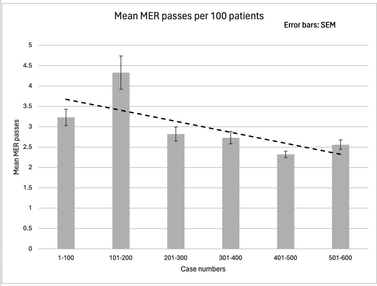
600 sequential patients that underwent DBS for PD from 2001 to 2023 at an average of 27 patients per year. The mean age of patients at the time of DBS insertion was 64 years. 142 patients, or 23.7% of our surgical cohort were >70 years of age at the time of implantation and 3 (0.5%) patients were over the age of 80. 63% of patients were men and 37% women. The mean number of MER passes for this cohort was three per patient, with a mean of 1.4 passes in the right brain and 1.6 passes in the left brain. There is a reduction in the average of MER passes per patient in the second half of the series (2.5) when compared with the first half (3.5). Variance also decreased, noting a reduction in SEM per 100 surgeries performed from 0.201 and 0.41 in the first and second 100 patients, to 0.08 and 0.12 over the fifth and sixth hundred patients, respectively (figure 4).
Figure 4. Mean MER passes per 100 patients. MER, microelectrode recording.
Indicators of DBS benefit
An audit of the first 55 patients treated in our centre was undertaken in an open-label fashion with postoperative assessment undertaken after a mean of 20 months post implantation (range 6–46 months). Follow-up data was available on 50 patients (4 patients unavailable, 1 deceased from other causes at time of assessment). Mean patient age at time of surgery was 58 years (range 29–77) and average time from PD diagnosis to surgery was 11.2 years (range 3–26 years). Mean UPDRS III (Unified Parkinson’s Disease Rating Scale III) off medication improvement=54% (8–86%). Dyskinesia was not disabling in 32% of the cohort preoperatively and 74% postoperatively. This data is consistent with published open-label efficacy data.30 31 Revision of DBS lead placement has not been required in the second 300 patients in the series.
We now routinely perform postoperative CT to preoperative MRI fusion using Stim XT (Boston Scientific), which we use to confirm lead placement and at times to guide stimulation programming and perform regular audit regarding DBS steering since changing to steerable electrodes. At the last review (March 2024) we had implanted 136 bilateral steering DBS systems, 130 of which were for PD. Vertical steering is used routinely throughout the cohort to optimise rostrocaudal stimulation configuration. Horizontal steering is in use in 30% of electrodes. This distribution is consistent with published series.32 33
Intracranial haemorrhages
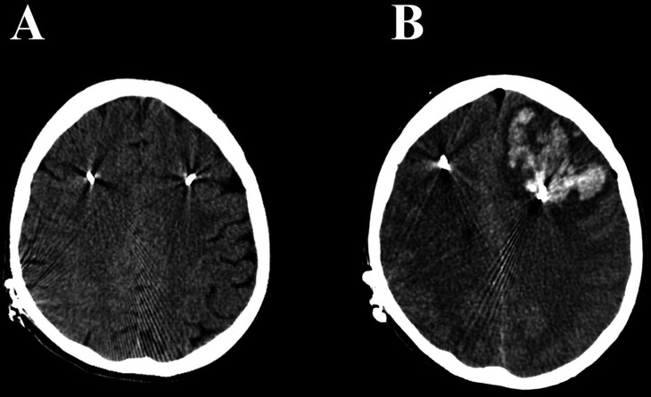
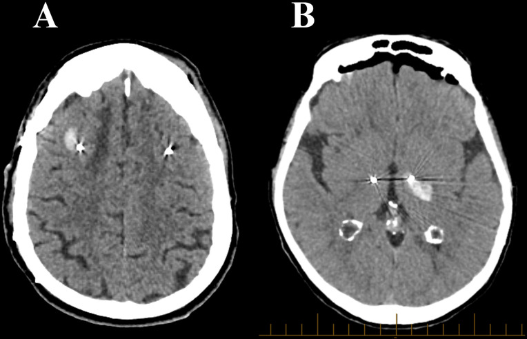
A total of three patients (0.5% per bilateral surgery; 0.25% per lead implantation) experienced vascular complications. All three cases of ICH occurred intra or acutely postoperatively (within 36 hours of implantation) and all three were symptomatic. Based on routine postoperative CT imaging, no cases of asymptomatic ICH occurred in this series. All three cases were women. The mean age at the time of DBS insertion was 68.5 years. Of the three patients, one was a haemorrhagic venous infarct (figure 5), one a subcortical haemorrhage (figure 6 Panel A) and one an internal capsule haemorrhage as shown on CT (figure 6 Panel B).
Figure 5. Panel A: case 1 CTB immediately postop. Panel B: case 1 CTB 36 hours postop.
CTB, Computed Tomography Brain Scan.
Figure 6. Panel A: case 2 CTB showing subcortical haemorrhage. Panel B: case 3 CTB showing internal capsule haemorrhage. CTB, Computed Tomography Brain Scan.
Case 1—venous infarct
Normal perioperative course with unremarkable postoperative CTB (Computed Tomography Brain Scan). Two MER passes were made on the uncomplicated side. One MER pass was made on the side complicated by venous infarction. Expressive dysphasia and right-sided hemiparesis developed 36 hours postoperatively, at which time CT brain showed a large left frontal ICH with midline shift. At craniotomy for evacuation of haemorrhage, a retrograde thrombosis from a cortical vein diathermied in the original operation was noted, resulting in patchy venous infarction with associated thrombosis. This patient’s right-sided hemiparesis later improved, with a long-term functional outcome of independent mobility using a four-wheel walker. Dysphasia persisted.
Case 2—implantation through gliotic tumour bed
The electrode unavoidably traversed a meningioma resection cavity/gliotic tumour bed where a small subcorital haemorrhage occurred adjacent to and tracking down the right DBS electrode. One MER pass was made on either side. This patient experienced visuospatial and left sided neglect with mild left lower limb weakness postoperatively. Long-term, this patient recovered to independent mobility using a walking stick.
Case 3—thalamocapsular haemorrhage
This patient became symptomatic with decreased alertness intraoperatively, and was transferred to ICU (Intensive Care Unit). CT imaging revealed a left thalamocapsular haemorrhage. Postoperatively, the patient was noted to have significant truncal and right upper limb ataxia and right sided hemiparesis. One MER pass was made on both sides. There was no identified anatomic variant or vascular anomaly. Long-term, weakness resolved and the patient returned to independent mobility with a four-wheel walker.
Discussion
The vascular complication rate in this series is 0.5%. This compares with vascular complications rates of 0.49–8%412,26 in the published literature. This literature identifies vascular risk as multifactorial including both patient and operative factors. Identified patient considerations include increased age and pre-existing comorbidities. Surgical considerations include surgical experience, the presence or absence of trajectory planning and utilisation of MER, including attendant number of passes.
Patient considerations
Age over 60, general frailty, hypertension and presence of additional comorbidities have been identified as patient risk factors for ICH.24 While it is notable that all three patients who suffered vascular complications were >65 years of age at time of implantation, none of our patients >70 or 80 years experienced vascular complications, suggesting advanced age was not a significant factor in our series.
Pre-existing hypertension and associated cerebrovascular consequences has also been noted in the literature as risk factor. This risk may be as high as 2.5 times in patients with a history of hypertension when compared with patients without hypertension.25 34 Pathological changes to the vasculature in brains of patients with chronic hypertension has been proposed to cause a decreased tolerance for multiple passes or incursions.34 Conversely, none of the cases of haemorrhage from our cohort had pre-existing hypertension, and none were hypertensive intraoperatively.
Implantation through the previous meningioma bed was likely a significant contributor to haemorrhage given changes in the brain consistency in Case 2. Previous lesions/surgery in the DBS track may represent an additional factor that increases the risk of vascular complications.
Operative considerations
Trajectory planning
At the commencement of our series, MRI imaging did not facilitate trajectory planning. Initially, only region of interest imaging was available and the implantation site was selected in proximity to the coronal suture with burr hole entry lateral enough to avoid a ventricular breach with any subsequently inserted electrode. Subsequently, our adoption of Brainlab iPlan software prior to 2010 permitted the inclusion of whole brain trajectory planning using CT imaging. The subsequent availability of 3D MRI imaging, again introduced prior to 2010, allowed the addition of T1 whole brain CT/MRI fusion, which established clearer visualisation of cortical sulci and deep sulci. A more recent 2022 upgrade of the 3D imaging T2 sequence, inclusion of the FGATIR sequence in pallidal and thalamic cases and utilisation of Brainlab Elements software currently provide additional perspectives in trajectory planning and target visualisation.
In combination, these imaging modalities offer visualisation of the planned trajectory in respect of skull thickness and irregularities (such as hyperostosis), cortical anatomy, proximity to the dura, dural and cortical veins, arteriovenous anomalies and the lateral ventricle.
The importance of planning for dural and cortical veins is highlighted in the venous infarct in Case 1, similar to that reported by Tani N, et al, where venous infarction occurred secondary to diathermy of a venous lacuna during dural opening.35 In these cases, knowledge of the cortical vascular anatomy may have permitted an alternate entry site and trajectory.
Trajectory mapping through the brain parenchyma has been demonstrated to reduce the risk of vascular complications. The series of Elias W.J, et al reported a vascular complication rate of 10.1% when there was sulcal penetration or close traverse to a sulcus as opposed to 0.7% when trajectories were clearly positioned within a gyrus.36 They additionally demonstrated that avoiding the traversing of the lateral ventricle reduces haemorrhagic risk.36 Further, Wang X, et al37 demonstrated a significant reduction in symptomatic haemorrhage when trajectory planning was introduced into their surgical targeting paradigm.
Brain shift secondary to CSF egress may be an additional risk factor for haemorrhage, given variations in electrode trajectory when the brain has moved with respect to the imaged position, increasing the risk of turning a projected gyrus track into a sulcal traverse.19 Brain shift is a major potential confound to reaching the intended target, but also increases the MER pass requirement to satisfactorily localise the nucleus.
Strategies we employed to minimise brain shift variance have included entry point planning, utilisation of a more supine operating position (limiting brain shift to the anteroposterior plane rather than both the anteroposterior and rostrocaudal plane) and the use of fibrin glue after microelectrode implantation to 10 mm above the planned target.
Trajectory variance may also be introduced through errors in CT/MRI fusion, MRI image quality or human factors. These possibilities are reduced in our unit through the simultaneous use of two different targeting techniques, CT planning based on atlas coordinates corrected for measurements undertaken on the MRI by the neurosurgeon and direct workstation planning from the MRI by the neurologist. Both sets of coordinates are visualised on the Brainlab planning station, the trajectory then agreed on with potential hazards identified.
MER passes and the use of a ‘Naked’ MER
Multiple studies have suggested a relationship between the use of MER and vascular adverse events.19 38 A meta-analysis conducted by Rasiah N.P, et al demonstrates a correlation between the number of MER passes and the risk of ICH occurrence once the MER passes are greater than one.24 This has been attributed to the physical insult caused to the parenchyma and potential physical insult to underlying vessels.39
Notably in our series, only one MER pass was made on the side of vascular complication in all three cases. Equally, in some of our earlier cases, multiple MER passes were performed on one side prior to satisfactory target localisation without vascular complications.
Here, our use of a ‘naked MER’—a technique where the combined microelectrode/macroelectrode is introduced directly via the MicroDrive to the brain without prior introduction of a rigid guiding brain cannula, may be relevant. The enclosed microelectrode tip is only exposed 10 mm above the planned target, diminishing the risk of vascular injury due to the finer MER tip as opposed to that of the rounded macroelectrode.
The total diameter of the FHC stimulating macroelectrode is 0.77 mm, compared with the diameter of the typical brain cannula of 1.47–1.83 mm(FHC manual). The surface area () of the brain impacted by the ‘naked’ MER of 0.46 mm2, is approximately one-fifth to one-quarter of the brain surface area impacted when using a brain cannula (1.7 mm2 to 2.64 mm2). This supports the assertion of other authors that the use of rigid cannula/guide tube system could contribute to an increased rate of ICH.40
As the FHC microelectrode is inserted within the external macrostimulator sheath, this reduces the risk of injury to the microelectrode during passage through the brain. It is therefore reasonable to conclude the risk of insult to brain tissue and vasculature reduces with the decreased brain surface area impacted.
One might argue that any potential risk reduction by not implanting a brain cannula is offset by the introduction of the thermistor rod (1.6 mm) prior to permanent electrode implantation, however, it is important to note that the thermistor rod is introduced only once per side, when the target and trajectory is finalised and confirmed to be satisfactory on the basis of MER and macrostimulation responses. This distinction may be particularly relevant in situations where multiple passes are required in which the brain cannula is moved, necessitating a much larger footprint on the cortical surface and increased risk of haemorrhage, particularly from deep sulci which may not be directly visible to the surgeon.
Single surgical team/experience
Over the course of the series, we performed an average of 27 cases per year. With the exception of the first five cases, all operations in our cohort were performed by a single neurosurgeon/neurosurgical team with a small, consistent group of nurses and radiographers. This consistency of case load and operating team are likely relevant considering that the rate of vascular complications has been shown to correlate with the experience of the surgical team19 38 and be reduced with an institutional annual DBS caseload of greater than 20.24
Conclusion
This report details a vascular complication rate (0.5% per patient, 0.25% per electrode) in a large unselected consecutive cohort of 600 patients treated with DBS for PD by a single neurosurgeon/neurologist team over 20 years. In only one case, the cause of the haemorrhage remains unclear. If the case in which the patient had undergone previous neurosurgery in the region of the DBS implantation trajectory is excluded, the vascular complication rate in our series for ‘de novo’ DBS surgery in PD is 0.33% per patient (0.16% per electrode). The benefit of this for patients is self-evident, but directly impacts the risk/benefit evaluation process for patients considering the procedure for management of PD.
We note the evolution of our surgical technique assisted by evolving technology in mitigating surgical risk. In particular optimised imaging, routine trajectory planning and management of CSF egress and consequent brain shift, contributing to a reducing number of required MER passes for satisfactory lead localisation.
While elements of the procedure have evolved over time, the use of a ‘naked microelectrode’ implantation which does not rely on a brain parenchymal cannula has been a consistent feature, highlighting the possibility that this technique in and of itself, may be helpful in reducing the risk of vascular complications in DBS procedures.
Footnotes
Funding: A grant number is N/A. North Shore Private Hospital (Ramsay Health Care) provided funding for a part-time research assistant position to assist with this manuscript, with the value less than $A5000. This was paid directly from North Shore Private Hospital Staff Payroll to the research assistant over the period of the past 12 months.
Patient consent for publication: Not applicable.
Ethics approval: This study involves human participants and was approved by Ramsay Health Care Human Research Ethics Committee A HREC Reference Number: 2023/ETH/0053, Project ID:2023/PID/0116. As this paper is a retrospective case series involving existing data and conducting a literature review, according to the National Statement on Ethical Conduct in Human Research (2007), this falls under the negligible risk classification which determines: ‘The expression ‘negligible risk research’ describes research in which there is no foreseeable risk of harm or discomfort; and any foreseeable risk is no more than inconvenience’. Additionally, as case study/series propose to use existing personal health information obtained during standard care, they are classed as negligible risk and qualify for exemption from ethical review in accordance with section 5.1.22 of the National Statement on Ethical Conduct in Human Research (2007). The cases involved also span 23 years and 600 patients, making the seeking of individual consent impractical and in some cases not possible. While three people who underwent deep brain stimulation insertion suffered vascular complications and this will be reflected in the paper, information regarding these patients has been de-identified and anonymised. In accordance to Statutory Guidelines on Research for the Health Records and Information Privacy Act 2002 (NSW), the requirements are met for ‘Research exemption’ for the use of disclosing de-identified health information for the secondary purpose of research and statistics. All four requirements for a research exemption are met for this retrospective study in accordance with the IPC Statutory Guidelines on Research Section 1.2. This specifies that health information can be disclosed without the consent of the person, for the secondary purpose of research if: Criteria 1: The use or disclosure is reasonably necessary for research, or the compilation or analysis of statistics, in the public interest. Criteria 2: You have taken reasonable steps to de-identify the information, or the purpose of the research cannot be served by using or disclosing de-identified information and it is impracticable to seek the consent of the person to the use or disclosure. Criteria 3: If the information could reasonably be expected to identify individuals, the information is not published in a generally available publication. Criteria 4: The use or disclosure of the health information is in accordance with the statutory guidelines on research.
Provenance and peer review: Not commissioned; internally peer reviewed.
Contributor Information
Raymond Cook, Email: raymondjohncook@bigpond.com.au.
Nyssa Chennell Dutton, Email: nyssa.chennelldutton@gmail.com.
Peter A Silburn, Email: p.silburn@neurosciencesqld.com.au.
Linton J Meagher, Email: meagherl@ramsayhealth.com.au.
George Fracchia, Email: gozwoz@optusnet.com.au.
Nathan Anderson, Email: nathan@ando.id.au.
Glen Cooper, Email: glencoops@gmail.com.
Hoang-Mai Dinh, Email: Hoangmai.dinh@health.nsw.gov.au.
Stuart J Cook, Email: stuartcook1508@gmail.com.
Paul Silberstein, Email: paul@silberstein.com.au.
Data availability statement
All data relevant to the study are included in the article or uploaded as supplementary information.
References
- 1.Fox SH, Katzenschlager R, Lim S-Y, et al. International Parkinson and movement disorder society evidence-based medicine review: Update on treatments for the motor symptoms of Parkinson’s disease. Mov Disord. 2018;33:1248–66. doi: 10.1002/mds.27372. [DOI] [PubMed] [Google Scholar]
- 2.Lee DJ, Lozano CS, Dallapiazza RF, et al. Current and future directions of deep brain stimulation for neurological and psychiatric disorders. J Neurosurg. 2019;131:333–42. doi: 10.3171/2019.4.JNS181761. [DOI] [PubMed] [Google Scholar]
- 3.Schuepbach WMM, Rau J, Knudsen K, et al. Neurostimulation for Parkinson’s disease with early motor complications. N Engl J Med. 2013;368:610–22. doi: 10.1056/NEJMoa1205158. [DOI] [PubMed] [Google Scholar]
- 4.Hamani C, Richter E, Schwalb JM, et al. Bilateral subthalamic nucleus stimulation for Parkinson’s disease: a systematic review of the clinical literature. Neurosurgery. 2005;56:1313–21. doi: 10.1227/01.neu.0000159714.28232.c4. [DOI] [PubMed] [Google Scholar]
- 5.Deuschl G, Antonini A, Costa J, et al. European Academy of Neurology/Movement Disorder Society-European Section Guideline on the Treatment of Parkinson’s Disease: I. Invasive Therapies. Mov Disord . 2022;37:1360–74. doi: 10.1002/mds.29066. [DOI] [PubMed] [Google Scholar]
- 6.Das S, Matias CM, Ramesh S, et al. Capturing Initial Understanding and Impressions of Surgical Therapy for Parkinson’s Disease. Front Neurol. 2021;12:605959. doi: 10.3389/fneur.2021.605959. [DOI] [PMC free article] [PubMed] [Google Scholar]
- 7.Lange M, Mauerer J, Schlaier J, et al. Underutilization of deep brain stimulation for Parkinson’s disease? A survey on possible clinical reasons. Acta Neurochir (Wien) 2017;159:771–8. doi: 10.1007/s00701-017-3122-3. [DOI] [PubMed] [Google Scholar]
- 8.Hamberg K, Hariz GM. The decision-making process leading to deep brain stimulation in men and women with parkinson’s disease - an interview study. BMC Neurol. 2014;14:89. doi: 10.1186/1471-2377-14-89. [DOI] [PMC free article] [PubMed] [Google Scholar]
- 9.Dinkelbach L, Möller B, Witt K, et al. How to improve patient education on deep brain stimulation in Parkinson’s disease: the CARE Monitor study. BMC Neurol. 2017;17:36. doi: 10.1186/s12883-017-0820-7. [DOI] [PMC free article] [PubMed] [Google Scholar]
- 10.Lee JI. The Current Status of Deep Brain Stimulation for the Treatment of Parkinson Disease in the Republic of Korea. J Mov Disord. 2015;8:115–21. doi: 10.14802/jmd.15043. [DOI] [PMC free article] [PubMed] [Google Scholar]
- 11.Kim M-R, Yun JY, Jeon B, et al. Patients’ reluctance to undergo deep brain stimulation for Parkinson’s disease. Parkinsonism Relat Disord. 2016;23:91–4. doi: 10.1016/j.parkreldis.2015.11.010. [DOI] [PubMed] [Google Scholar]
- 12.Umemura A, Oka Y, Yamamoto K, et al. Complications of subthalamic nucleus stimulation in Parkinson’s disease. Neurol Med Chir (Tokyo) 2011;51:749–55. doi: 10.2176/nmc.51.749. [DOI] [PubMed] [Google Scholar]
- 13.Lyons KE, Wilkinson SB, Overman J, et al. Surgical and hardware complications of subthalamic stimulation: a series of 160 procedures. Neurology (ECronicon) 2004;63:612–6. doi: 10.1212/01.wnl.0000134650.91974.1a. [DOI] [PubMed] [Google Scholar]
- 14.Vergani F, Landi A, Pirillo D, et al. Surgical, medical, and hardware adverse events in a series of 141 patients undergoing subthalamic deep brain stimulation for Parkinson disease. World Neurosurg. 2010;73:338–44. doi: 10.1016/j.wneu.2010.01.017. [DOI] [PubMed] [Google Scholar]
- 15.Morishita T, Okun MS, Burdick A, et al. Cerebral venous infarction: a potentially avoidable complication of deep brain stimulation surgery. Neuromodulation. 2013;16:407–13. doi: 10.1111/ner.12052. [DOI] [PMC free article] [PubMed] [Google Scholar]
- 16.Videnovic A, Metman LV. Deep brain stimulation for Parkinson’s disease: prevalence of adverse events and need for standardized reporting. Mov Disord. 2008;23:343–9. doi: 10.1002/mds.21753. [DOI] [PubMed] [Google Scholar]
- 17.Mainardi M, Ciprietti D, Pilleri M, et al. Deep brain stimulation of globus pallidus internus and subthalamic nucleus in Parkinson’s disease: a multicenter, retrospective study of efficacy and safety. Neurol Sci. 2024;45:177–85. doi: 10.1007/s10072-023-06999-z. [DOI] [PMC free article] [PubMed] [Google Scholar]
- 18.Servello D, Galbiati TF, Iess G, et al. Complications of deep brain stimulation in Parkinson’s disease: a single-center experience of 517 consecutive cases. Acta Neurochir (Wien) 2023;165:3385–96. doi: 10.1007/s00701-023-05799-w. [DOI] [PubMed] [Google Scholar]
- 19.Seijo F, Alvarez de Eulate Beramendi S, Santamarta Liébana E. Surgical adverse events of deep brain stimulation in the subthalamic nucleus of patients with Parkinson’s disease. The learning curve and the pitfalls. Acta Neurochir (Wien) 2014;156:1505–12. doi: 10.1007/s00701-014-2082-0. [DOI] [PubMed] [Google Scholar]
- 20.Chang WS, Kim HY, Kim JP, et al. Bilateral subthalamic deep brain stimulation using single track microelectrode recording. Acta Neurochir. 2011;153:1087–95. doi: 10.1007/s00701-011-0953-1. [DOI] [PubMed] [Google Scholar]
- 21.Levi V, Carrabba G, Rampini P, et al. Short term surgical complications after subthalamic deep brain stimulation for Parkinson’s disease: does old age matter? BMC Geriatr. 2015;15:116. doi: 10.1186/s12877-015-0112-2. [DOI] [PMC free article] [PubMed] [Google Scholar]
- 22.Lachenmayer ML, Mürset M, Antih N, et al. Subthalamic and pallidal deep brain stimulation for Parkinson’s disease-meta-analysis of outcomes. NPJ Parkinsons Dis. 2021;7:77. doi: 10.1038/s41531-021-00223-5. [DOI] [PMC free article] [PubMed] [Google Scholar]
- 23.Xu S, Wang W, Chen S, et al. Deep Brain Stimulation Complications in Patients With Parkinson’s Disease and Surgical Modifications: A Single-Center Retrospective Analysis. Front Hum Neurosci. 2021;15:684895. doi: 10.3389/fnhum.2021.684895. [DOI] [PMC free article] [PubMed] [Google Scholar]
- 24.Rasiah NP, Maheshwary R, Kwon C-S, et al. Complications of Deep Brain Stimulation for Parkinson Disease and Relationship between Micro-electrode tracks and hemorrhage: Systematic Review and Meta-Analysis. World Neurosurg. 2023;171:e8–23. doi: 10.1016/j.wneu.2022.10.034. [DOI] [PubMed] [Google Scholar]
- 25.Xiaowu H, Xiufeng J, Xiaoping Z, et al. Risks of intracranial hemorrhage in patients with Parkinson’s disease receiving deep brain stimulation and ablation. Parkinsonism Relat Disord. 2010;16:96–100. doi: 10.1016/j.parkreldis.2009.07.013. [DOI] [PubMed] [Google Scholar]
- 26.Yang C, Qiu Y, Wang J, et al. Intracranial hemorrhage risk factors of deep brain stimulation for Parkinson’s disease: a 2-year follow-up study. J Int Med Res. 2020;48:300060519856747. doi: 10.1177/0300060519856747. [DOI] [PMC free article] [PubMed] [Google Scholar]
- 27.Stoehr K, Pazira K, Bonnet K, et al. Deep Brain Stimulation in Early-Stage Parkinson’s Disease: Patient Experience after 11 Years. Brain Sci. 2022;12 doi: 10.3390/brainsci12060766. [DOI] [PMC free article] [PubMed] [Google Scholar]
- 28.Cook RJ, Jones L, Fracchia G, et al. Globus pallidus internus deep brain stimulation as rescue therapy for refractory dyskinesias following effective subthalamic nucleus stimulation. Stereotact Funct Neurosurg. 2015;93:25–9. doi: 10.1159/000365223. [DOI] [PubMed] [Google Scholar]
- 29.Conti A, Gambadauro NM, Mantovani P, et al. A Brief History of Stereotactic Atlases: Their Evolution and Importance in Stereotactic Neurosurgery. Brain Sci. 2023;13:830. doi: 10.3390/brainsci13050830. [DOI] [PMC free article] [PubMed] [Google Scholar]
- 30.Limousin P, Krack P, Pollak P, et al. Electrical stimulation of the subthalamic nucleus in advanced Parkinson’s disease. N Engl J Med. 1998;339:1105–11. doi: 10.1056/nejm199810153391603. [DOI] [PubMed] [Google Scholar]
- 31.Schüpbach WMM, Chastan N, Welter ML, et al. Stimulation of the subthalamic nucleus in Parkinson’s disease: a 5 year follow up. J Neurol Neurosurg Psychiatry . 2005;76:1640–4. doi: 10.1136/jnnp.2005.063206. [DOI] [PMC free article] [PubMed] [Google Scholar]
- 32.Karl JA, Joyce J, Ouyang B, et al. Long-Term Clinical Experience with Directional Deep Brain Stimulation Programming: A Retrospective Review. Neurol Ther. 2022;11:1309–18. doi: 10.1007/s40120-022-00381-5. [DOI] [PMC free article] [PubMed] [Google Scholar]
- 33.Zitman FMP, Janssen A, van der Gaag NA, et al. The actual use of directional steering and shorter pulse width in selected patients undergoing deep brain stimulation. Parkinsonism Relat Disord. 2021;93:58–61. doi: 10.1016/j.parkreldis.2021.11.009. [DOI] [PubMed] [Google Scholar]
- 34.Gorgulho A, De Salles AAF, Frighetto L, et al. Incidence of hemorrhage associated with electrophysiological studies performed using macroelectrodes and microelectrodes in functional neurosurgery. J Neurosurg. 2005;102:888–96. doi: 10.3171/jns.2005.102.5.0888. [DOI] [PubMed] [Google Scholar]
- 35.Tani N, Yaegaki T, Kishima H. A Case Report: Hemorrhagic Venous Infarction after Deep Brain Stimulation Surgery Probably Due to Coagulation of Intradural Veins. NMC Case Rep J . 2021;8:315–8. doi: 10.2176/nmccrj.cr.2020-0305. [DOI] [PMC free article] [PubMed] [Google Scholar]
- 36.Elias WJ, Sansur CA, Frysinger RC. Sulcal and ventricular trajectories in stereotactic surgery. JNS . 2009;110:201–7. doi: 10.3171/2008.7.17625. [DOI] [PubMed] [Google Scholar]
- 37.Wang X, Li N, Li J, et al. Optimized Deep Brain Stimulation Surgery to Avoid Vascular Damage: A Single-Center Retrospective Analysis of Path Planning for Various Deep Targets by MRI Image Fusion. Brain Sci. 2022;12:967. doi: 10.3390/brainsci12080967. [DOI] [PMC free article] [PubMed] [Google Scholar]
- 38.Doshi PK, Rai N, Das D. Surgical and Hardware Complications of Deep Brain Stimulation-A Single Surgeon Experience of 519 Cases Over 20 Years. Neuromodulation. 2022;25:895–903. doi: 10.1111/ner.13360. [DOI] [PubMed] [Google Scholar]
- 39.Chou Y-C, Lin S-Z, Hsieh WA, et al. Surgical and hardware complications in subthalamic nucleus deep brain stimulation. J Clin Neurosci. 2007;14:643–9. doi: 10.1016/j.jocn.2006.02.016. [DOI] [PubMed] [Google Scholar]
- 40.Park CK, Jung NY, Kim M, et al. Analysis of Delayed Intracerebral Hemorrhage Associated with Deep Brain Stimulation Surgery. World Neurosurg. 2017;104:537–44. doi: 10.1016/j.wneu.2017.05.075. [DOI] [PubMed] [Google Scholar]
Associated Data
This section collects any data citations, data availability statements, or supplementary materials included in this article.
Data Availability Statement
All data relevant to the study are included in the article or uploaded as supplementary information.


