Abstract
Background
Bronchial artery embolization (BAE) is a common and important way to manage hemoptysis. This study’s purpose was to summarize the efficacy, safety, and related factors of BAE in the treatment of hemoptysis.
Methods
From January 2010 to August 2023, a systematic literature search was conducted in PubMed, EMBASE, Web of Science, and Cochrane Library databases. Original studies with BAE for hemoptysis were included, with no restrictions on language. The outcomes of interest were technical success rate, clinical success rate, recurrence rate, mortality rate, and major complication rate. Pooled proportions with 95% confidence intervals (CIs) were calculated using random-effects models. The Newcastle-Ottawa Scale (NOS) was employed for quality assessment. Factors such as publication year, region, sample size, amount of hemoptysis, etiology, and embolization materials were extracted for subgroup analyses. Additionally, sensitivity analyses and test for publication bias were conducted.
Results
A total of 32 studies, including 6,032 patients, met our inclusion criteria. 27 studies were of high quality, while five of moderate quality. The results indicated the prevalence of technical success was 97.2% (95% CI: 95.1–98.8%) and 93.2% (95% CI: 90.3–95.7%) in clinical success. Hemoptysis recurrence and mortality rates after BAE were 24.8% (95% CI: 20.5–29.4%) and 2.3% (95% CI: 1.1–3.8%), respectively. Moreover, the pooled prevalence of major complication was 0.1% (95% CI: 0.0–0.4%). Subgroup analysis revealed that studies published after 2017 demonstrated a higher technical success rate and a lower recurrence rate. Massive hemoptysis showed a higher technical success rate but a lower clinical success rate. BAE also demonstrated superior efficacy in patients with bronchiectasis. The clinical success rate was significantly higher in patients with benign diseases than those with malignancies. Gelatin sponge (GS) showed poor embolization efficacy. N-butyl-2-cyanoacrylate (NBCA) and coils exhibited reduced recurrence rates, while NBCA displayed an even lower recurrence rate than non-absorbable particles. The study by Ishikawa et al. influenced the stability of the pooled major complication rate, and the sensitivity analysis confirmed the robustness of the remaining results.
Conclusions
BAE is safe and effective in treating different degrees of hemoptysis caused by benign and malignant lesions. Promising clinical efficacy was observed with NBCA as an embolic material for the treatment of hemoptysis. However, further conclusions should be investigated using evidence-based medicine.
Keywords: Bronchial artery embolization (BAE), hemoptysis, embolization material, systematic review, meta-analysis
Highlight box.
Key findings
• Bronchial artery embolization (BAE) is a safe and effective treatment for various degrees of hemoptysis caused by benign and malignant lesions. N-butyl cyanoacrylate (NBCA) shows promising therapeutic benefits without an associated higher risk of complications during BAE.
What is known, and what is new?
• BAE is considered the first-line treatment for massive hemoptysis and recurrent hemoptysis.
• The selection of embolization materials for BAE varies among different centers, as there is no consensus on the best option.
• NBCA demonstrates a lower recurrence rate of hemoptysis in comparison to other embolization materials.
What is the implication, and what should change now?
• With the increasing standardization of practice among interventional radiologists, the future application of NBCA in BAE processes is anticipated to become more extensive. Further large-scale studies should be performed to confirm these findings.
Introduction
Hemoptysis is an expulsion of blood originating from the lower respiratory tract (1). In certain chronic diseases, newly formed thin-walled and fragile anastomotic branches between the pulmonary and bronchial arteries are prone to rupture under elevated systemic arterial pressure (2-4). The bronchial arterial system is the predominant source of hemoptysis (90%), with a minority of cases involving the aorta or non-bronchial systemic collaterals. Hemoptysis can also arise from pulmonary vessels (5%), like the erosion of malignant lesions or the rupture of pseudoaneurysms (3,5). Regional variations exist regarding the most common causes of hemoptysis. In France, cryptogenic hemoptysis (50%) was the most prominent case of hemoptysis requiring hospitalization, followed by infections (22%), cancer (17%), and bronchiectasis (7%) (6). Two Italian studies found that lung malignancies were the most prevalent cause, accounting for a similar proportion (approximately 19%), followed by pneumonia, bronchiectasis, acute bronchitis, or inflammatory exacerbation of chronic pulmonary disease (7,8). Bronchiectasis was a major cause of bloody sputum and hemoptysis (18.3%) in Japan (9). In areas with a high incidence of tuberculosis, tuberculosis-related hemoptysis was the most frequent cause and even represents 79.2% of all causes in India (10).
The management of hemoptysis included conservative treatments such as administering of hemostatic agents (antifibrinolytic and vasoconstrictive agents) and anti-inflammatory or anti-infective therapy. Invasive techniques include bronchoscopy, bronchial artery embolization (BAE), and surgical intervention. In deciding the proper course of treatment, it is important to consider the volume and timing of hemoptysis. In patients with large volume or rapid hemoptysis, conservative treatment may be insufficient, with mortality rates of critically ill patients using only conservative therapy reaching 50% (11). Bronchoscopy can effectively control bronchial bleeding, remove the blood clots, and identify the cause of hemoptysis by revealing proximal bronchial abnormalities. However, excessive blood accumulation in the airway can create challenges for timely clearance and airway observation. In cases with vascular malformations, bronchoscopy is often avoided entirely (2,5,12-14). Open surgical intervention is also associated with a poor prognosis, with nearly 40% of patients with massive hemoptysis succumbing to mortality (15). Additionally, surgical intervention is less efficacious in patients with diffuse lesions and compromised cardiopulmonary function.
BAE was first proposed by Remy et al. in 1973 (16). It has gained widespread acceptance due to its advantages of being minimally invasive while being able to control massive hemoptysis and improve patient prognosis (17). The advances in embolic materials and embolization techniques have facilitated BAE improvements. Commonly employed embolization materials encompass gelatin sponge (GS), polyvinyl alcohol (PVA), microspheres, coils, and N-butyl-2-cyanoacrylate (NBCA) etc. These materials exhibit variations in their degree of embolization, recirculation rate, and safety. The CIRSE Standards of Practice recommends the utilization of PVA particles with a diameter of 355–500 µm (18). However, the selection of materials in each center is primarily influenced by personal preferences or policies implemented by countries and hospitals without reaching a consensus. A nationwide study involving 8,563 patients in Japan (19) compared the safety of different embolization materials for BAE. The findings revealed that coil (0.06%) exhibited a lower incidence of spinal cord embolism compared to GS (0.18%) and NBCA (0.72%). Unfortunately, PVA and microspheres were not included in the evaluation as they were not used for BAE in Japan, despite PVA being globally the most used embolic material. The recurrence rate of hemoptysis has been reported to range from 10% to 57% following BAE, and the major complications, including spinal cord ischemia and cerebral infarction, occurred in 0–4.6% (20-22). Therefore, further understanding the therapeutic effect of BAE in different diseases is necessary to optimize the treatment plan, improve the efficacy, and reduce the recurrence rate and complications. This meta-analysis was designed to investigate the efficacy and safety of BAE in the treatment of hemoptysis. Meanwhile, the related factors affecting the results were also analyzed. We present this article in accordance with the PRISMA reporting checklist (available at https://cdt.amegroups.com/article/view/10.21037/cdt-24-157/rc).
Methods
PROSPERO’s registration number was CRD42023480571.
Search strategy
The PubMed, EMBASE, Web of Science, and the Cochrane Library databases between January 2010 and August 2023 were searched, and two evaluators critically reviewed each included study; the consultation of a third party was conducted in the event of a disagreement. The following search terms were used: ((((bronchial artery) OR (artery embolization)) OR (bronchial artery embolization)) OR (transcatheter embolization)) AND (hemoptysis) (Appendix 1). References from eligible studies were also reviewed to identify potential studies missed by the electronic search.
Selection criteria
A priori, all eligible studies would be included; exclusion criteria were as follows: (I) duplicates; (II) guidelines, consensus, comments, letters, conference papers, animal experiments, case reports, reviews, systematic review, meta-analysis; (III) outcomes of interest were irrelevant to the impact of BAE on the hemoptysis; (IV) group treated by BAE in hemoptysis with less than 50 participants; (V) full-texts could not be retrieved; (VI) cause of hemoptysis was not indicated; (VII) procedure of BAE was not specific; (VIII) no short-term clinical outcome indicators; (IX) no follow-up information or mean follow-up was less than 3 months; and (X) data overlap among studies: When multiple studies involved the same patient population treated with BAE, we only included the most comprehensive follow-up information or the most recently published one.
Data extraction
The data were extracted as follows: first author, publication year, region, number of patients, patient sex, degree of hemoptysis, etiology, embolic materials, technical success rate, clinical success rate, recurrence rate, mortality rate, and major complication rate.
Following the commonly used classification criteria in the literature and clinical practice, we categorized patients with hemoptysis into three groups (18): mild hemoptysis (<100 mL/24 h), moderate hemoptysis (100–300 mL/24 h), and massive or severe hemoptysis (>300 mL/24 h or any amount resulting in a decrease in hemoglobin >1 g/dL, hematocrit >5%, respiratory failure due to obstruction or other causes (PaO2 <60 mmHg), or hypotension with systolic blood pressure <90 mmHg).
The successful embolization of all culprit vessels defined technical success. Clinical success was defined as a cessation or clinically significant reduction of hemoptysis volume by more than 50% following BAE (18,20,23,24). Recurrence referred to clinically significant re-hemoptysis requiring treatment. The hemoptysis-related mortality rate refers to the proportion of patients for whom hemoptysis directly led to death. Major complications were characterized by hospital admission for therapy, prolonged hospitalization, permanent adverse sequelae, or death (25).
Study quality assessment
The Newcastle-Ottawa Scale (NOS) comprises eight items: selection, comparability, and exposure. Ratings on the NOS range from 0 to 9 stars, where scores of 3 or lower indicate poor quality, scores of 4 to 6 denote moderate quality, and scores of 7 to 9 signify high quality (26).
Statistical analysis
The STATA statistical software (version 17.0; StataCorp, College Station, TX, USA) was employed to analyze the data. The technical success rates, clinical success rates, recurrence rates, mortality, and major complication rates were extracted from each study for meta-analysis. Pooled statistics were expressed as proportions at 95% confidence intervals (CIs). Only a random-effects model was employed. I2 statistics were used to assess the extent of heterogeneity of the studies quantitatively. I2>50% was considered as statistically significant heterogeneity (27-30). The funnel plot and Egger’s test were performed to evaluate the presence of publication bias. Sensitivity analyses were performed by sequentially omitting one study at a time and recalculating the pooled proportion for the remaining studies. A two-sided P<0.05 was considered statistically significant. Subgroup analysis was performed by classification by publication year (during or after 2017 vs. before 2017) (20), region (Asia vs. Europe vs. America vs. Africa), sample size (≥100 vs. <100), the amount of hemoptysis (massive vs. non-massive), etiology including bronchiectasis (yes vs. no), tuberculosis (yes vs. no) and type of disease (benign vs. malignant), embolization materials including NBCA (yes vs. no), coils (yes vs. no), non-absorbable particles (yes vs. no), GS (yes vs. no) and classification by single embolization material (NBCA vs. non-absorbable particles).
Results
Study selection and participants’ characteristics
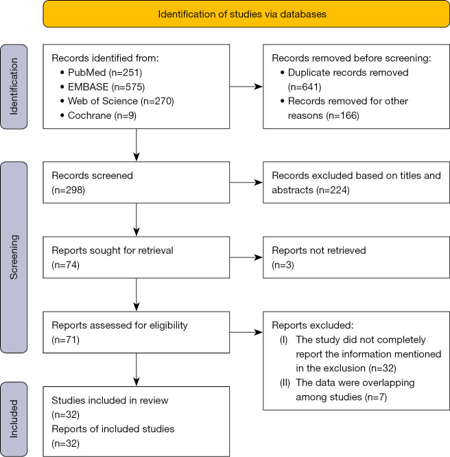
A total of 1,105 studies were screened, and 32 studies with 6,032 patients were finally included (Figure 1), which included 11 retrospective cohort studies (12,23,24,31-38), 20 retrospective single-arm studies (11,21,22,39-55) and one prospective single-arm study (17). Table 1 shows the characteristics of the included studies. The sample sizes ranged from 50 to 588 individuals. According to NOS, 27 studies were classified as high quality, while five were considered moderate quality.
Figure 1.
The flowchart of the search strategy.
Table 1. Characteristics of studies included in this meta-analysis.
| First author [year] | Region | Study design | Male, n (%) | Etiology | Grade of hemoptysis (%) | Embolic materials (%) | Major complication [n] | NOS score |
|---|---|---|---|---|---|---|---|---|
| Yan [2023] (31) | China | Retrospective cohort study | 231 (70.86) | Mixed | Mild: 39.0; moderate: 41.4; massive: 19.6 | PVA: 80.0; microspheres: 17.2; GS particles: 2.8 | Cerebral infarction [1] | 8 |
| Wang [2023] (32) | China | Retrospective cohort study | 312 (53.06) | Bronchiectasis | Moderate-massive: 100.0 | PVA: 100.0 | – | 9 |
| Park [2023] (23) | Korea | Retrospective cohort study | 93 (67.39) | Mixed | Mild: 100.0 | PVA: 70.3; microspheres: 6.5; mixed: 23.2 | – | 7 |
| Le Tat [2023] (21) | France | Retrospective single-arm study | 47 (75.81) | Malignancy | Moderate-massive: 100.0 | Not specified | Paraparesis [1]; regressive stroke [1]; acute pancreatitis [1] | 6 |
| García Jurado [2023] (39) | Spain | Retrospective single-arm study | 35 (63.64) | Mixed | Mild: 25.4; moderate: 56.4; massive: 18.2 | NBCA: 100.0 | – | 7 |
| Zhang [2022] (33) | China | Retrospective cohort study | 301 (71.67) | Mixed | Mild: 33.8; moderate: 41.4; massive: 24.8 | Not specified | – | 8 |
| Lee [2022] (34) | Korea | Retrospective cohort study | 103 (84.43) | Malignancy | Mild: 27.9; moderate: 54.1; massive: 18.0 | NBCA: 47.5; PVA: 52.5 | – | 9 |
| Omachi [2021] (17) | Japan | Prospective single-arm study | 24 (39.34) | Mixed | Not specified | Metallic platinum coils: 100.0 | – | 6 |
| Hwang [2021] (40) | Korea | Retrospective single-arm study | 143 (61.37) | Mixed | Mild: 74.2; moderate: 25.8 | Not specified | – | 7 |
| Fu [2021] (24) | China | Retrospective cohort study | 93 (61.18) | Mixed | Mild: 71.7; moderate-massive: 28.3 | Microspheres: 40.8; PVA: 59.2 | – | 8 |
| Tayal [2020] (41) | India | Retrospective single-arm study | 32 (64.00) | Mixed | Mild: 20.0; moderate-massive: 80.0 | Not specified | Transverse myelitis [1] | 6 |
| Takeda [2020] (42) | Japan | Retrospective single-arm study | 27 (25.47) | Bronchiectasis | Mild-moderate: 78.3; massive: 21.7 | Detachable or pushable coils: 100.0 | Mediastinal hematoma [1] | 9 |
| Sarioglu [2020] (35) | Turkey | Retrospective cohort study | 66 (74.16) | Mixed | Moderate: 53.9; massive: 46.1 | Microspheres: 100.0 | – | 7 |
| Peng [2019] (43) | China | Retrospective single-arm study | 178 (85.99) | Tuberculosis | Moderate: 13.0; massive: 87.0 | Not specified | Contrast media-related renal failure [1] | 9 |
| Han [2019] (44) | Korea | Retrospective single-arm study | 71 (84.52) | Malignancy | Mild: 25.0; moderate: 40.5; massive: 34.5 | PVA: 61.4; gelfoam: 18.1; mixed: 20.5 | – | 8 |
| Kucukay [2018] (45) | Turkey | Retrospective single-arm study | 80 (45.98) | Mixed | Massive: 100.0 | Microspheres: 100.0 | – | 8 |
| Choi [2018] (36) | Korea | Retrospective cohort study | 41 (57.75) | Mixed | Mild: 100.0 | PVA: 97.2; mixed: 2.8 | – | 8 |
| Ishikawa [2017] (46) | Japan | Retrospective single-arm study | 227 (46.42) | Mixed | Not specified | Not specified | Aortic dissection [1]; cerebellar infarctions [2]; mediastinal hematoma [5] | 9 |
| Pathak [2016] (22) | USA | Retrospective single-arm study | 30 (60.00) | Mixed | Not specified | Not specified | Stroke [1]; paraplegia [1] | 6 |
| Dabó [2016] (47) | Portugal | Retrospective single-arm study | 47 (70.15) | Mixed | Not specified | PVA: 94.0; mixed: 6.0 | – | 7 |
| Tom [2015] (48) | USA | Retrospective single-arm study | 44 (63.77) | Mixed | Not specified | Not specified | – | 6 |
| Bhalla [2015] (49) | India | Retrospective single-arm study | 255 (76.35) | Mixed | Mild: 20.9; moderate: 58.4; massive: 20.7 | Not specified | – | 8 |
| Pei [2014] (50) | China | Retrospective single-arm study | 98 (87.50) | Tuberculosis | Massive: 100.0 | Gelfoam: 100.0 | – | 8 |
| Garcia-Olivé [2014] (51) | Spain | Retrospective single-arm study | 132 (75.00) | Mixed | Moderate-massive: 100.0 | Not specified | Spinal cord ischemia [1] | 7 |
| Chen [2014] (37) | China | Retrospective cohort study | 121 (78.57) | Mixed | Mild: 23.4; moderate: 41.5; massive: 35.1 | GS particles: 7.8; PVA: 44.2; mixed: 48.0 | Ischemic colitis [1] | 9 |
| Racil [2013] (52) | Tunisia | Retrospective single-arm study | 38 (71.70) | Mixed | Mild: 1.9; moderate: 77.4; massive: 20.7 | Gelatine: 82.6; microspheres: 13.0; mixed: 4.4 | Cerebral ischemia [1] | 8 |
| Cornalba [2013] (53) | Italy | Retrospective single-arm study | 295 (61.84) | Mixed | Not specified | Not specified | Stroke [1]; transient ischaemic attack [1]; immediate transient tetraplegia [1] | 9 |
| Woo [2013] (12) | Korea | Retrospective cohort study | 242 (59.61) | Mixed | Mild: 29.6; moderate-massive: 70.4 | PVA: 72.2; NBCA: 27.8 | Lower extremity weakness [1] | 9 |
| Anuradha [2012] (54) | India | Retrospective single-arm study | 46 (79.31) | Mixed | Not specified | PVA: 62.5; gelfoam: 14.1; mixed: 23.4 | – | 7 |
| Shin [2011] (55) | Korea | Retrospective single-arm study | 129 (76.33) | Tuberculosis | Mild: 41.1; moderate: 33.1; massive: 25.8 | Not specified | Severe dyspnoea [1] | 8 |
| Hahn [2010] (38) | Korea | Retrospective cohort study | 246 (57.21) | Mixed | Mild-moderate: 63.5; massive: 36.5 | Gelfoam: 17.2; PVA: 82.8 | – | 9 |
| Chun [2010] (11) | UK | Retrospective single-arm study | 24 (48.00) | Mixed | Not specified | PVA: 100.0 | False aneurysm of femoral artery [1]; limb weakness [1] | 8 |
NOS, Newcastle-Ottawa Scale; PVA, polyvinyl alcohol; GS, gelatin sponge; NBCA, N-butyl-2-cyanoacrylate.
Overall meta-analysis
Technical success rates
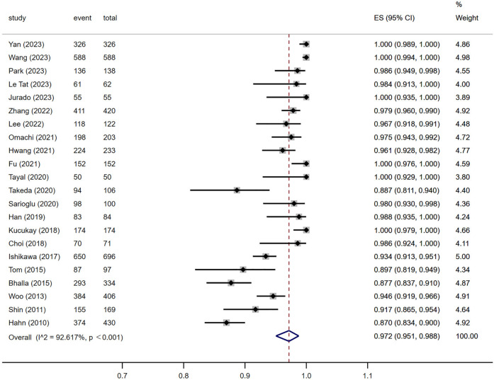
The technical success rates of BAE were reported in 22 articles, ranging from 87.0% to 100.0%. The pooled prevalence was 97.2% (95% CI: 95.1–98.8%) (Figure 2) with significant heterogeneity (I2=92.617%, P<0.001).
Figure 2.
Technical success rates. ES, estimate effect; CI, confidence interval.
Clinical success rates
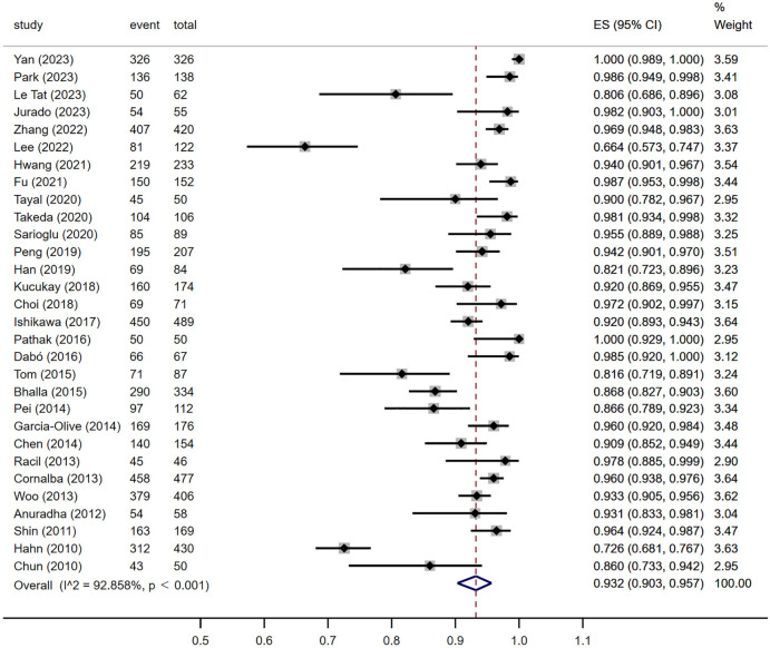
The clinical success rates were reported in 30 articles, ranging from 66.4% to 100.0%. The pooled prevalence was 93.2% (95% CI: 90.3–95.7%) (Figure 3) with significant heterogeneity (I2=92.858%, P<0.001).
Figure 3.
Clinical success rates. ES, estimate effect; CI, confidence interval.
Recurrence rates
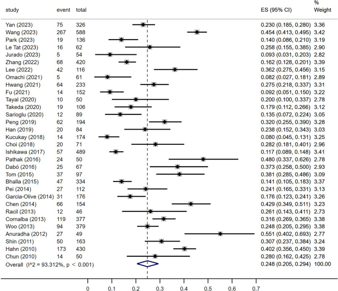
All 32 articles reported the recurrence rates ranging from 8.0% to 55.1%. The pooled prevalence was 24.8% (95% CI: 20.5–29.4%) (Figure 4) with significant heterogeneity (I2=93.312%, P<0.001).
Figure 4.
Recurrence rates. ES, estimate effect; CI, confidence interval.
Mortality rates
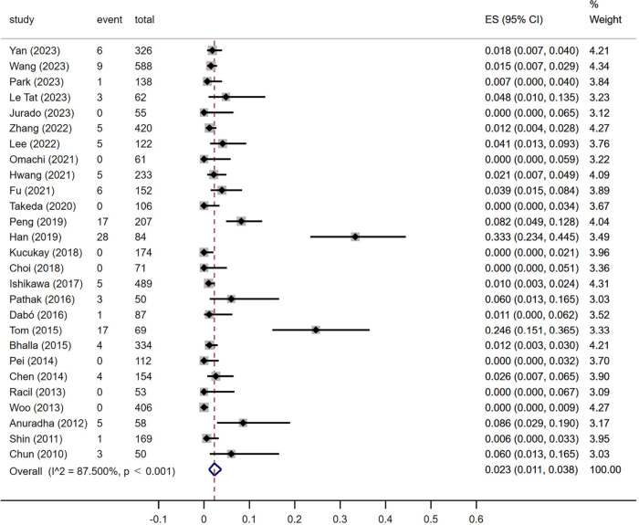
The mortality rates ranging from 0.0% to 33.3% were reported in 27 articles. The pooled prevalence was 2.3% (95% CI: 1.1–3.8%) (Figure 5) with significant heterogeneity (I2=87.500%, P<0.001).
Figure 5.
Mortality rates. ES, estimate effect; CI, confidence interval.
Major complication rates
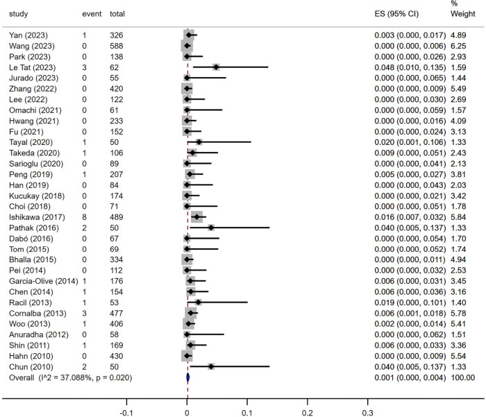
Major complication rates were recorded in all articles and ranged from 0.0% to 4.8%. The pooled prevalence was 0.1% (95% CI: 0.0–0.4%) (Figure 6). The results of the heterogeneity analysis indicated an I2 value of 37.088% with a P value of 0.02.
Figure 6.
Major complication rates. ES, estimate effect; CI, confidence interval.
Publication bias and sensitivity analysis
The Funnel plot and Egger’s test suggested publication bias in terms of technical success (P=0.04), clinical success (P<0.001), recurrence (P=0.050), mortality (P<0.001), and major complication (P<0.001) (Figures 7-11). The sensitivity analysis of the pooled technical success rate, clinical success rate, recurrence rate, and mortality rate indicated that the results of the meta-analysis were stable (Tables S1-S4). However, the sensitivity analysis of the pooled major complication rate, after excluding the study by Ishikawa et al., revealed significant differences compared to the other studies. The study by Ishikawa et al. (46), which reported a high incidence of mediastinal hematoma (5 cases, 62.5%), appeared to be the primary source of heterogeneity in the comparisons of major complication rates (Table S5).
Figure 7.
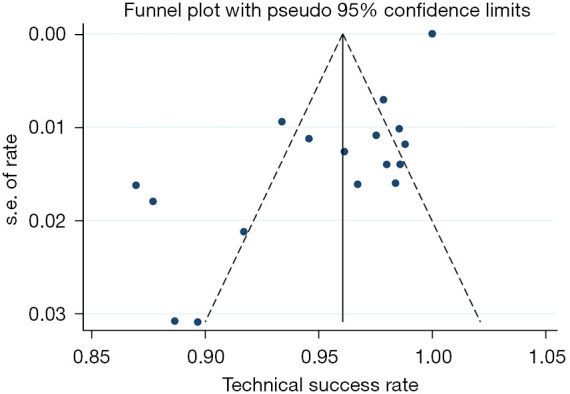
Funnel plot for technical success.
Figure 8.
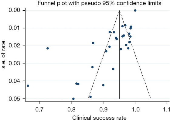
Funnel plot for clinical success.
Figure 9.
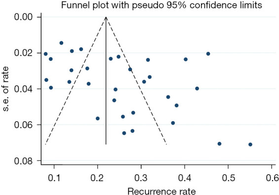
Funnel plot for recurrence.
Figure 10.
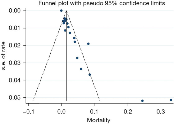
Funnel plot for mortality.
Figure 11.
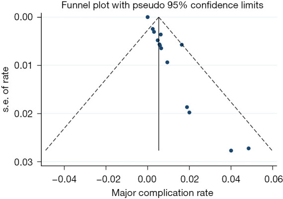
Funnel plot for major complication.
Subgroup analysis
Subgroup analysis could not find the source of heterogeneity (the subgroup analysis of technical success, clinical success, recurrence, mortality, and major complication were shown in Tables S6-S10, respectively.).
Subgroup analysis by publication year
BAE, after 2017, achieved higher technical success [98.6% (95% CI: 97.0–99.6%) vs. 97.2% (95% CI: 95.1–98.8%), P<0.001] and lower recurrence rates [19.8% (95% CI: 14.3–25.9%) vs. 31.7% (95% CI: 25.7–37.9%), P=0.006] than before. No statistically significant differences were observed in clinical success, mortality, and major complication rates.
Subgroup analysis by region
The largest number of included studies came from Asia, followed by Europe, America, and Africa. The technical success (P=0.001), recurrence (P=0.002), mortality (P<0.001), and major complication rates (P=0.02) were statistically significant among subgroups, while there was no significant difference in clinical success rate.
Subgroup analysis by amount of hemoptysis
Patients with massive hemoptysis exhibited a higher technical success rate [100.0% (95% CI: 97.9–100.0%) vs. 97.5% (95% CI: 95.7–98.8%), P=0.003] but a lower clinical success rate [90.6% (95% CI: 86.7–93.9%) vs. 96.6% (95% CI: 93.1–99.0%), P=0.01] than those with non-massive hemoptysis. At the same time, no significant differences were observed in other outcomes.
Subgroup analysis by etiology
Compared to other etiologies, patients with bronchiectasis-related hemoptysis showed higher technical success rate [99.7% (95% CI: 99.1–100.0%) vs. 97.4% (95% CI: 94.0–99.5%), P=0.02], higher clinical success rate [98.1% (95% CI: 93.4–99.8%) vs. 87.4% (95% CI: 79.4–93.7%), P=0.003], lower mortality rate [1.1% (95% CI: 0.4–2.0%) vs. 6.0% (95% CI: 1.1–13.9%), P=0.04], and lower major complication rate [0.0% (95% CI: 0.0–0.3%) vs. 0.3% (95% CI: 0.0–1.1%), P=0.050], but a higher recurrence rate [40.8% (95% CI: 37.2–44.5%) vs. 30.4% (95% CI: 24.8–36.3%), P=0.003] following BAE. The curative effect in tuberculosis patients did not surpass that of other diseases. Patients with hemoptysis caused by benign diseases showed a higher clinical success rate compared to those with malignant diseases [96.0% (95% CI: 91.9–98.7%) vs. 76.3% (95% CI: 65.2–85.8%), P<0.001]. However, no significant differences were observed between the two subgroups regarding technical success, recurrence, mortality, and major complication rates.
Subgroup analysis by embolization materials
Subgroup analysis revealed that GS exhibited decreased technical success [85.1% (95% CI: 75.0–92.3%) vs. 97.7% (95% CI: 94.6–99.6%), P=0.001], clinical success [81.2% (95% CI: 75.2–86.5%) vs. 91.2% (95% CI: 83.8–96.5%), P=0.03], and increased recurrence [35.9% (95% CI: 29.2–43.0%) vs. 19.7% (95% CI: 11.6–29.3%), P=0.007] of hemoptysis. NBCA [12.6% (95% CI: 8.4–17.5%) vs. 25.4% (95% CI: 20.1–31.2%), P=0.001] and coils [12.5% (95% CI: 8.3–17.3%) vs. 23.8% (95% CI: 17.3–31.0%), P=0.006] showed superior outcomes compared to other embolic materials in the recurrence of hemoptysis, with NBCA exhibiting a lower recurrence rate than non-absorbable particles [12.6% (95% CI: 8.4–17.5%) vs. 26.5% (95% CI: 15.7–39.0%), P=0.02]. The utilization of coils showed a higher incidence of major complication rate [1.1% (95% CI: 0.3–2.2%) vs. 0.1% (95% CI: 0.0–0.4%), P=0.007], while non-absorbable particles showed a reduced occurrence of major complication rate [0.0% (95% CI: 0.0–0.2%) vs. 0.4% (95% CI: 0.1–1.1%), P=0.02].
Discussion
BAE has been recognized as the first-line treatment for massive hemoptysis and recurrent hemoptysis. Hemostasis can be effectively achieved in most patients through BAE, with a low incidence of hemoptysis-related mortality and procedure-related major complications. However, the high postoperative recurrence rate remains unresolved. Although there have been several pertinent meta-analysis regarding BAE for the treatment of hemoptysis (56,57), the research on the factors influencing its efficacy and safety remains limited. Therefore, we conducted a comprehensive subgroup analysis to address this research gap.
The subgroup analysis showed that BAE after 2017 had a better technical success rate and lower recurrence rate than before, which was attributed to the increased utilization of preoperative contrast-enhanced computed tomography (CECT) or computed tomography angiography (CTA), proficient implementation of embolization techniques and optimized selection of appropriate embolization materials. Studies showed that the accuracy of CECT in detecting bronchial arteries was 97.5% (58) while CTA could be up to 98.8% accurate (59), which greatly reduced the BAE procedure time and X-ray exposure (59,60). Super-selective embolization was associated with improved efficacy in achieving complete embolization, reduced recanalization rates and a decreased risk of ectopic embolism. The selection of the approach and catheter was crucial for improving technical success. Research indicated that the radial or brachial artery approach could be viable options for culprit arteries originating from the subclavian or axillary arteries (46,61). Additionally, the JL4 catheter showed superior adaptability to the aortic arch morphology, providing enhanced stability for selecting ectopic bronchial arteries originate from this specific location (62).
Out of the 32 studies, 26 included patients were with massive and non-massive hemoptysis but did not report outcomes based on the amount of hemoptysis. Only six studies were available for subgroup analysis. The results showed that patients with massive hemoptysis could achieve better technical success and lower clinical success than those with non-massive hemoptysis. However, in the subgroup analysis of the technical success rate, only the study by Kucukay et al. (45) met the inclusion criteria for the group with massive hemoptysis, achieving a technical success rate of 100%. This limited representation affects the reliability of the overall study results. For patients with non-massive hemoptysis, the technical failure of BAE primarily arised from challenges in superselection due to orifice stenosis. Additional factors contributing to this failure include the presence of arterial dissection and the inability to localize a source of bleeding. Extravasation of the contrast agent was more likely to occur in patients with massive hemoptysis, facilitating the identification and embolization of the culprit artery (60,63). However, there was currently a lack of research examining the differences in bronchial artery tortuosity and hypertrophy between patients with massive and non-massive hemoptysis. Further investigation was warranted. The study conducted by Park et al. (23) demonstrated that BAE treatment in patients with non-massive hemoptysis yielded significant short-term efficacy, and early embolization (<24 h) not only reduced the length of hospital stay but also significantly decreased the rate of early recurrence (<3 months) compared to delayed embolization. A second embolization was still effective in patients with recurrent hemoptysis (40). Moreover, the state of hemoptysis exhibited instability and carried the likelihood of progression to life-threatening hemoptysis. Yan et al. (64) compared BAE and conservative treatment in patients with non-massive hemoptysis caused by bronchiectasis and revealed that BAE demonstrated superior long-term control of hemoptysis. Fartoukh et al. (65) found that the combination of BAE and medication was significantly more efficacious than medication alone in preventing the recurrence of mild hemoptysis at both 30 days (recurrence rate: 11.8% vs. 44.7%) and 90 days (hemoptysis-free survival: 91.2% vs. 60.2%). Therefore, BAE could be considered in non-massive hemoptysis patients who have shown a poor response to conservative treatment alone, exhibited indications for interventional therapy, presented with long-term hemoptysis affecting quality of life, and patients with a risk of progression to massive hemoptysis.
Bronchiectasis was one of the most common causes of hemoptysis. The US Bronchiectasis Research Registry reported that 23% of patients with bronchiectasis have a history of hemoptysis and exhibit a notable recurrence rate (66). The results showed that bronchiectasis resulted in better short-term outcomes, albeit with a higher recurrence rate. However, it is noteworthy that the follow-up duration for studies on hemoptysis caused by bronchiectasis exceeded that of other studies, which might have influenced the outcomes. Furthermore, the severity of bronchiectasis and its association with infection were significant predictors of hemoptysis recurrence and hospitalization mortality (32,67). Therefore, it was crucial to implement clinical interventions to delay the progression of bronchiectasis, particularly in patients presenting with hemoptysis. As such, BAE may be a viable treatment option for managing bronchiectasis.
Tuberculosis was also a prevalent cause of hemoptysis, especially in developing countries. The results showed that tuberculosis and its sequelae do not exhibit superior therapeutic outcomes compared to other etiologies. Nevertheless, it was undeniable that BAE remained a safe and productive approach for managing tuberculosis-related hemoptysis. Research demonstrated that active tuberculosis was associated with a lower recurrence rate of hemoptysis when compared to tuberculosis sequelae due to the effective anti-tuberculosis therapy provided alongside BAE. Diabetes mellitus, pulmonary cavity formation accompanied by fungal infection, and aggressive pleural thickening were associated with an elevated recurrence rate, and should be well managed. Additionally, surgical intervention should be considered in cases of aggressive pleural thickening (43,50).
BAE for hemoptysis was extensively discussed in patients with benign lung diseases, and BAE was typically considered a secondary treatment option following the failure of other therapies in patients with lung malignancies. Compared to other diseases, lung malignancies exhibited distinctive characteristics, including a significant prevalence of non-bronchial artery hypertrophy and pulmonary artery hemoptysis, propensity for collateral circulation formation, and challenges in follow-up management (21,51,68-70). Three studies for lung malignancies in the meta-analysis were all published within the past 5 years, indicating an increasing focus on BAE in patients with lung malignancies. The clinical success rate of BAE treatment for benign lung diseases was superior to malignancies, while no statistical difference was found for other outcomes. The potential sources of bias in the results might include the following: firstly, the etiology of the included studies was heterogeneous, with a limited number of studies and a small sample size available for subgroup analysis; secondly, advancements in super-selective embolization technology have augmented the efficacy of culprit artery embolization for malignant tumors; thirdly, there were significant variations in follow-up duration among the included studies; fourthly, the rapid progression of malignancies might impact patients’ primary cause of death. Le Tat et al. (21) observed that the one-year survival rate was significantly higher in the clinically successful group than in the clinically unsuccessful group (37% vs. 8%); BAE exhibited potential for both short- and long-term efficacy in patients with malignancies. In addition to simple embolization, some scholars have explored the efficacy of bronchial artery chemoembolization (BACE) in patients with lung malignancies accompanied by hemoptysis in recent years. In a study involving 187 patients with hemoptysis due to lung cancer, Xiaobing et al. (71) reported that BACE achieved significant disease control (87.7%) and remission rates (73.8%). Another study conducted by Yu et al. (72) comparing chemoembolization with embolization alone also demonstrated that chemoembolization resulted in a higher disease control rate (50.0% vs. 31.0%), higher disease response rate (100% vs. 72.4%) and longer median overall survival time (28.5 vs. 22.5 months). Chemotherapy combined with embolization could effectively reduce lesion volume, alleviate atelectasis, and improve outcomes for patients with advanced disease and those with poor surgical candidates (73,74). Studies on the selection of chemotherapy drugs and their compatibility with embolic materials are currently limited; large-scale clinical controlled trials are still needed in the future.
As an absorbable embolic material, GS exhibited relatively low technical and clinical success rates, accompanied by a potential risk of early recurrence. Conversely, non-absorbable embolic agents demonstrated superior short-term outcomes, aligning with previous studies (19,51). However, its structural composition could facilitate platelet aggregation and fibrin deposition, thereby accelerating clot formation compared to alternative embolization materials. For cryptic hemoptysis, Nagano et al. (75) found that GS exhibited comparable effectiveness, a low risk of recurrence, and a minimal occurrence of serious complications compared to other non-absorbable materials because it was believed that cryptogenic hemoptysis had no underlying abnormal cause that could lead to repeated hemoptysis. However, with only 22 patients in the study and 11 of them receiving PVA, the small sample size introduces factors that might affect the reliability of the conclusions. Shimohira et al.’s study (76) on hemoptysis caused by pulmonary aspergilloma revealed that GS for BAE was ineffective. However, their study was still limited by its small sample size. Therefore, insufficient empirical evidence supports using a single GS as an effective material for hemoptysis embolization.
Coils showed a lower recurrence rate in our meta-analysis. However, the challenges associated with retreatment following the recurrence of hemoptysis compared to alternative embolization materials have hindered their widespread adoption as mainstream options in clinical practice and were selected as a sealing agent for cases involving evident artery hypertrophy, Rasmussen’s aneurysm, pseudoaneurysm or pulmonary arteriovenous malformation. Ishikawa et al.’s study (19) found that the proportion of coils for BAE decreased from 19% to 16% between 2011 and 2017. Nevertheless, several studies revealed that the efficacy and safety of the coil were comparable to those of other embolic materials (19,51). Even in patients with recurrent hemoptysis, repeated coil embolization could still achieve a high success rate (97.7%) (19), particularly following the emergence of micro coils (31,46). Additionally, it was important to note that all the studies using coils alone for embolization were from Japan, as PVA and microspheres were not approved for Japanese public health insurance. The effectiveness of coil embolization might differ by region. Coils were associated with a higher major complication rate in the subgroup analysis. However, among the eight patients experiencing major complications in Ishikawa et al.’s study (46), only one case was linked to the symptomatic cerebellar infarction caused by ectopic embolization of a postoperative thrombus near the vertebral artery using coils, which introduced interference with the study outcomes. This complication could also occur with other embolization materials (77,78). Hence, vigilance should be exercised regarding the risk of cerebellar infarction after embolization of arteries adjacent to the vertebral artery. Mediastinal hematoma (five cases), a significant component of serious complications in Ishikawa et al.’ study, could be resolved through coil embolization. Furthermore, as a flow-independent embolic agent, coils exhibited a lower incidence of spinal cord infarction (19), a critical complication necessitating thorough evaluation during the BAE procedure. The utilization of detachable coils even enhanced the precision and safety of embolization. Additionally, employing coils for sealing after distal embolization with non-absorbable particles may diminish the probability of recanalization and the risk of ectopic embolization resulting from reflux (31).
PVA has emerged as the most frequently employed embolic material, which exhibited excellent histocompatibility. However, its irregular shape posed a significant risk of catheter occlusion. Microspheres were adjusted to uniform, regular, non-deformable shapes with anti-aggregation properties, reducing the risk of catheter obstruction. After comparing PVA and microspheres, Fu et al. and Park et al. observed no significant differences in efficacy or safety (23,24). NBCA was semifluid and mixed with iodized oil (at ratios of approximately 1:2–1:5) to adjust the concentration according to the flow rate, diameter, and the distal advancement of the microcatheter of culprit arteries (12,34,39,79). The results revealed that NBCA was related to a lower recurrence rate due to its dense embolization of the distal part of the culprit vessel. The supplementary meta-analysis on non-absorbable particles and NBCA observed that NBCA demonstrated a more stable curative effect than non-absorbable particles and did not increase the risk of complications. Moreover, Lee et al. (34) found that NBCA had superior hemostatic efficacy and lower recurrence rates than PVA in patients of lung malignancies with coagulopathy due to its ability to achieve hemostasis independently of blood coagulation (80). NBCA showed significant potential for enhancing the effectiveness of BAE procedures. However, the application of NBCA in patients with hemoptysis is not as widespread as that of other embolization materials due to concerns regarding premature polymerization, adhesion of the microcatheter tip to the glue cast, and complications such as non-target embolization and tissue necrosis (79). Therefore, its use is generally not recommended for the treatment of hemoptysis for individuals needing more experience with NBCA in the CIRSE Standards of Practice (18). The study conducted by Shamseldin et al. (81) proposed the implementation of standardized operating procedures for NBCA, including a rapid flush with 5% dextrose solution followed by the slow injection of the mixture. Additionally, simultaneous extubation during withdrawal or injection mixture administration should be avoided to minimize complications and pitfalls. Kolu et al. (82) employed a mixture of NBCA and iodide at a ratio of 1:14 to achieve enhanced embolization of the distal vascular bed and minimize reflux. However, more studies are currently needed to investigate the efficacy and safety of mixtures with varying concentrations. With the increasing standardization of practice among interventional radiologists, the future application of NBCA in BAE processes is anticipated to become more extensive.
Systemic artery-to-pulmonary fistula (SA-PF) was uncommon and caused by the progression of chronic inflammation or infection, cancer, post-traumatic, or congenital disease (83-85). Several studies have suggested that the presence of SA-PF may contribute to hemoptysis recurrence and an increased risk of ectopic embolism, including pulmonary embolism and systemic arterial embolism (31,33,37,47,86,87) due to embolic material instability and dislocation under intravascular pressure differences (88). Large-diameter non-absorbable particles were found to facilitate embolization (33,35,89). Tayal et al. (41) used microspheres with a size range of 500–700 µm first and followed by 300–500 µm for prompt occlusion of systemic artery-to-pulmonary artery fistulas while minimizing subclinical embolization and severe ectopic embolization. Kucukay et al. (45) also used 700–900 µm microspheres to effectively embolize the culprit artery, and no recurrence was observed during the 6-month follow-up period. Therefore, large-diameter, non-absorbable particles could be considered in cases where SA-PF was detected on preoperative CTA or angiography.
This systematic review and meta-analysis had several limitations. Firstly, some subgroups had fewer studies due to inconsistent data on hemoptysis amount, etiology and embolization materials, which affected the results. Secondly, heterogeneity in defining clinical success rate and follow-up time across studies impacted result accuracy. Therefore, consensus on quantifying hemoptysis and defining outcome, along with larger-scale studies with increased sample sizes, is necessary to validate the efficacy and safety of BAE in managing hemoptysis. Nevertheless, the comprehensive subgroup analysis indicated the potential efficacy of BAE across different etiologies and highlighted the benefits of various embolization materials, supporting future treatment decisions for patients with hemoptysis.
Conclusions
BAE is safe and effective in treating of different degrees of hemoptysis caused by benign and malignant lesions. The use of GS embolization alone is currently not recommended; NBCA demonstrates a favorable therapeutic effect, and shows a decreased likelihood of recurrence compared to particle embolic agents without an increase in the risk of BAE. However, it is necessary to establish standardized training protocols for interventional radiologists.
Supplementary
The article’s supplementary files as
Acknowledgments
We would like to thank Ching-Hsuan Lin for his help in polishing our paper.
Funding: This work was partially supported by the Research Project of Zhejiang Chinese Medical University (No. 2022JKJNTZ20).
Ethical Statement: The authors are accountable for all aspects of the work in ensuring that questions related to the accuracy or integrity of any part of the work are appropriately investigated and resolved.
Footnotes
Reporting Checklist: The authors have completed the PRISMA reporting checklist. Available at https://cdt.amegroups.com/article/view/10.21037/cdt-24-157/rc
Conflicts of Interest: All authors have completed the ICMJE uniform disclosure form (available at https://cdt.amegroups.com/article/view/10.21037/cdt-24-157/coif). J.L. reports funding support from the Research Project of Zhejiang Chinese Medical University (No. 2022JKJNTZ20). The other authors have no conflicts of interest to declare.
References
- 1.Expert Panel on Thoracic Imaging ; Olsen KM, Manouchehr-Pour S, et al. ACR Appropriateness Criteria® Hemoptysis. J Am Coll Radiol 2020;17:S148-59. 10.1016/j.jacr.2020.01.043 [DOI] [PubMed] [Google Scholar]
- 2.Kathuria H, Hollingsworth HM, Vilvendhan R, et al. Management of life-threatening hemoptysis. J Intensive Care 2020;8:23. 10.1186/s40560-020-00441-8 [DOI] [PMC free article] [PubMed] [Google Scholar]
- 3.Yoon W, Kim JK, Kim YH, et al. Bronchial and nonbronchial systemic artery embolization for life-threatening hemoptysis: a comprehensive review. Radiographics 2002;22:1395-409. 10.1148/rg.226015180 [DOI] [PubMed] [Google Scholar]
- 4.Bruzzi JF, Rémy-Jardin M, Delhaye D, et al. Multi-detector row CT of hemoptysis. Radiographics 2006;26:3-22. 10.1148/rg.261045726 [DOI] [PubMed] [Google Scholar]
- 5.Sakr L, Dutau H. Massive hemoptysis: an update on the role of bronchoscopy in diagnosis and management. Respiration 2010;80:38-58. 10.1159/000274492 [DOI] [PubMed] [Google Scholar]
- 6.Abdulmalak C, Cottenet J, Beltramo G, et al. Haemoptysis in adults: a 5-year study using the French nationwide hospital administrative database. Eur Respir J 2015;46:503-11. 10.1183/09031936.00218214 [DOI] [PubMed] [Google Scholar]
- 7.Mondoni M, Carlucci P, Job S, et al. Observational, multicentre study on the epidemiology of haemoptysis. Eur Respir J 2018;51:1701813. 10.1183/13993003.01813-2017 [DOI] [PubMed] [Google Scholar]
- 8.Vanni S, Bianchi S, Bigiarini S, et al. Management of patients presenting with haemoptysis to a Tertiary Care Italian Emergency Department: the Florence Haemoptysis Score (FLHASc). Intern Emerg Med 2018;13:397-404. [DOI] [PubMed] [Google Scholar]
- 9.Fujii H, Hara Y, Obase Y, et al. Nationwide survey in Japan of the causative diseases of bloody sputum and hemoptysis in departments of respiratory medicine at university hospitals and core hospitals. Respir Investig 2024;62:395-401. 10.1016/j.resinv.2024.02.003 [DOI] [PubMed] [Google Scholar]
- 10.Singh SK, Tiwari KK. Etiology of hemoptysis: A retrospective study from a tertiary care hospital from northern Madhya Pradesh, India. Indian J Tuberc 2016;63:44-7. 10.1016/j.ijtb.2016.02.007 [DOI] [PubMed] [Google Scholar]
- 11.Chun JY, Belli AM. Immediate and long-term outcomes of bronchial and non-bronchial systemic artery embolisation for the management of haemoptysis. Eur Radiol 2010;20:558-65. 10.1007/s00330-009-1591-3 [DOI] [PubMed] [Google Scholar]
- 12.Woo S, Yoon CJ, Chung JW, et al. Bronchial artery embolization to control hemoptysis: comparison of N-butyl-2-cyanoacrylate and polyvinyl alcohol particles. Radiology 2013;269:594-602. 10.1148/radiol.13130046 [DOI] [PubMed] [Google Scholar]
- 13.Davidson K, Shojaee S. Managing Massive Hemoptysis. Chest 2020;157:77-88. 10.1016/j.chest.2019.07.012 [DOI] [PubMed] [Google Scholar]
- 14.Deshwal H, Sinha A, Mehta AC. Life-Threatening Hemoptysis. Semin Respir Crit Care Med 2021;42:145-59. 10.1055/s-0040-1714386 [DOI] [PubMed] [Google Scholar]
- 15.Fernando HC, Stein M, Benfield JR, et al. Role of bronchial artery embolization in the management of hemoptysis. Arch Surg 1998;133:862-6. 10.1001/archsurg.133.8.862 [DOI] [PubMed] [Google Scholar]
- 16.Remy J, Voisin C, Ribet M, et al. Treatment, by embolization, of severe or repeated hemoptysis associated with systemic hypervascularization. Nouv Presse Med 1973;2:2060. [PubMed] [Google Scholar]
- 17.Omachi N, Ishikawa H, Hara M, et al. The impact of bronchial artery embolisation on the quality of life of patients with haemoptysis: a prospective observational study. Eur Radiol 2021;31:5351-60. 10.1007/s00330-020-07533-x [DOI] [PMC free article] [PubMed] [Google Scholar]
- 18.Kettenbach J, Ittrich H, Gaubert JY, et al. CIRSE Standards of Practice on Bronchial Artery Embolisation. Cardiovasc Intervent Radiol 2022;45:721-32. 10.1007/s00270-022-03127-w [DOI] [PMC free article] [PubMed] [Google Scholar]
- 19.Ishikawa H, Ohbe H, Omachi N, et al. Spinal Cord Infarction after Bronchial Artery Embolization for Hemoptysis: A Nationwide Observational Study in Japan. Radiology 2021;298:673-9. 10.1148/radiol.2021202500 [DOI] [PubMed] [Google Scholar]
- 20.Panda A, Bhalla AS, Goyal A. Bronchial artery embolization in hemoptysis: a systematic review. Diagn Interv Radiol 2017;23:307-17. 10.5152/dir.2017.16454 [DOI] [PMC free article] [PubMed] [Google Scholar]
- 21.Le Tat T, Carlier R, Zhang N, et al. Endovascular Management of Life-Threatening Hemoptysis in Primary Lung Cancer: A Retrospective Study. Cardiovasc Intervent Radiol 2023;46:891-900. 10.1007/s00270-023-03488-w [DOI] [PubMed] [Google Scholar]
- 22.Pathak V, Stavas JM, Ford HJ, et al. Long-term outcomes of the bronchial artery embolization are diagnosis dependent. Lung India 2016;33:3-8. 10.4103/0970-2113.173059 [DOI] [PMC free article] [PubMed] [Google Scholar]
- 23.Park SJ, Lee S, Lee HN, et al. Early versus delayed bronchial artery embolization for non-massive hemoptysis. Eur Radiol 2023;33:116-24. 10.1007/s00330-022-08993-z [DOI] [PubMed] [Google Scholar]
- 24.Fu Z, Li X, Cai F, et al. Microspheres present comparable efficacy and safety profiles compared with polyvinyl alcohol for bronchial artery embolization treatment in hemoptysis patients. J Transl Med 2021;19:422. 10.1186/s12967-021-02947-7 [DOI] [PMC free article] [PubMed] [Google Scholar]
- 25.Angle JF, Siddiqi NH, Wallace MJ, et al. Quality improvement guidelines for percutaneous transcatheter embolization: Society of Interventional Radiology Standards of Practice Committee. J Vasc Interv Radiol 2010;21:1479-86. 10.1016/j.jvir.2010.06.014 [DOI] [PubMed] [Google Scholar]
- 26.Stang A. Critical evaluation of the Newcastle-Ottawa scale for the assessment of the quality of nonrandomized studies in meta-analyses. Eur J Epidemiol 2010;25:603-5. 10.1007/s10654-010-9491-z [DOI] [PubMed] [Google Scholar]
- 27.Higgins JP, Thompson SG. Quantifying heterogeneity in a meta-analysis. Stat Med 2002;21:1539-58. 10.1002/sim.1186 [DOI] [PubMed] [Google Scholar]
- 28.Cordero CP, Dans AL. Key concepts in clinical epidemiology: detecting and dealing with heterogeneity in meta-analyses. J Clin Epidemiol 2021;130:149-51. 10.1016/j.jclinepi.2020.09.045 [DOI] [PubMed] [Google Scholar]
- 29.Borenstein M, Hedges LV, Higgins JP, et al. A basic introduction to fixed-effect and random-effects models for meta-analysis. Res Synth Methods 2010;1:97-111. 10.1002/jrsm.12 [DOI] [PubMed] [Google Scholar]
- 30.DerSimonian R, Laird N. Meta-analysis in clinical trials revisited. Contemp Clin Trials 2015;45:139-45. 10.1016/j.cct.2015.09.002 [DOI] [PMC free article] [PubMed] [Google Scholar]
- 31.Yan HT, Lu GD, Liu J, et al. Does the presence of systemic artery-pulmonary circulation shunt during bronchial arterial embolization increase the recurrence of noncancer-related hemoptysis? A retrospective cohort study. Respir Res 2023;24:119. 10.1186/s12931-023-02427-0 [DOI] [PMC free article] [PubMed] [Google Scholar]
- 32.Wang LL, Lu HW, Li LL, et al. Pseudomonas aeruginosa isolation is an important predictor for recurrent hemoptysis after bronchial artery embolization in patients with idiopathic bronchiectasis: a multicenter cohort study. Respir Res 2023;24:84. 10.1186/s12931-023-02391-9 [DOI] [PMC free article] [PubMed] [Google Scholar]
- 33.Zhang CJ, Jiang FM, Zuo ZJ, et al. Clinical characteristics and postoperative outcomes of systemic artery-to-pulmonary vessel fistula in hemoptysis patients. Eur Radiol 2022;32:4304-13. 10.1007/s00330-021-08484-7 [DOI] [PubMed] [Google Scholar]
- 34.Lee JH, Yoon CJ, Jung YS, et al. Comparison of n-butyl-2-cyanoacrylate and polyvinyl alcohol particles for bronchial artery embolisation in primary lung cancer: a retrospective cohort study. Respir Res 2022;23:257. 10.1186/s12931-022-02183-7 [DOI] [PMC free article] [PubMed] [Google Scholar]
- 35.Sarioglu O, Capar AE, Yavuz MY, et al. Angiographic Findings and Outcomes of Bronchial Artery Embolization in Patients with Pulmonary Tuberculosis. Eurasian J Med 2020;52:126-31. 10.5152/eurasianjmed.2020.19221 [DOI] [PMC free article] [PubMed] [Google Scholar]
- 36.Choi J, Baik JH, Kim CH, et al. Long-term outcomes and prognostic factors in patients with mild hemoptysis. Am J Emerg Med 2018;36:1160-5. 10.1016/j.ajem.2017.11.053 [DOI] [PubMed] [Google Scholar]
- 37.Chen J, Chen LA, Liang ZX, et al. Immediate and long-term results of bronchial artery embolization for hemoptysis due to benign versus malignant pulmonary diseases. Am J Med Sci 2014;348:204-9. 10.1097/MAJ.0000000000000226 [DOI] [PubMed] [Google Scholar]
- 38.Hahn S, Kim YJ, Kwon W, et al. Comparison of the effectiveness of embolic agents for bronchial artery embolization: gelfoam versus polyvinyl alcohol. Korean J Radiol 2010;11:542-6. 10.3348/kjr.2010.11.5.542 [DOI] [PMC free article] [PubMed] [Google Scholar]
- 39.García Jurado PB, Pérez Montilla ME, Lombardo Galera MS, et al. Embolization of bronchial arteries and nonbronchial systemic arteries with n-butyl-cyanoacrylate in patients with hemoptysis: A retrospective single-center study. Radiologia (Engl Ed) 2023;65:99-105. 10.1016/j.rxeng.2020.12.003 [DOI] [PubMed] [Google Scholar]
- 40.Hwang JH, Kim JH, Park S, et al. Feasibility and outcomes of bronchial artery embolization in patients with non-massive hemoptysis. Respir Res 2021;22:221. 10.1186/s12931-021-01820-x [DOI] [PMC free article] [PubMed] [Google Scholar]
- 41.Tayal M, Chauhan U, Sharma P, et al. Bronchial artery embolization. What further we can offer? Wideochir Inne Tech Maloinwazyjne 2020;15:478-87. [DOI] [PMC free article] [PubMed] [Google Scholar]
- 42.Takeda K, Kawashima M, Masuda K, et al. Long-Term Outcomes of Bronchial Artery Embolization for Patients with Non-Mycobacterial Non-Fungal Infection Bronchiectasis. Respiration 2020;99:961-9. 10.1159/000511132 [DOI] [PubMed] [Google Scholar]
- 43.Peng Y, Zhu Y, Ao G, et al. Effect of bronchial artery embolisation on the management of tuberculosis-related haemoptysis. Int J Tuberc Lung Dis 2019;23:1269-76. 10.5588/ijtld.19.0135 [DOI] [PubMed] [Google Scholar]
- 44.Han K, Yoon KW, Kim JH, et al. Bronchial Artery Embolization for Hemoptysis in Primary Lung Cancer: A Retrospective Review of 84 Patients. J Vasc Interv Radiol 2019;30:428-34. 10.1016/j.jvir.2018.08.022 [DOI] [PubMed] [Google Scholar]
- 45.Kucukay F, Topcuoglu OM, Alpar A, et al. Bronchial Artery Embolization with Large Sized (700-900 µm) Tris-acryl Microspheres (Embosphere) for Massive Hemoptysis: Long-Term Results (Clinical Research). Cardiovasc Intervent Radiol 2018;41:225-30. 10.1007/s00270-017-1818-7 [DOI] [PubMed] [Google Scholar]
- 46.Ishikawa H, Hara M, Ryuge M, et al. Efficacy and safety of super selective bronchial artery coil embolisation for haemoptysis: a single-centre retrospective observational study. BMJ Open 2017;7:e014805. 10.1136/bmjopen-2016-014805 [DOI] [PMC free article] [PubMed] [Google Scholar]
- 47.Dabó H, Gomes R, Marinho A, et al. Bronchial artery embolisation in management of hemoptysis--A retrospective analysis in a tertiary university hospital. Rev Port Pneumol (2006) 2016;22:34-8. [DOI] [PubMed] [Google Scholar]
- 48.Tom LM, Palevsky HI, Holsclaw DS, et al. Recurrent Bleeding, Survival, and Longitudinal Pulmonary Function following Bronchial Artery Embolization for Hemoptysis in a U.S. Adult Population. J Vasc Interv Radiol 2015;26:1806-13.e1. 10.1016/j.jvir.2015.08.019 [DOI] [PubMed] [Google Scholar]
- 49.Bhalla A, Kandasamy D, Veedu P, et al. A retrospective analysis of 334 cases of hemoptysis treated by bronchial artery embolization. Oman Med J 2015;30:119-28. 10.5001/omj.2015.26 [DOI] [PMC free article] [PubMed] [Google Scholar]
- 50.Pei R, Zhou Y, Wang G, et al. Outcomes of bronchial artery embolization for life-threatening hemoptysis secondary to tuberculosis. PLoS One 2014;9:e115956. 10.1371/journal.pone.0115956 [DOI] [PMC free article] [PubMed] [Google Scholar]
- 51.Garcia-Olivé I, Sanz-Santos J, Centeno C, et al. Predictors of recanalization in patients with life-threatening hemoptysis requiring artery embolization. Arch Bronconeumol 2014;50:51-6. 10.1016/j.arbr.2014.01.004 [DOI] [PubMed] [Google Scholar]
- 52.Racil H, Rajhi H, Ben Naceur R, et al. Endovascular treatment of haemoptysis: medium and long-term assessment. Diagn Interv Imaging 2013;94:38-44. 10.1016/j.diii.2012.05.010 [DOI] [PubMed] [Google Scholar]
- 53.Cornalba GP, Vella A, Barbosa F, et al. Bronchial and nonbronchial systemic artery embolization in managing haemoptysis: 31 years of experience. Radiol Med 2013;118:1171-83. 10.1007/s11547-012-0866-y [DOI] [PubMed] [Google Scholar]
- 54.Anuradha C, Shyamkumar NK, Vinu M, et al. Outcomes of bronchial artery embolization for life-threatening hemoptysis due to tuberculosis and post-tuberculosis sequelae. Diagn Interv Radiol 2012;18:96-101. [DOI] [PubMed] [Google Scholar]
- 55.Shin BS, Jeon GS, Lee SA, et al. Bronchial artery embolisation for the management of haemoptysis in patients with pulmonary tuberculosis. Int J Tuberc Lung Dis 2011;15:1093-8. 10.5588/ijtld.10.0659 [DOI] [PubMed] [Google Scholar]
- 56.Zheng Z, Zhuang Z, Yang M, et al. Bronchial artery embolization for hemoptysis: A systematic review and meta-analysis. J Interv Med 2021;4:172-80. 10.1016/j.jimed.2021.08.003 [DOI] [PMC free article] [PubMed] [Google Scholar]
- 57.Karlafti E, Tsavdaris D, Kotzakioulafi E, et al. Which Is the Best Way to Treat Massive Hemoptysis? A Systematic Review and Meta-Analysis of Observational Studies. J Pers Med 2023;13:1649. 10.3390/jpm13121649 [DOI] [PMC free article] [PubMed] [Google Scholar]
- 58.Le HY, Le VN, Pham NH, et al. Value of multidetector computed tomography angiography before bronchial artery embolization in hemoptysis management and early recurrence prediction: a prospective study. BMC Pulm Med 2020;20:231. 10.1186/s12890-020-01271-y [DOI] [PMC free article] [PubMed] [Google Scholar]
- 59.Li PJ, Yu H, Wang Y, et al. Multidetector computed tomography angiography prior to bronchial artery embolization helps detect culprit ectopic bronchial arteries and non-bronchial systemic arteries originating from subclavian and internal mammary arteries and improve hemoptysis-free early survival rate in patients with hemoptysis. Eur Radiol 2019;29:1950-8. 10.1007/s00330-018-5767-6 [DOI] [PubMed] [Google Scholar]
- 60.Ittrich H, Klose H, Adam G. Radiologic management of haemoptysis: diagnostic and interventional bronchial arterial embolisation. Rofo 2015;187:248-59. [DOI] [PubMed] [Google Scholar]
- 61.Lee Y, Lee M, Hur S, et al. Bronchial and non-bronchial systemic artery embolization with transradial access in patients with hemoptysis. Diagn Interv Radiol 2022;28:359-63. 10.5152/dir.2022.201100 [DOI] [PMC free article] [PubMed] [Google Scholar]
- 62.An J, Dong Y, Niu H. Application of the 5F JL4 Catheter in Bronchial Artery Embolization With the Opening in the Inferior Wall of the Aortic Arch. Vasc Endovascular Surg 2023;57:379-85. 10.1177/15385744221149910 [DOI] [PubMed] [Google Scholar]
- 63.Lee MK, Kim SH, Yong SJ, et al. Moderate hemoptysis: recurrent hemoptysis and mortality according to bronchial artery embolization. Clin Respir J 2015;9:53-64. 10.1111/crj.12104 [DOI] [PubMed] [Google Scholar]
- 64.Yan HT, Lu GD, Zhang JX, et al. Comparison of Bronchial Artery Embolisation Versus Conservative Treatment for Bronchiectasis-Related Nonmassive Haemoptysis: A Single-Centre Retrospective Study. Cardiovasc Intervent Radiol 2023;46:369-76. 10.1007/s00270-023-03361-w [DOI] [PubMed] [Google Scholar]
- 65.Fartoukh M, Demoule A, Sanchez O, et al. Randomised trial of first-line bronchial artery embolisation for non-severe haemoptysis of mild abundance. BMJ Open Respir Res 2021;8:e000949. 10.1136/bmjresp-2021-000949 [DOI] [PMC free article] [PubMed] [Google Scholar]
- 66.Aksamit TR, O'Donnell AE, Barker A, et al. Adult Patients With Bronchiectasis: A First Look at the US Bronchiectasis Research Registry. Chest 2017;151:982-92. 10.1016/j.chest.2016.10.055 [DOI] [PMC free article] [PubMed] [Google Scholar]
- 67.Park Y, Yong SH, Leem AY, et al. Impact of non-cystic fibrosis bronchiectasis on critically ill patients in Korea: a retrospective observational study. Sci Rep 2021;11:15757. 10.1038/s41598-021-95366-z [DOI] [PMC free article] [PubMed] [Google Scholar]
- 68.Razazi K, Parrot A, Khalil A, et al. Severe haemoptysis in patients with nonsmall cell lung carcinoma. Eur Respir J 2015;45:756-64. 10.1183/09031936.00010114 [DOI] [PubMed] [Google Scholar]
- 69.Singer ED, Faiz SA, Qdaisat A, et al. Hemoptysis in Cancer Patients. Cancers (Basel) 2023;15:4765. 10.3390/cancers15194765 [DOI] [PMC free article] [PubMed] [Google Scholar]
- 70.Gershman E, Guthrie R, Swiatek K, et al. Management of hemoptysis in patients with lung cancer. Ann Transl Med 2019;7:358. 10.21037/atm.2019.04.91 [DOI] [PMC free article] [PubMed] [Google Scholar]
- 71.Xiaobing L, Meipan Y, Pengfei X, et al. Bronchial Artery Chemoembolization for Hemoptysis in Advanced Primary Lung Cancer. Clin Lung Cancer 2022;23:e203-9. 10.1016/j.cllc.2021.10.011 [DOI] [PubMed] [Google Scholar]
- 72.Yu G, Shen Y, Chen L, et al. Drug-eluting beads bronchial arterial chemoembolization vs. conventional bronchial arterial chemoembolization in the treatment of advanced non-small cell lung cancer. Front Med (Lausanne) 2023;10:1201468. 10.3389/fmed.2023.1201468 [DOI] [PMC free article] [PubMed] [Google Scholar]
- 73.Seki A, Shimono C. Transarterial chemoembolization for management of hemoptysis: initial experience in advanced primary lung cancer patients. Jpn J Radiol 2017;35:495-504. 10.1007/s11604-017-0659-2 [DOI] [PubMed] [Google Scholar]
- 74.Nezami N, Georgiades C, Hong KK, et al. Bronchial Artery Chemoembolization With Radiopaque Doxorubicin Eluding Beads in Patients With Malignant Hemoptysis from Metastatic Lung Cancer. Technol Cancer Res Treat 2022;21:15330338221131167. 10.1177/15330338221131167 [DOI] [PMC free article] [PubMed] [Google Scholar]
- 75.Nagano N, Suzuki M, Yamamoto S, et al. Short- and long-term efficacy of bronchial artery embolization using a gelatin sponge for the treatment of cryptogenic hemoptysis. Glob Health Med 2022;4:315-21. 10.35772/ghm.2022.01057 [DOI] [PMC free article] [PubMed] [Google Scholar]
- 76.Shimohira M, Ohta K, Nagai K, et al. Bronchial arterial embolization using a gelatin sponge for hemoptysis from pulmonary aspergilloma: comparison with other pulmonary diseases. Emerg Radiol 2019;26:501-6. 10.1007/s10140-019-01695-y [DOI] [PubMed] [Google Scholar]
- 77.Nisar T. A Rare Case of Posterior Circulation Stroke Caused by Bronchial Artery Embolization. J Stroke Cerebrovasc Dis 2018;27:e153-5. 10.1016/j.jstrokecerebrovasdis.2018.02.040 [DOI] [PubMed] [Google Scholar]
- 78.Yu L, Li X, Lin F, et al. Posterior circulation infarction after bronchial artery embolization. Acta Radiol Open 2023;12:20584601231168968. 10.1177/20584601231168968 [DOI] [PMC free article] [PubMed] [Google Scholar]
- 79.Mazıcan M, Karluka I, Fındıkcıoglu A, et al. Bronchial artery embolization in patients with life-threatening massive hemoptysis: comparison of the efficacy and safety of particulate embolizing agents and n-2-butyl-cyanoacrylate. Eur Rev Med Pharmacol Sci 2024;28:310-8. [DOI] [PubMed] [Google Scholar]
- 80.Kanematsu M, Watanabe H, Kondo H, et al. Postpartum hemorrhage in coagulopathic patients: preliminary experience with uterine arterial embolization with N-butyl cyanoacrylate. J Vasc Interv Radiol 2011;22:1773-6. 10.1016/j.jvir.2011.08.016 [DOI] [PubMed] [Google Scholar]
- 81.Shamseldin M, Kluge J, Bauer JU, et al. Efficacy and safety of treating acute haemoptysis using glue embolization: A retrospective observational study and comparison to the literature. J Med Imaging Radiat Oncol 2024;68:177-84. 10.1111/1754-9485.13611 [DOI] [PubMed] [Google Scholar]
- 82.Kolu M, Kurtuluş Ş, Dere O, et al. Embolization with more diluted glue-lipiodol in patients with massive hemoptysis: single center experience results. Eur Rev Med Pharmacol Sci 2022;26:1543-8. [DOI] [PubMed] [Google Scholar]
- 83.Jacheć W, Tomasik A, Kurzyna M, et al. The multiple systemic artery to pulmonary artery fistulas resulting in severe irreversible pulmonary arterial hypertension in patient with previous history of pneumothorax. BMC Pulm Med 2019;19:80. 10.1186/s12890-019-0832-8 [DOI] [PMC free article] [PubMed] [Google Scholar]
- 84.Riehl G, Chaffanjon P, Frey G, et al. Postoperative systemic artery to pulmonary vessel fistula: analysis of three cases. Ann Thorac Surg 2003;76:1873-7. 10.1016/S0003-4975(03)01056-7 [DOI] [PubMed] [Google Scholar]
- 85.Hearne SF, Burbank MK. Internal mammary artery-to-pulmonary artery fistulas. Case report and review of the literature. Circulation 1980;62:1131-5. 10.1161/01.CIR.62.5.1131 [DOI] [PubMed] [Google Scholar]
- 86.Baltacioğlu F, Cimşit NC, Bostanci K, et al. Transarterial microcatheter glue embolization of the bronchial artery for life-threatening hemoptysis: technical and clinical results. Eur J Radiol 2010;73:380-4. 10.1016/j.ejrad.2008.10.017 [DOI] [PubMed] [Google Scholar]
- 87.Shimmyo T, Omori T, Hirano A, et al. Secondary systemic artery to pulmonary artery and pulmonary vein fistulas following the video-assisted thoracic surgery for pneumothorax: a case report. Surg Case Rep 2018;4:1. 10.1186/s40792-017-0407-y [DOI] [PMC free article] [PubMed] [Google Scholar]
- 88.Lu GD, Zu QQ, Zhang JX, et al. Risk factors contributing to early and late recurrence of haemoptysis after bronchial artery embolisation. Int J Tuberc Lung Dis 2018;22:230-5. 10.5588/ijtld.17.0543 [DOI] [PubMed] [Google Scholar]
- 89.Frood R, Karthik S, Mirsadraee S, et al. Bronchial Artery Embolisation for Massive Haemoptysis: Immediate and Long-Term Outcomes-A Retrospective Study. Pulm Ther 2020;6:107-17. 10.1007/s41030-020-00112-x [DOI] [PMC free article] [PubMed] [Google Scholar]


