Correction: Acta Neuropathologica Communications (2024) 12:128 10.1186/s40478-024-01832-w
Following publication of the original article [1], the author found that the affiliation details for author Ilah Shin were incorrectly given as Ilah Shin1,2 but should have been Ilah Shin1. The second affiliation, “Department of Statistics and Data Science, Yonsei University, 50 Yonsei-ro, Sedaemun-gu, Seoul, 03722, Republic of Korea” has been deleted, and the affiliation order has been renumbered. And, there is a misalignment in Fig. 1 and the resolution of figures 2 and 3 needs to be improved.
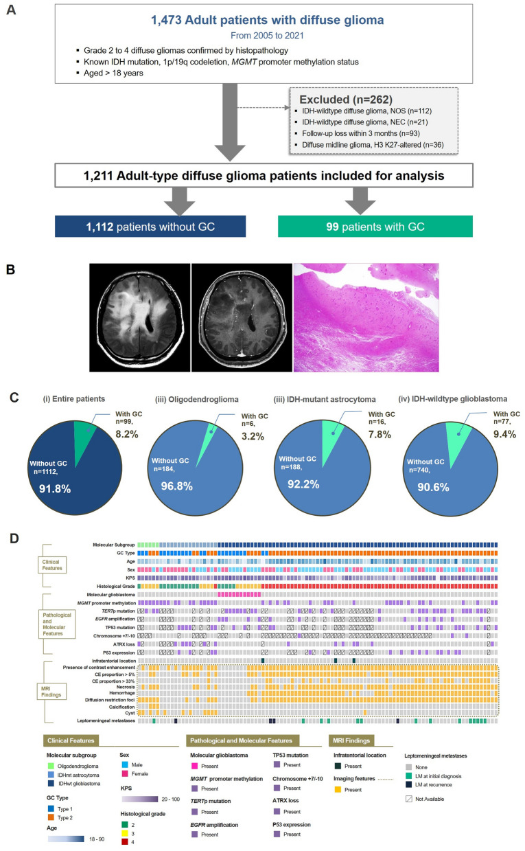
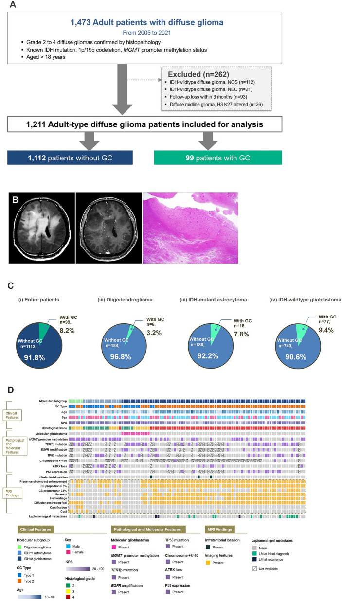
Fig. 1.
Patient characteristics of the study cohort of adult diffuse glioma patients of our institution. A Flow chart of patient inclusion. B Representative imaging and histologic findings in a patient with IDH-wildtype glioblastoma showing GC. On MRI, a diffuse infiltrative glioma involving bilateral cerebral hemispheres is seen on FLAIR image. Faint enhancement is seen in some areas on postcontrast T1-weighted image. On low-power view (H&E; × 1.25), glioma cells are diffusely infiltrated into the cerebral parenchyma, suggesting GC. C Pie charts summarizing the distribution of molecular types of the adult-type diffuse glioma in patients with and without GC. D Summary plot of the clinical, molecular and imaging findings of patients with GC. GC = gliomatosis cerebri; IDH = isocitrate dehydrogenase; MGMT = O6-methylguanine-methyltransferase, NOS = not otherwise specified, NEC = not elsewhere classified, CE = contrast-enhancing, TERTp = telomerase reverse transcriptase promoter, = epidermal growth factor receptor
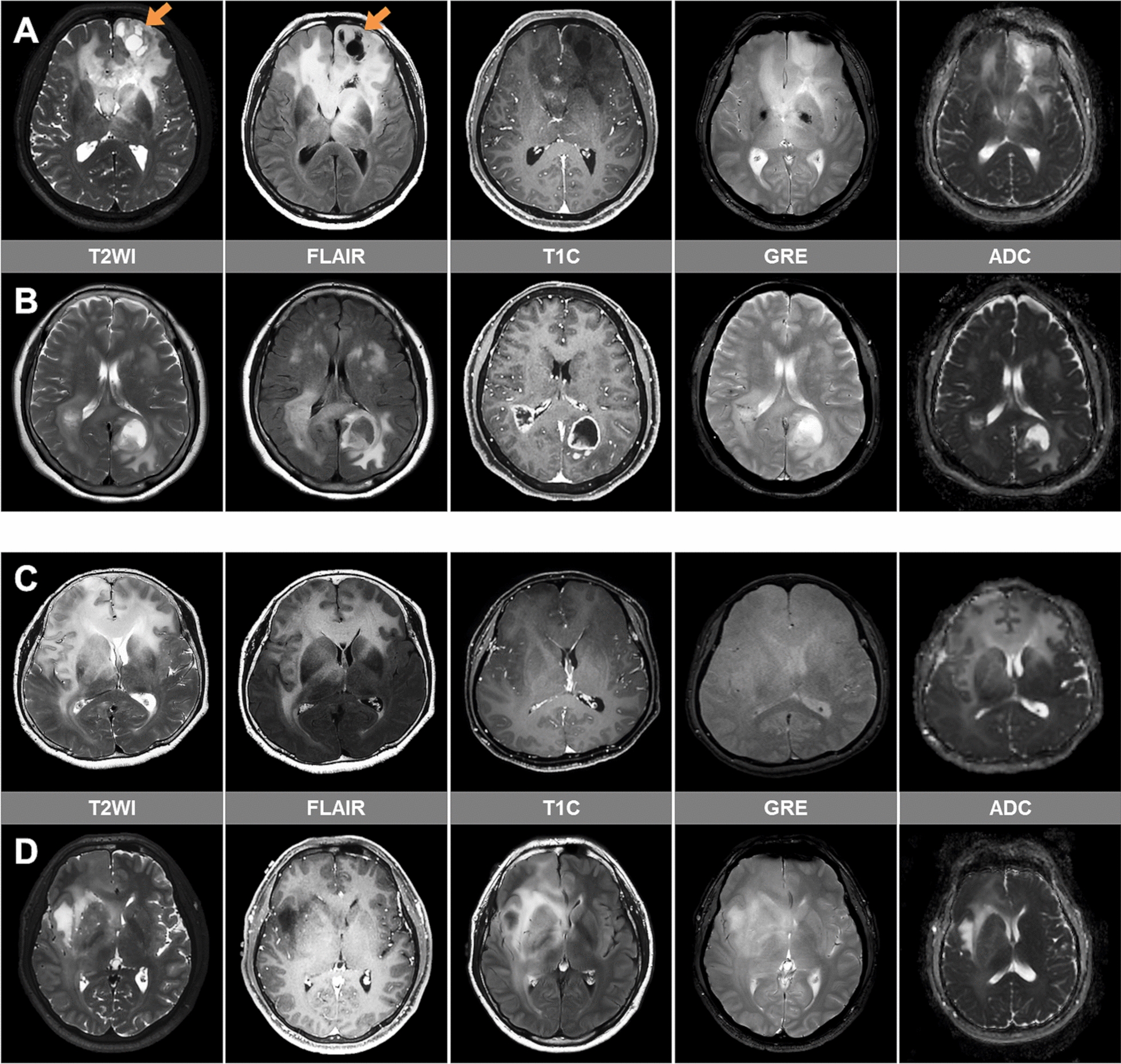
Fig. 2.

Representative imaging cases of GC cases with correctly A, B and incorrectly C, D predicted IDH mutation status according to multivariable model. A A 59-year-old male with IDH-mutant astrocytoma, CNS WHO grade 3. MRI shows a non-enhancing diffuse infiltrative tumor involving bilateral frontal lobes, left basal ganglia, and left thalamus. There is no discrete tumor mass, indicating type 1 GC. Cystic changes are seen at the left frontal lobe (arrows) on T2-weighted and FLAIR images. There is no hemorrhage on gradient recalled echo (GRE)-weighted image and no cellularity increase on apparent diffusion coefficient (ADC) map. B A 60-year-old female with IDH-wildtype glioblastoma, CNS WHO grade 4. MRI shows a non-enhancing dif-fuse infiltrative tumor involving the bilateral parietotemporooccipital lobes. There are obvious contrast-enhancing tumor masses, indicating type 2 GC. Contrast-enhancing necrotic tumor portions are seen at the right temporal and left parietotemporal lobes. There is a focal cellularity increase of solid enhancing tumor portions on ADC map. C A 65-year-old female with IDH-mutant astrocytoma, CNS WHO grade 2 showing a non-enhancing diffuse in-filtrative tumor without necrosis, cystic change, nor hemorrhage. D A 32-year-old male with IDH-wildtype glioblastoma, CNS WHO grade 4. This patient was histologically grade 2, but was classified as IDH-wildtype glioblastoma due to presence of TERTp mutation (molecular glioblastoma). This case also shows imaging finding of a non-enhancing diffuse infiltrative tumor without necrosis, cystic change, nor haemorrhage
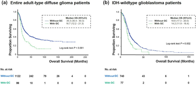
Fig. 3.
Kaplan–Meier curves of the OS of the according to the presence of GC in the a entire adult-type diffuse glioma patients and b IDH-wildtype glioblastoma patients. GC = gliomatosis cerebri; IDH = isocitrate dehydrogenase
The figures should have appeared as shown below.
The original article has been corrected.
Footnotes
Publisher's Note
Springer Nature remains neutral with regard to jurisdictional claims in published maps and institutional affiliations.
Reference
- 1.Shin I, Park YW, Sim Y et al (2024) Revisiting gliomatosis cerebri in adult-type diffuse gliomas: a comprehensive imaging, genomic and clinical analysis. Acta Neuropathol Commun 12:128. 10.1186/s40478-024-01832-w [DOI] [PMC free article] [PubMed] [Google Scholar]


