Abstract
Objective
To investigate the correlation between tumor size, tumor location, and prognosis in patients with early-stage endometrial cancer (EC) receiving adjuvant radiotherapy.
Methods
Data of patients who had been treated for stage I–II EC from March 1999 to September 2017 in 13 tertiary hospitals in China was screened. Cox regression analysis was performed to investigate associations between tumor size, tumor location, and other clinical or pathological factors with cancer-specific survival (CSS) and distant metastasis failure-free survival (DMFS). The relationship between tumor size as a continuous variable and prognosis was demonstrated by restricted cubic splines. Prognostic models were constructed as nomograms and evaluated by Harrell’s C-index, calibration curves and receiver operating characteristic (ROC) curves.
Results
The study cohort comprised 805 patients with a median follow-up of 61 months and a median tumor size of 3.0 cm (range 0.2–15.0 cm). Lower uterine segment involvement (LUSI) was found in 243 patients (30.2%). Tumor size and LUSI were identified to be independent prognostic factors for CSS. Further, tumor size was an independent predictor of DMFS. A broadly positive relationship between poor survival and tumor size as a continuous variable was visualized in terms of hazard ratios. Nomograms constructed and evaluated for CSS and DMFS had satisfactory calibration curves and C-indexes of 0.847 and 0.716, respectively. The area under the ROC curves for 3- and 5-year ROC ranged from 0.718 to 0.890.
Conclusion
Tumor size and LUSI are independent prognostic factors in early-stage EC patients who have received radiotherapy. Integrating these variables into prognostic models would improve predictive ability.
Keywords: Endometrial Neoplasms, Radiotherapy, Neoplasm Staging, Survival
Synopsis
This multicenter retrospective study demonstrated the prognostic value of tumor size and location. Prognostic models integrating tumor size and location showed satisfactory performances. Tumor size as a continuous variable displayed a broadly positive correlation with poor prognosis.
INTRODUCTION
Endometrial cancer (EC) is the third most common gynecological carcinoma worldwide following breast and cervical cancer [1]. In Asia, EC was estimated to have caused up to 167,310 new cases and 40,995 deaths in 2020, indicating a high burden of disease [2]. Decisions on whether to combine surgery with adjuvant therapies are based on evidence-based guidelines [3]. However, there is still a recurrence rate of 7%–10% in patients with International Federation of Gynecology and Obstetrics (FIGO) stage I–II EC [4,5,6,7]. It is reasonable to assume that additional tumor-related variables may facilitate prognostic risk stratification.
Tumor size, defined as the maximal diameter of all dimensions, has been reported to be associated with poor prognosis in various types of carcinomas [8,9,10]. Tumor location, defined as anatomical site, has also been used to predict treatment outcomes of cancer patients [11,12,13]. EC is often classified according to whether it involves the lower uterine segment (LUS), which is defined as the region of transition from endometrial to endocervical tissue [14]. Whether tumor size and lower uterine segment involvement (LUSI) should be recognized as significant prognostic factors for early-stage EC is controversial; meanwhile, there is no consensus on the optimal cutoff values or categories [15,16,17,18].
The aim of this study is to determine whether tumor size and location are independent prognostic risk factors in early-stage EC patients who have undergone radiotherapy. We hope to facilitate the prediction of poor prognosis by establishing a model incorporating factors with prognostic value.
MATERIALS AND METHODS
1. Patients
This retrospective study examined data of EC patients who had undergone surgery and adjuvant therapy at 13 grade A tertiary hospitals in China from March 1999 to September 2017. All patients were surgically staged I–II according to the FIGO 2009 staging system and received radiotherapy in accordance with European Society for Medical Oncology-European Society of Gynecological Oncology-European Society for Radiotherapy & Oncology (ESMO-ESGO-ESTRO) risk stratification. Tumor size was measured by pathologists on surgical specimens and defined as the maximal diameter in any dimension. LUSI was defined as present when tumor extension was detected within the LUS region by a combination of macroscopic and microscopic pathological assessment, along with preoperative magnetic resonance imaging (MRI) or ultrasound images. Patients were excluded if data on histological type or tumor size was missing and/or if they had been followed up for less than 3 months. This study was approved by the Ethics Review Committee of Peking Union Medical College Hospital, Chinese Academy of Medical Sciences (No. S-K139 and I-24PJ0314).
2. Treatment and follow-up
All patients had undergone total hysterectomy and bilateral salpingo-oophorectomy with or without lymphadenectomy. Pre- or post-operative pelvic and abdominal MRI, computed tomography (CT), or positron emission tomography (PET)/CT images were examined for any evidence of metastasis in patients who had not undergone lymph node dissection. Postoperatively, all patients received adjuvant radiotherapy, vaginal brachytherapy (VBT) or pelvic external beam radiotherapy (EBRT), with or without chemotherapy and endocrine therapy. VBT guided by 2- or 3-dimensional high-dose rate brachytherapy plan was delivered to the vaginal stump and upper half of the vagina. Clinical target volume of EBRT involved the vaginal stump, upper half of the vagina, and regional lymphatic drainage, including the para-uterine, presacral, obturator, internal iliac, external iliac and common iliac areas. EBRT was delivered by conventional 4-field box radiotherapy, 3-dimensional conformal radiotherapy, or intensity-modulated radiotherapy techniques. Intravenous concurrent or sequential adjuvant chemotherapy such as platinum-based regimen was administered according to pathological features, individual assessment by a physician, the patient’s physical condition and willingness to consent to this treatment.
Follow-up visits were conducted every 3 to 6 months for the first 2 years, every 6 months for the following 3 years, and then annually. Follow-up consisted of physical gynecological examination, lab tests, including the biomarker CA125, and imaging techniques, including chest CT and pelvic and abdominal ultrasound or CT. Pelvic and abdominal MRI and PET/CT were performed if clinically indicated.
3. Primary outcomes and statistical analysis
The primary outcomes were cancer-specific survival (CSS) and distant metastasis failure-free survival (DMFS). Death from EC was defined as cancer-specific death (CSD). CSS was defined as the time from hysterectomy to the date of CSD or last follow-up. DMFS was calculated from surgery to the date of diagnosing the first distant metastasis (DM) or last follow-up.
Receiver operating characteristic (ROC) curves were performed to calculate the optimal cut-off value for tumor size regarding CSD and DM by Youden index. The Kaplan–Meier method and log-rank test were used to illustrate survival differences between patients divided by the cut-off value for tumor size. Multivariate Cox (MVC) proportional hazards regression modeling was performed to identify independent prognostic factors of survivals in early-stage EC patients. Hazard ratios (HRs) with corresponding 95% confidence interval (CI) were estimated. Two-tailed p-values below 0.05 were considered to denote statistical significance. The relationship between tumor size as a continuous variable and prognosis was demonstrated by restricted cubic splines. Nomograms were plotted to visualize prognostic models for CSS and DMFS. Calibration plots, ROC curves and Harrell’s C-index were used to evaluate the models’ performances. Statistical analyses were performed using R version 4.2.2 (R Foundation for Statistical Computing, Vienna, Austria).
RESULTS
1. Patient characteristics
We retrospectively screened data of 1,277 patients with early-stage EC who had received radiotherapy in 13 China’s grade A tertiary hospitals between March 1999 and September 2017 and enrolled the 805 patients who met the eligibility criteria for this study (Fig. S1, Table S1). The median duration of follow-up was 61.0 months (interquartile range, 42.3–87.0). By the last follow-up, 53 (6.6%) of the 805 patients had developed distant metastases. EC was the cause of death in 27 (45%) of the 60 patients who had deceased. Baseline patient characteristics, pathological findings, treatment patterns and other clinical features are summarized in Table 1.
Table 1. Patient demographics and tumor-related characteristics.
| Characteristics | Values (n=805) | ||
|---|---|---|---|
| Age (yr) | 57 (51–62) | ||
| Pathology | |||
| Endometrioid carcinoma | 750 (93.2) | ||
| Non-endometrioid carcinoma | 55 (6.8) | ||
| Tumor size (cm) | 3.0 (2.0–4.0) | ||
| MI | |||
| <1/2 | 445 (55.3) | ||
| ≥1/2 | 360 (44.7) | ||
| LVSI | |||
| No | 663 (82.4) | ||
| Yes | 142 (17.6) | ||
| LUSI | |||
| No | 562 (69.8) | ||
| Yes | 243 (30.2) | ||
| FIGO grade* | |||
| Grade 1 | 281 (34.9) | ||
| Grade 2 | 333 (41.4) | ||
| Grade 3 | 191 (23.7) | ||
| FIGO 2009 stage | |||
| I | 731 (90.8) | ||
| II | 74 (9.2) | ||
| ESMO-ESGO-ESTRO risk classification | |||
| Low risk | 258 (32.0) | ||
| Intermediate risk | 219 (27.2) | ||
| High-intermediate risk | 160 (19.9) | ||
| High risk | 168 (20.9) | ||
| Radiotherapy mode | |||
| VBT alone | 457 (56.8) | ||
| EBRT±VBT | 348 (43.2) | ||
| Chemotherapy† | |||
| No | 668 (83.0) | ||
| Yes | 137 (17.0) | ||
| Platinum + Paclitaxel | 33 (4.1) | ||
| Platinum + Anthracycline ± Cyclophosphamide | 8 (1.0) | ||
| Unknown | 96 (11.9) | ||
Values are presented as median (interquartile range) or number (%).
EBRT, external beam radiotherapy; ESMO-ESGO-ESTRO, European Society for Medical Oncology-European Society of Gynecological Oncology-European Society for Radiotherapy & Oncology; FIGO, International Federation of Gynecology and Obstetrics; LUSI, lower uterine segment involvement; LVSI, lymphatic vascular space invasion; MI, myometrial invasion; VBT, vaginal brachytherapy.
*Poorly differentiated EC including serous, clear cell, undifferentiated and mixed cell carcinoma were classified into FIGO grade 3.
†Carboplatin and cisplatin were indicated as platinum. Doxorubicin and epirubicin were indicated as anthracycline.
2. Screening for prognostic factors
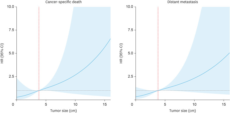
Age and tumor size were subjected to MVC regression analysis as continuous variables, whereas myometrial invasion (MI), lymphatic vascular space invasion (LVSI), LUSI, FIGO grade, FIGO 2009 stage, risk classification, radiotherapy mode and chemotherapy were explored as categorical variables. This analysis identified tumor size and LUSI as independent risk factors for CSS. Tumor size was also an independent prognostic factor for DMFS, as shown in Table 2. Kaplan–Meier survival analysis curves for CSS and DMFS rates according to each independent risk factor are shown in Figs. S2 and S3. After adjusting the covariables (age, LUSI and FIGO grade), restricted cubic splines were used to demonstrate the broadly positive relationships between the 2 survivals and tumor size in terms of HRs (Fig. 1). Reference values, defined by ROC analysis of 5-year CSS and DMFS, were both calculated as 3.8 cm.
Table 2. Multivariate Cox regression analysis of prognostic factors in early-stage EC patients.
| Variable | Cancer-specific death | Distant metastasis | |||
|---|---|---|---|---|---|
| HR (95% CI) | p-value | HR (95% CI) | p-value | ||
| Age (yr) | |||||
| Continuous variable | 1.095 (1.048–1.145) | <0.001 | 1.053 (1.021–1.086) | 0.001 | |
| Tumor size (cm) | |||||
| Continuous variable | 1.234 (1.034–1.473) | 0.020 | 1.156 (1.018–1.314) | 0.026 | |
| MI | |||||
| <1/2 | Reference | Reference | |||
| ≥1/2 | 1.374 (0.582–3.245) | 0.468 | 1.113 (0.618–2.004) | 0.721 | |
| LVSI | |||||
| No | Reference | Reference | |||
| Yes | 1.015 (0.293–3.515) | 0.981 | 1.417 (0.590–3.403) | 0.435 | |
| LUSI | |||||
| No | Reference | Reference | |||
| Yes | 2.716 (1.208–6.107) | 0.016 | 1.719 (0.959–3.081) | 0.069 | |
| FIGO grade* | |||||
| Grade 1 | Reference | Reference | |||
| Grade 2 | 3.817 (1.033–14.104) | 0.045 | 2.169 (1.021–4.607) | 0.044 | |
| Grade 3 | 4.251 (0.818–22.091) | 0.085 | 2.367 (0.838–6.688) | 0.104 | |
| FIGO 2009 stage | |||||
| I | Reference | Reference | |||
| II | 1.088 (0.281–4.208) | 0.902 | 1.681 (0.623–4.533) | 0.305 | |
| ESMO-ESGO-ESTRO risk classification | |||||
| Low/intermediate risk | Reference | Reference | |||
| High intermediate/high risk | 1.730 (0.329–9.096) | 0.518 | 1.074 (0.3529–3.269) | 0.900 | |
| Radiotherapy mode | |||||
| VBT alone | Reference | Reference | |||
| EBRT+VBT | 0.851 (0.344–2.101) | 0.726 | 1.087 (0.572–2.064) | 0.799 | |
| Chemotherapy | |||||
| No | Reference | Reference | |||
| Yes | 1.931 (0.739–5.048) | 0.180 | 1.547 (0.771–3.103) | 0.219 | |
CI, confidence interval; EBRT, external beam radiotherapy; EC, endometrial cancer; ESMO-ESGO-ESTRO, European Society for Medical Oncology-European Society of Gynecological Oncology-European Society for Radiotherapy & Oncology; FIGO, International Federation of Gynecology and Obstetrics; HR, hazard ratio; LUSI, lower uterine segment involvement; LVSI, lymphatic vascular space invasion; MI, myometrial invasion; VBT, vaginal brachytherapy.
*Poorly differentiated EC including serous, clear cell, undifferentiated and mixed cell carcinoma were classified into FIGO grade 3.
Fig. 1. Estimates of the dependence of cancer-specific death and distant metastasis on tumor size in early-stage EC patients who had undergone radiotherapy. Reference value=3.8 cm.
CI, confidence interval; EC, endometrial cancer; HR, hazard ratio.
3. Development and assessment of prognostic models
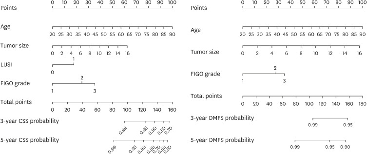
The independent risk factors identified by MVC regression analysis were integrated to develop prognostic models for CSS and DMFS in early-stage EC patients (Fig. 2). The sum of the scores assigned to each variable could be unitized to predict the probability of 3- and 5-year CSS and DMFS in early-stage EC patients who had received radiotherapy. Harrell’s C-indexes of prognostic models for CSS and DMFS were 0.847 (95% CI=0.784–0.910) and 0.716 (95% CI=0.649–0.783), respectively, indicating satisfactory discrimination levels. Similar C-indexes were achieved on internal validation using bootstrap to obtain a dataset of 1,000 samples. The C-index was estimated to be 0.847 (95% CI=0.785–0.907) for CSS and 0.716 (95% CI=0.645–0.774) for DMFS.
Fig. 2. Nomograms of prognostic models for CSS and DMFS in early-stage EC patients who had undergone radiotherapy.
CSS, cancer-specific survival; DMFS, distant metastasis failure-free survival; EC, endometrial cancer; FIGO, International Federation of Gynecology and Obstetrics; LUSI, lower uterine segment involvement.
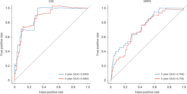
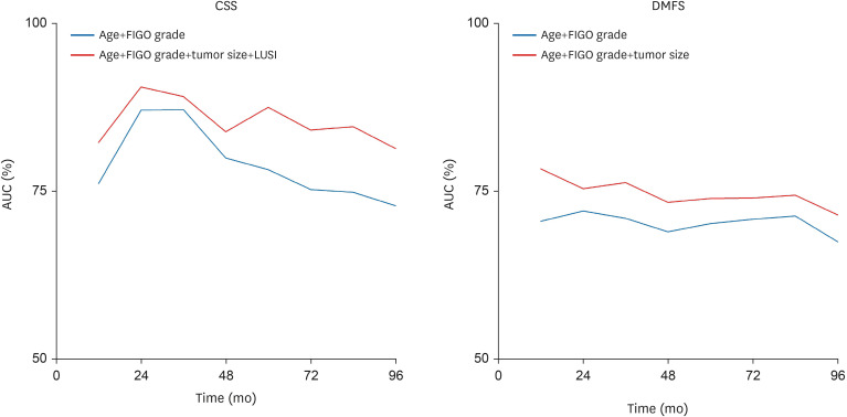
Calibration plots and ROC curves were constructed to illustrate model performances. According to calibration plots (Fig. S4), the predicted probabilities of both models were close to the actual 5-year CSS and DMFS probabilities. As shown by ROC curves (Fig. 3), the area under the ROC curves for predicting 3- and 5-year CSS as well as DMFS ranged from 0.718 to 0.890 in the prognostic models. These values were generally higher than those obtained from predictions based solely on age and FIGO grade. Time-dependent ROC curves over 8 years of follow-up indicated that prognostic models incorporating tumor size and location exhibited superior performance (Fig. 4).
Fig. 3. Three- and 5-year ROC curves of the prognostic models for CSS and DMFS.
AUC, area under the ROC curve; CSS, cancer-specific survival; DMFS, distant metastasis failure-free survival; ROC, receiver operating characteristic.
Fig. 4. Time-dependent ROC curves of the prognostic models for CSS and DMFS over a follow-up period of 8 years.
AUC, area under the ROC curve; CSS, cancer-specific survival; DMFS, distant metastasis failure-free survival; FIGO, International Federation of Gynecology and Obstetrics; LUSI, lower uterine segment involvement; ROC, receiver operating characteristic.
DISCUSSION
In this study, independent prognostic factors of CSS and DMFS were analyzed in early-stage EC patients who had undergone radiotherapy. Older age, larger tumor size and higher FIGO grade were associated with poor CSS and DMFS. LUSI was associated with poor CSS. In particular, tumor size was explored as a continuous variable. We plotted nomograms to illustrate the combined effect of all prognostic factors on CSS and DMFS. Both models showed satisfactory performances according to ROC curves and calibration plots. In the National Comprehensive Cancer Network guidelines [19], tumor size and location are considered potential adverse risk factors when making decisions about adjuvant therapy for stage I EC patients. However, it is unclear how to make optimal use of these factors in clinical practice. According to the ESMO-ESGO-ESTRO consensus [20], whether to incorporate tumor size into risk stratification remains controversial and requires further studies with adequate sample sizes. In the revised FIGO 2023 staging system, tumor size and location are not considered as references [21]. The results of this study are expected to demonstrate the value of tumor size and location in predicting survival and disease progression.
Although tumor size has not been formally included in the risk assessment guidelines for EC, it is recognized as an essential factor in the Tumor, Node, Metastasis staging of various tumors, such as breast and cervical cancer, and contributes to deciding on disease management [22,23]. From a pathophysiological perspective, the size of the primary tumor and disease progression may be correlated because of common gene expressions or regulations [24,25]. Several studies have explored the prognostic significance of tumor size [15,26,27,28]. Shah et al. [15] found that patients of all stages with tumor sizes ≤2 cm had significantly lower risk of nodal metastasis than patients with tumors >2 cm (6.3% vs. 26.3%, p<0.005). However, the cutoff value of 2 cm was chosen arbitrarily, and multivariate analysis did not identify tumor size as an independent prognostic factor [15]. Chattopadhyay et al. [26] investigated tumor size in 216 patients diagnosed with FIGO (1998) stage I endometroid EC, 44 of whom (26%) had received adjuvant radiotherapy. They found that tumor size independently predicted distant failure and disease-related death [26]. In that study, a cutoff of 3.75 cm was determined by ROC curve analyses [26]. Mahdi et al. [16] studied 19,692 early-stage endometrioid EC patients drawn from the Surveillance, Epidemiology, and End Results (SEER) dataset from 1988 to 2007, 4,303 (21.8%) of whom had undergone radiation therapy. They found that tumor size >5 cm was a predictor of lymph node metastasis and disease-specific survival [16]. The cutoff values in the above studies are relatively close to 3.8 cm, the cut-off value that we determined in our study. Hou et al. identified 52,208 EC patients of all stages in the SEER database from 2004 to 2018 and found that tumor size had prognostic significance as a continuous variable for CSS, the cutoff value in that study being 4.0 cm [27]. The relationship between tumor size and prognosis initially tended to increase rapidly and then more slowly, the turning point being 7.5 cm [27]. However, treatment history was not included in analysis, potentially introducing bias into the findings. In comparison, we analyzed the prognostic effect of tumor size on CSS and DMFS in a relatively large sample of early-stage EC patients from multiple centers, thus largely avoiding the confounding effects of stage, ethnicity, and adjuvant therapy. It is worth noting that HRs for poor prognosis inclined sharply with increasing tumor size, especially when distant from the reference value of 3.8 cm. The impact of tumor size on the prognosis of early-stage EC patients may surpass the range of values investigated in this study. We further analyzed the survival curve of 17 patients who had been excluded from this study because diffuse tumor involvement of the uterine cavity prevented accurate measurement of tumor size (Fig. S5). The prognosis of patients with diffuse tumor involvement was significantly worse than that of the other two groups.
The prognostic value of LUSI has been explored in several studies [17,18,28,29]. The impact of LUSI on prognosis may be related to different lymphatic drainage pathways from different locations in the uterus [30]. Besides, LUSI may be associated with a greater risk of tumor invasion of the cervix because of the anatomical proximity, which would further influence prognosis. In a single-center study, Kizer et al. [17] found a correlation between LUSI and poor prognosis in early-stage endometrioid EC patients. In contrast, Erkaya et al. [18] found that LUSI was not an independent prognostic factor for overall survival, but was associated with lymph node metastasis. In another study, Miyoshi et al. [28] found that LUS carcinoma was independently associated with progression-free survival, but not with overall survival, in patients with disease of all FIGO stages. Hochreiter et al. [29] found that LUSI had an impact on disease-free survival in stage IB grade 2–3 EC patients treated with VBT. Most findings presented above were derived from relatively small, single-center studies. In our study, LUSI was independently associated with CSS but not significantly associated with DMFS, the HR being 1.719 (95% CI=0.959–3.081). Although the p value for CSS calculated by the log-rank method was 0.057 (Fig. S2), the p-value calculated by MVC analysis was 0.016. Given that multiple factors are considered in clinical practice, LUSI was determined as an independent prognostic factor according to MVC results. We hope that our study would provide insight into the prognostic impact of LUSI and reduce the possible influences of different stages and treatment modalities.
As stated earlier, EC has caused a massive burden of disease globally. Further research into the prognostic factors for EC is needed to facilitate clinical decision-making. Incorporating tumor size and location into risk stratification may result in stratification migration for some patients, possibly indicating the need for treatment changes. We constructed a HR scoring system using the prognostic factors identified in this study (Fig. S6) and compared aspects of the new group set and ESMO-ESGO-ESTRO risk classification. Regarding CSS, 19.23% of patients in this study who did not receive EBRT and were initially categorized as intermediate or high-intermediate risk by ESMO-ESGO-ESTRO risk classification had HR scores above the 75th percentile of the entire cohort when our findings were applied. Meanwhile, 10.95% of patients who had undergone EBRT and were in the ESMO-ESGO-ESTRO high risk category had HR scores below the 25th percentile. The nonnegligible stratification deviations suggest the necessity of correctly identifying prognostic factors when deciding on appropriate treatment. Thus, we recommend that tumor size and location should be explicitly and quantitatively described in the pathology report of each EC patient’ surgical specimen to provide reference information. Clinicians should consider these characteristics when deciding treatment strategies. For example, postoperative adjuvant radiotherapy should be considered in patients at low or intermediate risk with large tumors or LUSI, despite these patients have no other risk factors such as MI, non-endometrial histology type and high FIGO grade. More aggressive treatment plans should also be considered for patients at high-intermediate risk with large tumors or LUSI.
The current study has several limitations. This is a retrospective study with censors. Selection bias may have resulted from exclusion of patients whose tumor characteristics were incompletely recorded or missing. A prospective validation cohort is needed to evaluate the validity of generalizing our findings. Furthermore, the factors considered were limited. More variables, such as molecular subtypes, radiomics features and radiotherapy sensitivity index, need to be simultaneously included in multivariate analysis to enable construction of a more accurate and robust prognostic model. Nevertheless, our findings reveal the significance of tumor size and location in early-stage EC patients who have undergone radiotherapy. The prognostic models constructed using factors that are easily accessible in routine EC clinical practice performed satisfactorily.
ACKNOWLEDGEMENTS
The authors wish to acknowledge Dr. Zi Liu from the First Affiliated Hospital of Xi’an Jiaotong University, Dr. Jianli He from the General Hospital of Ningxia Medical University, Dr. Xiaoge Sun from the Affiliated Hospital of Inner Mongolia Medical University, Dr. Wei Zhong from the Affiliated Cancer Hospital of Xinjiang Medical University, Dr. Fengjv Zhao from Gansu Provincial Cancer Hospital, Dr. Xiaomei Li from Peking University First Hospital, Dr. Sha Li from the 940th Hospital of Joint Logistics Support force of Chinese People’s Liberation Army, Dr. Hong Zhu from Xiangya Hospital Central South University and Dr. Zhanshu Ma from the Affiliated Hospital of Chi Feng University for their help in resources collection and data curation of this study.
Footnotes
Funding: This work was supported by National High Level Hospital Clinical Research Funding (grant number: 2022-PUMCH-A-036).
Conflict of Interest: No potential conflict of interest relevant to this article was reported.
- Conceptualization: J.S., H.X.
- Data curation: W.L., Z.L., W.T.
- Investigation: J.S.
- Funding acquisition: H.X.
- Supervision: H.K., Z.F.
- Writing - original draft: J.S.
- Writing - review & editing: W.L., Z.L., W.T., H.K., Z.F., H.X.
SUPPLEMENTARY MATERIALS
Enrollment distribution across multiple centers
Participant flow diagram.
Kaplan–Meier curves of CSS.
Kaplan–Meier curves of DMFS.
Five-year calibration plots of prognostic model for CSS and DMFS.
Kaplan–Meier curves of different tumor size groups in CSS and DMFS.
Risk scores based on hazard ratios for cancer-specific death and distant metastasis. Each dot represents a patient. Red dots represent patients who have experienced cancer-specific death or distant metastasis.
References
- 1.Siegel RL, Miller KD, Wagle NS, Jemal A. Cancer statistics, 2023. CA Cancer J Clin. 2023;73:17–48. doi: 10.3322/caac.21763. [DOI] [PubMed] [Google Scholar]
- 2.Sung H, Ferlay J, Siegel RL, Laversanne M, Soerjomataram I, Jemal A, et al. Global cancer statistics 2020: GLOBOCAN estimates of incidence and mortality worldwide for 36 cancers in 185 countries. CA Cancer J Clin. 2021;71:209–249. doi: 10.3322/caac.21660. [DOI] [PubMed] [Google Scholar]
- 3.Concin N, Matias-Guiu X, Vergote I, Cibula D, Mirza MR, Marnitz S, et al. ESGO/ESTRO/ESP guidelines for the management of patients with endometrial carcinoma. Radiother Oncol. 2021;154:327–353. doi: 10.1016/j.radonc.2020.11.018. [DOI] [PubMed] [Google Scholar]
- 4.Francis SR, Ager BJ, Do OA, Huang YJ, Soisson AP, Dodson MK, et al. Recurrent early stage endometrial cancer: patterns of recurrence and results of salvage therapy. Gynecol Oncol. 2019;154:38–44. doi: 10.1016/j.ygyno.2019.04.676. [DOI] [PubMed] [Google Scholar]
- 5.Nwachukwu C, Baskovic M, Von Eyben R, Fujimoto D, Giaretta S, English D, et al. Recurrence risk factors in stage IA grade 1 endometrial cancer. J Gynecol Oncol. 2021;32:e22. doi: 10.3802/jgo.2021.32.e22. [DOI] [PMC free article] [PubMed] [Google Scholar]
- 6.Randall ME, Filiaci V, McMeekin DS, von Gruenigen V, Huang H, Yashar CM, et al. Phase III trial: adjuvant pelvic radiation therapy versus vaginal brachytherapy plus paclitaxel/carboplatin in high-intermediate and high-risk early stage endometrial cancer. J Clin Oncol. 2019;37:1810–1818. doi: 10.1200/JCO.18.01575. [DOI] [PMC free article] [PubMed] [Google Scholar]
- 7.Bendifallah S, Ouldamer L, Lavoue V, Canlorbe G, Raimond E, Coutant C, et al. Patterns of recurrence and outcomes in surgically treated women with endometrial cancer according to ESMO-ESGO-ESTRO Consensus Conference risk groups: results from the FRANCOGYN study Group. Gynecol Oncol. 2017;144:107–112. doi: 10.1016/j.ygyno.2016.10.025. [DOI] [PubMed] [Google Scholar]
- 8.Dai W, Mo S, Xiang W, Han L, Li Q, Wang R, et al. The critical role of tumor size in predicting prognosis for T1 colon cancer. Oncologist. 2020;25:244–251. doi: 10.1634/theoncologist.2019-0469. [DOI] [PMC free article] [PubMed] [Google Scholar]
- 9.Joseph RW, Elassaiss-Schaap J, Kefford R, Hwu WJ, Wolchok JD, Joshua AM, et al. Baseline tumor size is an independent prognostic factor for overall survival in patients with melanoma treated with pembrolizumab. Clin Cancer Res. 2018;24:4960–4967. doi: 10.1158/1078-0432.CCR-17-2386. [DOI] [PMC free article] [PubMed] [Google Scholar]
- 10.Zhan G, Peng H, Zhou L, Jin L, Xie X, He Y, et al. A web-based nomogram model for predicting the overall survival of hepatocellular carcinoma patients with external beam radiation therapy: a population study based on SEER database and a Chinese cohort. Front Endocrinol (Lausanne) 2023;14:1070396. doi: 10.3389/fendo.2023.1070396. [DOI] [PMC free article] [PubMed] [Google Scholar]
- 11.Benesch MG, Mathieson A, O’Brien SB. Effects of tumor localization, age, and stage on the outcomes of gastric and colorectal signet ring cell adenocarcinomas. Cancers (Basel) 2023;15:714. doi: 10.3390/cancers15030714. [DOI] [PMC free article] [PubMed] [Google Scholar]
- 12.Weng S, Zhu N, Li D, Chen Y, Tan Y, Chen J, et al. Clinical characteristics, treatment, and prognostic factors of patients with primary extramammary Paget’s disease (EMPD): a retrospective analysis of 44 patients from a single center and an analysis of data from the Surveillance, Epidemiology, and End Results (SEER) database. Front Oncol. 2020;10:1114. doi: 10.3389/fonc.2020.01114. [DOI] [PMC free article] [PubMed] [Google Scholar]
- 13.Mickevicius NJ, Carle AB, Bluemel T, Santarriaga S, Schloemer F, Shumate D, et al. Location of brain tumor intersecting white matter tracts predicts patient prognosis. J Neurooncol. 2015;125:393–400. doi: 10.1007/s11060-015-1928-5. [DOI] [PMC free article] [PubMed] [Google Scholar]
- 14.Mayr NA, Wen BC, Benda JA, Sorosky JI, Davis CS, Fuller RW, et al. Postoperative radiation therapy in clinical stage I endometrial cancer: corpus, cervical, and lower uterine segment involvement--patterns of failure. Radiology. 1995;196:323–328. doi: 10.1148/radiology.196.2.7617840. [DOI] [PubMed] [Google Scholar]
- 15.Shah C, Johnson EB, Everett E, Tamimi H, Greer B, Swisher E, et al. Does size matter? Tumor size and morphology as predictors of nodal status and recurrence in endometrial cancer. Gynecol Oncol. 2005;99:564–570. doi: 10.1016/j.ygyno.2005.06.011. [DOI] [PubMed] [Google Scholar]
- 16.Mahdi H, Munkarah AR, Ali-Fehmi R, Woessner J, Shah SN, Moslemi-Kebria M. Tumor size is an independent predictor of lymph node metastasis and survival in early stage endometrioid endometrial cancer. Arch Gynecol Obstet. 2015;292:183–190. doi: 10.1007/s00404-014-3609-6. [DOI] [PubMed] [Google Scholar]
- 17.Kizer NT, Gao F, Guntupalli S, Thaker PH, Powell MA, Goodfellow PJ, et al. Lower uterine segment involvement is associated with poor outcomes in early-stage endometrioid endometrial carcinoma. Ann Surg Oncol. 2011;18:1419–1424. doi: 10.1245/s10434-010-1454-9. [DOI] [PMC free article] [PubMed] [Google Scholar]
- 18.Erkaya S, Öz M, Topçu HO, Şirvan AL, Güngör T, Meydanli MM. Is lower uterine segment involvement a prognostic factor in endometrial cancer? Turk J Med Sci. 2017;47:300–306. doi: 10.3906/sag-1602-137. [DOI] [PubMed] [Google Scholar]
- 19.Abu-Rustum N, Yashar C, Arend R, Barber E, Bradley K, Brooks R, et al. Uterine neoplasms, version 1.2023, NCCN Clinical Practice Guidelines in Oncology. J Natl Compr Canc Netw. 2023;21:181–209. doi: 10.6004/jnccn.2023.0006. [DOI] [PubMed] [Google Scholar]
- 20.Colombo N, Creutzberg C, Amant F, Bosse T, González-Martín A, Ledermann J, et al. ESMO-ESGO-ESTRO Consensus Conference on endometrial cancer: diagnosis, treatment and follow-up. Int J Gynecol Cancer. 2016;26:2–30. doi: 10.1097/IGC.0000000000000609. [DOI] [PMC free article] [PubMed] [Google Scholar]
- 21.Berek JS, Matias-Guiu X, Creutzberg C, Fotopoulou C, Gaffney D, Kehoe S, et al. FIGO staging of endometrial cancer: 2023. Int J Gynaecol Obstet. 2023;162:383–394. doi: 10.1002/ijgo.14923. [DOI] [PubMed] [Google Scholar]
- 22.Olawaiye AB, Baker TP, Washington MK, Mutch DG. The new (Version 9) American Joint Committee on Cancer tumor, node, metastasis staging for cervical cancer. CA Cancer J Clin. 2021;71:287–298. doi: 10.3322/caac.21663. [DOI] [PubMed] [Google Scholar]
- 23.Giuliano AE, Connolly JL, Edge SB, Mittendorf EA, Rugo HS, Solin LJ, et al. Breast cancer-major changes in the American Joint Committee on Cancer eighth edition cancer staging manual. CA Cancer J Clin. 2017;67:290–303. doi: 10.3322/caac.21393. [DOI] [PubMed] [Google Scholar]
- 24.You T, Gao W, Wei J, Jin X, Zhao Z, Wang C, et al. Overexpression of LIMK1 promotes tumor growth and metastasis in gastric cancer. Biomed Pharmacother. 2015;69:96–101. doi: 10.1016/j.biopha.2014.11.011. [DOI] [PubMed] [Google Scholar]
- 25.Dillenburg-Pilla P, Maria AG, Reis RI, Floriano EM, Pereira CD, De Lucca FL, et al. Activation of the kinin B1 receptor attenuates melanoma tumor growth and metastasis. PLoS One. 2013;8:e64453. doi: 10.1371/journal.pone.0064453. [DOI] [PMC free article] [PubMed] [Google Scholar]
- 26.Chattopadhyay S, Cross P, Nayar A, Galaal K, Naik R. Tumor size: a better independent predictor of distant failure and death than depth of myometrial invasion in International Federation of Gynecology and Obstetrics stage I endometrioid endometrial cancer. Int J Gynecol Cancer. 2013;23:690–697. doi: 10.1097/IGC.0b013e31828c85c6. [DOI] [PubMed] [Google Scholar]
- 27.Hou X, Yue S, Liu J, Qiu Z, Xie L, Huang X, et al. Association of tumor size with prognosis in patients with resectable endometrial cancer: a SEER database analysis. Front Oncol. 2022;12:887157. doi: 10.3389/fonc.2022.887157. [DOI] [PMC free article] [PubMed] [Google Scholar]
- 28.Miyoshi A, Kanao S, Naoi H, Otsuka H, Yokoi T. Investigation of the clinical features of lower uterine segment carcinoma: association with advanced stage disease and indication of poorer prognosis. Arch Gynecol Obstet. 2018;297:193–198. doi: 10.1007/s00404-017-4576-5. [DOI] [PubMed] [Google Scholar]
- 29.Hochreiter A, Kelly JR, Young MR, Litkouhi B, Black JD, Stromberger C, et al. Outcomes and relapse patterns of stage IB grade 2 or 3 endometrial cancer treated with adjuvant vaginal brachytherapy. Int J Gynecol Cancer. 2020;30:48–55. doi: 10.1136/ijgc-2019-000675. [DOI] [PubMed] [Google Scholar]
- 30.Geppert B, Lönnerfors C, Bollino M, Arechvo A, Persson J. A study on uterine lymphatic anatomy for standardization of pelvic sentinel lymph node detection in endometrial cancer. Gynecol Oncol. 2017;145:256–261. doi: 10.1016/j.ygyno.2017.02.018. [DOI] [PubMed] [Google Scholar]
Associated Data
This section collects any data citations, data availability statements, or supplementary materials included in this article.
Supplementary Materials
Enrollment distribution across multiple centers
Participant flow diagram.
Kaplan–Meier curves of CSS.
Kaplan–Meier curves of DMFS.
Five-year calibration plots of prognostic model for CSS and DMFS.
Kaplan–Meier curves of different tumor size groups in CSS and DMFS.
Risk scores based on hazard ratios for cancer-specific death and distant metastasis. Each dot represents a patient. Red dots represent patients who have experienced cancer-specific death or distant metastasis.


