Abstract
Background
Mutations in TBL1X, part of the NCOR1/SMRT corepressor complex, were identified in patients with hereditary X-linked central congenital hypothyroidism and associated hearing loss. The role of TBL1X in thyroid hormone (TH) action, however, is incompletely understood. The aim of the present study was to investigate the role of TBL1X on T3-regulated gene expression in two human liver cell models.
Methods
A human hepatoma cell line (HepG2) wherein TBL1X was downregulated using siRNAs, and human-induced pluripotent stem cell-derived hepatocytes (iHeps) generated from individuals with a TBL1X N365Y mutation. Both cell types were treated with increasing concentrations of T3. The expression of T3-regulated genes was measured by qPCR.
Results
KLF9, CPT1A, and PCK1 mRNA expression were higher upon T3 stimulation in the HepG2 cells with decreased TBL1X expression compared to controls, while DIO1 mRNA expression was lower. Hemizygous TBL1X N365Y iHeps exhibited decreased expression of CPT1A, G6PC1, PCK1, FBP1, and ELOVL2 compared to cells with the heterozygous TBL1X N365Y allele, but KLF9 and HMGCS2 expression was unaltered.
Conclusion
Downregulation of TBL1X in HepG2 cells and the TBL1X N365Y variant in iHeps have differential effects on T3-regulated gene expression. This suggests that TBL1X may play a gene context role in TH action.
Keywords: coregulators, TBL1X, thyroid hormone action, WD40 repeat-containing proteins
Introduction
Isolated central congenital hypothyroidism (CH) is characterized by decreased serum thyroid hormone (TH) concentrations in combination with a normal, low or slightly elevated serum thyroid stimulating hormone (TSH) due to a defect at the level of the pituitary and/or the hypothalamus (1). Isolated central CH is associated with pathogenic variants in a small number of genes involved in signaling pathways of the hypothalamus-pituitary-thyroid (HPT) axis (2, 3, 4, 5). In 2016, we described mutations in Transducin Beta Like X-Linked (TBL1X, encoding Transducin-Beta-Like Protein 1, X-Linked (TBL1X), a WD40 repeat-containing protein) in families with isolated central CH and hearing loss (1). More recently García M et al. reported a truncating TBL1X mutation in a patient with central hypothyroidism, hypoacusia, attention-deficit/hyperactivity disorder (ADHD), gastrointestinal dysmotility, and a Chiari malformation type I (CMI) (6). All mutations (N365Y, W369R, H453Y, R339X) reported to date are located in the highly conserved WD40-repeat domain of the protein, influencing its expression and thermal stability (1, 6). The molecular mechanism behind the association between central CH and TBL1X mutations remains unexplained.
TBL1X is a WD40 repeat-containing protein (1, 7) and part of the nuclear receptor corepressor (NCOR1)/silencing mediator of retinoic and thyroid hormone receptors (SMRT) corepressor complex that interacts with nuclear hormone receptors (NRs) and other transcription factors (TFs) (8). Critically, the NCOR1/SMRT corepressor complex is recruited to the thyroid hormone receptor (TR) isoforms to suppress the expression of positively regulated target genes via histone deacetylation in the absence (or presence of low levels) of triiodothyronine (T3, the active TH) (9). NCOR1 is the major TR corepressor involved in hepatic and systemic T3-regulated gene expression (10). Indeed, the expression of a mutant NCOR1 allele that cannot interact with the TRs leads to the de-repression of positively regulated targets in the hypothyroid state (11, 12). NCOR1 is also thought to be important in determining TH sensitivity and the set point of the HPT-axis (13, 14, 15). In contrast, SMRT (or NCOR2) seems to play a minor role in TH action. This notion was based on the observation that the expression of positively regulated T3 target genes in liver-specific SMRT knockout mice remained unchanged compared to control animals. Moreover, postnatal global disruption of SMRT in mice did not dysregulate the HPT axis (10). Interestingly, our group demonstrated that a postnatal deletion strategy to disrupt both NCOR1 and SMRT in mice is rapidly lethal anticipated by metabolic abnormalities including hypoglycemia, hypothermia, and weight loss (16).
The role of TBL1X in the NCOR1/SMRT corepressor complex and thus TH action is incompletely understood. However, the presence of central CH in patients carrying the various TBL1X mutations suggests an essential role of TBL1X in TH signaling. Currently, patients with central CH with a TBL1X mutation are treated with levothyroxine, although an increased sensitivity to TH could be hypothesized. The liver is an important T3 target organ with known effects on beta-oxidation, gluconeogenesis, and cholesterol metabolism (17). Interestingly, mice that lack TBL1X in hepatocytes have increased steatosis and decreased beta-oxidation more consistent with hypothyroidism, but also consistent with the effects of NCOR1 deletion in the liver (18).
The aim of our study was therefore to clarify the effects of i) knockdown of TBL1X; and ii) the mutation TBL1X N365Y on the expression of T3-regulated genes in liver cells as a first step in understanding the role of TBL1X in TH-signaling in vivo. To this end, we used both a human hepatoma cell line (HepG2) wherein TBL1X was knocked down, and human-induced pluripotent stem cell-derived hepatocytes (iHeps) developed from a hemizygous affected father and heterozygous daughter carrying the X-linked TBL1X N365Y mutation (1).
Materials and methods
Statement regarding sex as a biological variable
Our study examined male and female human samples from father and daughter carrying the X-linked TBL1X N365Y mutation, and different effects were reported. The father is hemizygous and diagnosed with central hypothyroidism, while the daughter is heterozygous and presents normal TSH and fT4 levels. mRNA expression of T3 responsive genes was done in both male and female human iPSCs-derived hepatocytes samples and different findings associated with T3 sensitivity were identified for both sexes investigated. Our study examined mRNA expression of T3-responsive genes in HepG2 derived from the liver tissue of a 15-year-old Caucasian American male (19).
In vitro experiments with the human hepatoma cell line HepG2
The human hepatoma cell line HepG2 (ATCC, Rockville, USA) was cultured in Dulbecco's modified Eagle's medium with glucose (1 g/L) (Gibco), supplemented with 10% fetal bovine serum (FBS, Sigma) and 1% penicillin- streptomycin-neomycin (Sigma). The cells were cultured in a medium with low T3 concentrations (Dulbecco’s modified Eagle’s medium, 10% charcoal-stripped FBS, and 1% penicillin-streptomycin-neomycin) prior to the experiment for 3 days reaching 80% confluence. Knockdown of TBL1X was performed by introducing small interference RNA (siRNA) using Lipofectamine™ RNAiMAX (Invitrogen) according to the manufacturer’s protocol. Three specific siRNAs of TBL1X (siRNA1:IDs13823, siRNA2:IDs13824, siRNA3:IDs13825) and negative control siRNAs (scrambled siRNAs with matching GC content) were predesigned by Ambion. The knockdown efficiency of the TBL1X gene was determined by measuring TBL1X mRNA expression in HepG2 cells by qPCR.
Approximately 2.9 × 10⁶ cells/well were seeded in 6-well plates in a total volume of 2 mL. After transfection, cells were rested for 24 h at 37°C and 5% CO2 until T3 administration.
Twenty four hours after transfection, the medium was changed and T3 (Sigma) was added in increasing concentrations (0 nM, 0.1 nM, 1 nM, 10 nM, 100 nM) for 24h and subsequently cells were harvested for RNA isolation. Three independent experiments were performed each containing a technical triplicate.
Induced pluripotent stem cells (iPSCs) and the generation of iHeps
Peripheral blood mononuclear cells (PBMCs) of a male patient (case ID A.III.8) with the TBL1X N365Y variant (c.1246A>T, N365Y) and his daughter (case ID A.IV.2), who carries the TBL1X N365Y variant but does not have central hypothyroidism, were collected and frozen until further use. Clinical details of the family have been reported elsewhere (1). Both patients provided written informed consent before enrollment. iPSCs lines MTSTH001-1 and MTSTH002 were generated by reprogramming PBMCs of the male patient (case ID A.III.8) and his daughter (case ID A.IV.2). PBMCs were reprogrammed using the Sendai virus Cyto Tune2 Reprogramming Kit (Thermo Fisher Scientific) as we have previously detailed (20, 21). iPSCs were maintained on growth factor reduced Matrigel (Corning) in mTeSR-1 media (StemCell Technologies) and verified to be free of mycoplasma and karyotypically normal as determined by G-band karyotyping analysis from 20 metaphases. Detailed protocols for iPSC derivation and culture are available for download at https://crem.bu.edu/cores-protocols/.
Directed differentiation into the hepatic lineage (iHeps)
iPSCs-directed differentiation to iHeps was performed using the previously published protocol (21, 22, 23). In brief, undifferentiated iPSCs were passaged at 1 × 10⁶ cells per well of a Matrigel-coated 6-well plate, and placed into hypoxic conditions (5% O2, 5% CO2, 90% N2) for the remainder of the differentiation. Cells were patterned into definitive endoderm using the STEMdiff Definitive Endoderm Kit per manufacturer’s instructions (StemCell Technologies) for 4 days with endoderm efficiency confirmed via cell surface staining for CXCR4 and cKit. Cells were subsequently grown in serum free base media supplemented with stage-specific growth factors to specify the hepatic lineage and induce maturation. At day 19 iHeps were treated for 24h with escalating concentrations of T3 (Sigma; 0 nM, 0.1 nM, 1 nM, 10 nM, and 100 nM). A detailed protocol for the derivation of hepatocytes from iPSCs is available for free download from https://crem.bu.edu/cores-protocols/. The iHep experiment was repeated 4 times and the total number of samples per group in each line (TBL1X 002, and TBL1X 001) was combined as T3 0 nM n = 8, T3 0.1 nM n = 4, T3 1 nM n = 8, T3 10 nM n = 4, and T3 100 nM n = 8. However, some samples were omitted from the analysis secondary to technical failure with RNA quality or PCR amplification.
Flow cytometry
Definitive endoderm (DE) efficiency was quantified at day 5 using anti-human CD184 (CXCR4)-PE (StemCell Technologies) and anti-human CD117(CKIT)-APC (Thermo Fisher Scientific) conjugated monoclonal antibodies. Cells were harvested, then the pellet was resuspended in PBS+ (2% FBS in PBS), and incubated with CKIT and CXCR4 antibodies for 30 min on ice. To confirm the cells differentiated into hepatocytes at day 20 we analyzed alpha-fetoprotein (AFP) presence through flow cytometry. iHEPs were fixed in 1.6% paraformaldehyde (VWR) for 20 min at 37°C, then permeabilized for 5 min in permeabilization wash buffer (BioLegend). Cells were incubated for 30 min at room temperature with AFP (Abcam) antibody, followed by incubation with anti-rabbit IgG-AlexaFluor488 (Thermo Fisher Scientific) secondary antibody for 30 min at room temperature. Data analysis was performed using FlowJo software (Becton, Dickinson & Company). For all flow cytometry analysis, gating was determined using isotype-stained controls.
RNA isolation and qPCR
Total RNA from HepG2 cells was isolated using the High Pure RNA isolation kit (Roche). RNA yield was determined using the Nanodrop (Nanodrop) and cDNA was synthesized with equal RNA input with the Transcriptor First Strand cDNA Synthesis Kit (Roche) for qPCR using oligo-d (T) primers (Roche Molecular Biochemicals). As a control for genomic DNA contamination, a cDNA synthesis reaction without reverse transcriptase was included. Quantitative PCR was performed using the SensiFAST SYBR No-ROX Kit (Bioline). The primers used for qPCR are listed in Table 1. Quantification was performed using the LinReg software. PCR efficiency was checked individually and samples with a deviation of more than 5% of the mean were excluded from the analysis. Calculated values were related to the geometric mean expression of the reference genes eukaryotic translation elongation factor 1 alpha 1 (EEF1A1), TATA-box binding protein (TBP), and hypoxanthine phosphoribosyltransferase 1 (HPRT), all showing stable expression under the experimental conditions.
Table 1.
Primers used in HepG2 cells.
| Gene | Accession number | Sequences (5′-3′) | Product length | |
|---|---|---|---|---|
| Forward | Reverse | |||
| TBL1X | NM_005647.4 | AACAGGCTCTGTGATGGCTG | GGGATTACAAAGTTGCGCGT | 216 |
| TBL1XR1 | NM_024665.7 | CCATGGCCAGTCCACTACAG | TCCAGCACTTGGTGAACAGA | 126 |
| HPRT | NM_000194.3 | CCTGCTGGATTACATCAAAGCACTG | TCCAACACTTCGTGGGGTCCT | 289 |
| EEF1A1 | NM_001402.6 | TTTTCGCAACGGGTTTGCC | TTGCCCGAATCTACGTGTCC | 120 |
| TBP | NM_001172085.2 | CCCGAAACGCCGAATATAATCC | AATCAGTGCCGTGGTTCGTG | 80 |
| KLF9 | NM_001206.4 | CCTCCCATCTCAAAGCCCATT | CGCCTTTTTCGATCGCTTGAT | 248 |
| DIO1 | NM_001039716.3 | TGGTTCGTCTTGAAGGTCCG | AAATTCAGCACCAGTGGCCT | 149 |
| THRSP | NM_003251.4 | CGAGAAAGCCCAGGAGGTGA | AGCATCCCGGAGAACTGAGC | 204 |
| CPT1A | NM_001876.4 | TGTGCTGGATGGTGTCTGTCTC | CGTCTTTTGGGATCCACGATT | 100 |
| G6PC1 | NM_000151.4 | GACTGGCTCAACCTCGTCTT | CGTAGTATACACCTGCTGTGCC | 181 |
| PCK1 | NM_002591.4 | GCTGGTGTCCCTCTAGTCTATG | GGTATTTGCCGAAGTTGTAG | 166 |
Total RNA from iHeps was isolated at day 20 using the miRNeasy kit (Qiagen) according to the manufacturer’s instruction. 250 ng of RNA was reverse transcribed into cDNA using a kit (SuperScript VILO; Invitrogen). qPCR was performed in duplicate using QuantStudio 6 Pro system (Thermo Fisher). TaqMan assays for KLF transcription factor 9 (KLF9), carnitine palmitoyltransferase 1A (CPT1A), glucose-6-phosphatase catalytic subunit 1 (G6PC1), phosphoenolpyruvate carboxykinase 1 (PCK1), fructose-bisphosphatase 1 (FBP1), ELOVL fatty acid elongase 2 (ELOVL2), and 3-hydroxy-3-methylglutaryl-CoA synthase 2 (HMGCS2) were used to quantify the transcripts. The Taqman assays used are listed in Table 2. Relative mRNA levels were calculated using standard-curve methods and normalized to the expression level of Eukaryotic 18S rRNA Endogenous Control (VIC™ ⁄ TAMRA Probe – Applied Biosystems™).
Table 2.
Taqman primers used in Iheps.
| Gene symbol | Reference |
|---|---|
| CPT1A | Hs00912671_m1 |
| ELOVL2 | Hs00214936_m1 |
| FBP1 | Hs00983323_m1 |
| G6PC | Hs02802676_m1 |
| HMGCS2 | Hs00985427_m1 |
| KLF9 | Hs00230918_m1 |
| PCK1 | Hs00159918_m1 |
Statistical analysis
Results are expressed as mean ± s.d. Data from three repeated HepG2 experiments were combined. Data from the HepG2 experiments was normalized to the mean value of the control transfected group without T3 stimulation (T3 0 nM) per experiment. The effects of knockdown and T3 administration were analyzed by two-way ANOVA using Graphpad Prism 9.0 software with two grouping factors (knockdown and T3 administration) followed by Tukey post-hoc analysis. Statistical analysis of iHeps data was performed by two-way ANOVA using GraphPad Prism 10 software with two grouping factors (mutation and T3 administration) followed by Tukey’s post-hoc test. P values < 0.05 were considered statistically significant.
Study approval
The study was approved by the Medical Ethical Committee of the Academic Medical Centre, Amsterdam UMC (number NL66178.018.18). The experiments involving the differentiation of human iPSCs lines were performed with the approval of Boston University Institutional Review Board (BUMC IRB protocol H33122).
Results
Effects of TBL1X knockdown on T3 regulated gene expression in HepG2 cells
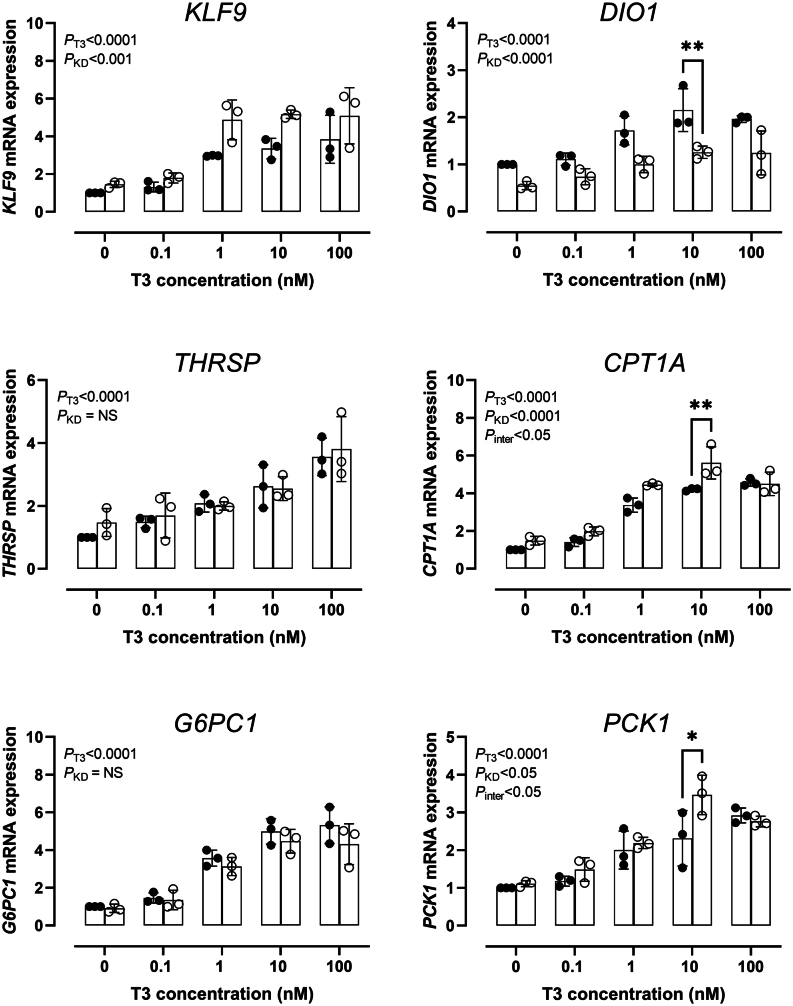
The knockdown efficiency of TBL1X in the three consecutive experiments was 42%, 46%, and 66% respectively and a combined knockdown efficiency of TBL1X was shown in Supplementary Figure 1 (see section on supplementary materials given at the end of this article). To evaluate the effect of TBL1X knockdown in HepG2 cells on T3-regulated gene expression, mRNA expression of DIO1, KLF9, THRSP, CPT1A, G6PC1, and PCK1 was measured (Fig. 1). The mRNA expression of all these genes increased to varying extent upon increasing T3 concentrations. TBL1X knockdown increased the expression of KLF9, CPT1A,and PCK1 compared to the expression in cells of the control transfected groups. Interestingly, the knockdown of TBL1X markedly decreased the expression of DIO1 (PANOVA < 0.0001). Knockdown of TBL1X did not have an effect on THRSP and G6PC1 expression. The interaction between T3 stimulation and TBL1X knockdown on the expression of KLF9, DIO1, G6PC1, and THRSP was not significant while there is an interaction effect for CPT1A and PCK1 (both P < 0.05). In addition, we measured mRNA expression of FBP1, ELOVL2, and HMGCS2 (all T3 negatively regulated genes) in HepG2 cells, FBP1 and HMGCS2 were not expressed in HepG2 cells (data not shown) while ELOVL2 was not responsive to T3 (Supplementary Figure 2).
Figure 1.
Effects of TBL1X knockdown on T3 regulated genes in HepG2 cells. Controls are represented by black dots and TBL1X knockdown by open circles. Cells are stimulated with increasing concentration of T3. mRNA expression is normalized to the control group without T3 which is set at 1. Mean values ± s.d. of three independent experiments are shown, and each experiment consists of 3 values per group (technical triplicates). P-values for two-way analysis of variance are indicated on the top-left corner of each figure panel. Post hoc analysis (Tukey) P-values are indicated by asterisks above the corresponding bars: *(P < 0.05), **(P <0.01).
Characterization of iPSC derived hepatic cells derived from TBL1X N365Y mutation iPSC lines
Flow cytometry was used to analyze the expression of definitive endoderm-specific markers such as CKIT and CXCR4, and the hepatocyte maker AFP. Flow cytometric results demonstrated no differences in endodermal (Supplementary Figure 3A and 3B) or hepatocyte specification (Supplemental Figure 3C and 3D) between the father and the daughter carrying the TBL1X N365Y mutation lines. Additionally, we confirmed the presence of iHeps through mRNA expression of AFP and Hepatocyte Nuclear Factor 4 (HNF4). The expression of these hepatocyte-specific markers was similar between father and daughter lines, confirming the efficiency of our protocol in differentiating iPSC into fetal hepatocytes (Supplementary Figure 4).
The effect of TBL1X N365Y on T3 regulated genes in iHeps
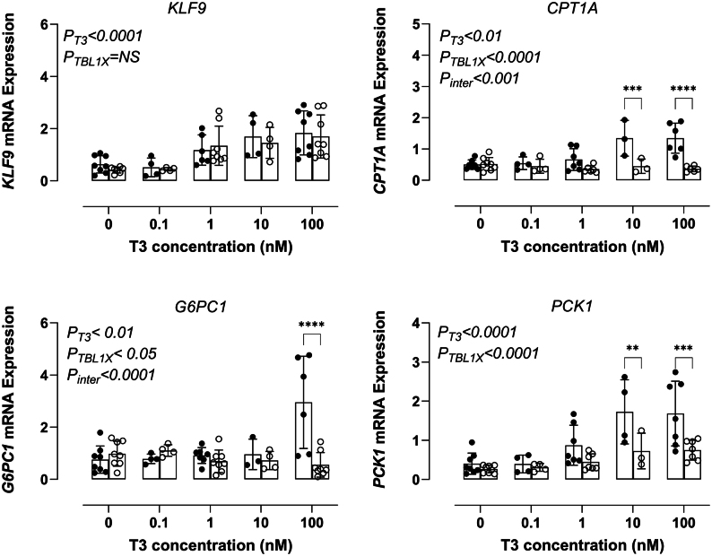
To evaluate the effect of TBL1X N365Y on T3-regulated gene expression in iHeps from the affected father and the daughter carrying the TBL1X N365Y mutation (on one X chromosome), mRNA expression of KLF9, CPT1A, G6PC1,and PCK1 was measured (Fig. 2). The mRNA expression of all these genes increased to varying extent upon increasing T3 concentrations. The mRNA expression of CPT1A, G6PC1, and PCK1 is lower in the cells of the father who is hemizygous for TBL1X N365Y compared to the expression in iHeps of the daughter who is a carrier. TBL1X N365Y did not affect KLF9 expression which is similar in both father and daughter. Additionally, mRNA expression of three T3 negatively regulated genes, FBP1, ELOVL2, and HMGCS2 was measured (Fig. 3). The mRNA expression of all these genes decreased to varying extent upon increasing T3 concentrations. Hemizygous TBL1X N365Y decreased the expression of FBP1 and ELOVL2 compared to the expression in iHeps of the daughter who is a carrier. HMGCS2 expression did not differ between the patient and the carrier.
Figure 2.
Effects of TBL1X N365Y on T3 positively regulated genes in iHeps cells. The daughter mutation is represented by black dots, the father mutation by open dots. Cells were stimulated with increasing concentration of T3. Mean values ± s.d. consists of T3 0 nM n = 8, T3 0.1 nM n = 4, T3 1 nM n = 8, T3 10 nM n = 4, and T3 100 nM n = 8 per group. P-values for two-way analysis of variance are indicated on the top-left corner of each figure panel. Post hoc analysis (Tukey) P-values are indicated by asterisks above the corresponding bars: **(P < 0.01), ***(P < 0.001), ****(P < 0.0001).
Figure 3.
Effects of TBL1X N365Y on T3 negatively regulated genes in iHeps cells. The daughter mutation is represented by black dots, the father mutation by open dots. Cells were stimulated with increasing concentration of T3. Mean values ± S.D. consists of T3 0 nM n = 8, T3 0.1 nM n = 4, T3 1 nM n = 8, T3 10 nM n = 4, and T3 100 nM n = 8 per group. P-values for two-way analysis of variance are indicated on the top-left corner of each figure panel. Post hoc analysis (Tukey) P-values are indicated by asterisks above the corresponding bars: *(P < 0.05), **(P < 0.01), ***(P < 0.001), ****(P < 0.0001).
Discussion
Mutations in the WD40 repeat domain of TBL1X have been identified as one of the genetic causes of central CH (1, 6). Central hypothyroidism is defined as TH deficiency due to insufficient stimulation of the thyroid gland by the pituitary and/or hypothalamus (1). Central CH associated with mutations in TBL1X is accompanied by impaired hearing (1, 6). TBL1X is a component of the NCOR1/SMRT corepressor complex which represses gene expression mediated by unliganded TR. All known mutations are located at the WD40 repeat domain which is involved in nuclear protein-protein interactions. We used two human cellular models, the HepG2 cell line and iHeps, to investigate the role of TBL1X in TH action.
We found that TBL1X knockdown in HepG2 cells results in increased expression of KLF9, CPT1A, and PCK1 compared to control cells, which is consistent with the role of TBL1X in the corepressor complex. Disturbed corepressor function is supposed to lead to an increase in T3-regulated gene expression. This is consistent with the observation of Takamizawa et al. (24) who showed that TBL1X N365Y significantly inhibited the TR/NCOR1 mediated activity of the TRH promoter in n-1 cells, even in the presence of wild-type TBL1X. However, the fact that TBL1X knockdown markedly repressed the T3-induced expression of DIO1 in HepG2 cells but did not affect the T3-induced expression of G6PC1 and THRSP, indicates a differential role of TBL1X in T3 action that is gene context-specific. Of note, the knockdown of TBL1XR1, a close homolog of TBL1X, in HepG2 cells similarly repressed the expression of DIO1 markedly (25), while an impaired NCOR1 complex significantly activates the expression of liver DIO1 in mice (12). This indicates that each component of the NCOR1/SMRT corepressor complex has unique roles in the regulation of T3 genes.
Given the importance of T3 in regulating liver metabolic pathways, and to further explore whether TBL1X mutations change T3 signaling we analyzed the expression of T3 target genes in human iHeps derived from a father and daughter who both carry the TBL1X N365Y mutation. The hemizygous father was diagnosed with central CH at the age of 2 weeks while the heterozygous daughter had normal TSH and fT4 concentrations (1). We showed before that TBL1X N365Y protein was poorly expressed compared with wild-type protein (1). TBL1XN365Y markedly repressed the T3-induced expression of CPT1A, G6PC1, and PCK1 in the cells derived from the father, suggesting that TBL1X acts as a coactivator. Surprisingly, in the father’s iHeps T3 upregulated the expression of KLF9 to a similar extent as observed in the daughter. The decreased sensitivity of CPT1A, G6PC1, and PCK1 to TH is in line with mice with impaired hepatic expression of TBL1X which show increased steatosis and decreased fatty acid oxidation (18), both symptoms of hypothyroidism. We next assessed the expression of genes negatively regulated by T3 such as FBP1, ELOVL2, and HMGCS2. The expression of FBP1 and ELOVL2 upon T3 was lower in the father cells compared to the daughter while HMGCS2 mRNA expression did not differ. Collectively, these results suggest that patients carrying the TBL1X mutation mainly have a decreased sensitivity to T3 which is dependent on the gene evaluated and the sample’s features (hemizygous or carrier). While the effects of T3 in the father’s iHeps were observed in the context of CPT1A, G6PC1, and PCK1, and in the negatively regulated T3 target genes FBP1 and ELOVL2, the carrier’s iHeps displayed more pronounced T3 sensitivity to the genes analyzed. The data suggest that the TBL1X mutation could play a role in determining T3 sensitivity in human hepatocytes. Nevertheless, further studies are necessary to explore these differential effects.
The difference in TH action between HepG2 and iHeps may be related to the difference between a TBL1X knockdown and a TBL1X mutation. We showed earlier that the latter seems to have a deleterious effect on protein folding (1) while the knockdown of the TBL1X gene results in less protein. However, both models show a differential effect on T3-related genes.
Alternatively, the difference in TH action between HepG2 and iHeps could also be explained by the expression levels of TRα and TRβ. TRβ1 is the main TR isoform in hepatocytes (26). THRB expression is present at low levels in HepG2 cells (27) as well as THRA (28). iHeps express both THRA and THRB mRNA, with THRB being the dominant receptor, which is similar to the expression pattern observed in human hepatocytes (29). Therefore, it is possible that iHeps more accurately replicate the in vivo hepatocyte environment compared to HepG2 cells. It is tempting to speculate that the differences in the response to T3 between HepG2 and iHeps are the result of differences in the expression of TRα and TRβ. However, the lack of reliable antibodies against TRα and TRβ makes it difficult to determine the expression levels of both receptors in both models. Alternatively, the nature of the cell model (tumor cell line or differentiated iPSC) could be the reason for the differential responses (30, 31). In addition to the role of TBL1X in TR function, TBL1X interacts with other transcription factors such as the estrogen receptor (ER), the androgen receptor (AR), and PPARγ, which may exert an influence on the gene expression as well (8).
In conclusion, we studied the role of TBL1X in TH action in HepG2 cells and iHeps and found a differential effect of T3 on the expression of a variety of T3-responsive genes. TBL1X acts as a corepressor in the regulation of KLF9, CPT1A, and PCK1 in HepG2 cells while it acts as a coactivator in the regulation of DIO1. In iHeps, TBL1X appears to function as a coactivator as the expression of T3 positively regulated genes was repressed by the mutation in TBL1X. However, the observation that T3 negatively regulated genes in iHeps were repressed by mutated TBL1X indicates that TBL1X indeed acts as a corepressor. Further research is needed to unravel the mechanisms involved, but based on the results of this study it can be concluded that a mutation in TBL1X does not cause general T3 hyper- or hypo-sensitivity while knockdown of TBL1X in HepG2 cells seems to strengthen the sensitivity to T3 selectively.
Supplementary Materials
Declaration of interest
EB receives consultancy fees from Madrigal and Aligos.
AB is an editorial board member of the European Thyroid Journal. She was not involved in the editorial or peer review process for this paper.
Funding
This work was supported by a grant (to YH) from the Chinese Scholarship Council (CSC) (no. 201907720056), ANH and AW were supported by NIH grant DK117940.
Author contribution statement
YH: conducting experiments, acquiring and analyzing data, writing the manuscript; LSDO: conducting experiments, acquiring and analyzing data, writing the manuscript; KF: conducting experiments, acquiring data; ASPT: collecting patients and reviewing the manuscript; EF: reviewing the manuscript, supervision; JEK: conducting experiments, acquiring data; AAW: designing research studies, reviewing the manuscript; ANH: designing research studies, providing reagents, reviewing the manuscript; EB: designing research studies, reviewing the manuscript, supervision; AB: designing research studies, providing reagents, reviewing the manuscript, supervision.
References
- 1.Heinen CA, Losekoot M, Sun Y, Watson PJ, Fairall L, Joustra SD, Zwaveling-Soonawala N, Oostdijk W, van den Akker ELT, Alders M, et al.Mutations in TBL1X are associated with central hypothyroidism. Journal of Clinical Endocrinology and Metabolism 20161014564–4573. ( 10.1210/jc.2016-2531) [DOI] [PMC free article] [PubMed] [Google Scholar]
- 2.Heinen CA, de Vries EM, Alders M, Bikker H, Zwaveling-Soonawala N, van den Akker ELT, Bakker B, Hoorweg-Nijman G, Roelfsema F, Hennekam RC, et al.Mutations in IRS4 are associated with central hypothyroidism. Journal of Medical Genetics 201855693–700. ( 10.1136/jmedgenet-2017-105113) [DOI] [PMC free article] [PubMed] [Google Scholar]
- 3.Hayashizaki Y Hiraoka Y Endo Y Miyai K & Matsubara K. Thyroid-stimulating hormone (TSH) deficiency caused by a single base substitution in the CAGYC region of the beta-subunit. EMBO Journal 198982291–2296. ( 10.1002/j.1460-2075.1989.tb08355.x) [DOI] [PMC free article] [PubMed] [Google Scholar]
- 4.Collu R, Tang J, Castagné J, Lagacé G, Masson N, Huot C, Deal C, Delvin E, Faccenda E, Eidne KA, et al.A novel mechanism for isolated central hypothyroidism: inactivating mutations in the thyrotropin-releasing hormone receptor gene. Journal of Clinical Endocrinology and Metabolism 1997821561–1565. ( 10.1210/jcem.82.5.3918) [DOI] [PubMed] [Google Scholar]
- 5.Sun Y, Bak B, Schoenmakers N, van Trotsenburg ASP, Oostdijk W, Voshol P, Cambridge E, White JK, le Tissier P, Gharavy SNM, et al.Loss-of-function mutations in IGSF1 cause an X-linked syndrome of central hypothyroidism and testicular enlargement. Nature Genetics 2012441375–1381. ( 10.1038/ng.2453) [DOI] [PMC free article] [PubMed] [Google Scholar]
- 6.García M Barreda-Bonis AC Jiménez P Rabanal I Ortiz A Vallespín E Del Pozo Á Martínez-San Millán J González-Casado I & Moreno JC. Central hypothyroidism and novel clinical phenotypes in hemizygous truncation of TBL1X. Journal of the Endocrine Society 20193119–128. ( 10.1210/js.2018-00144) [DOI] [PMC free article] [PubMed] [Google Scholar]
- 7.Heinen CA, Jongejan A, Watson PJ, Redeker B, Boelen A, Boudzovitch-Surovtseva O, Forzano F, Hordijk R, Kelley R, Olney AH, et al.A specific mutation in TBL1XR1 causes Pierpont syndrome. Journal of Medical Genetics 201653330–337. ( 10.1136/jmedgenet-2015-103233) [DOI] [PMC free article] [PubMed] [Google Scholar]
- 8.Hu Y Bruinstroop E Hollenberg AN Fliers E & Boelen A. The role of WD40 repeat-containing proteins in endocrine (dys)function. Journal of Molecular Endocrinology 202371. ( 10.1530/JME-22-0217) [DOI] [PubMed] [Google Scholar]
- 9.Astapova I & Hollenberg AN. The in vivo role of nuclear receptor corepressors in thyroid hormone action. Biochimica et Biophysica Acta 201318303876–3881. ( 10.1016/j.bbagen.2012.07.001) [DOI] [PMC free article] [PubMed] [Google Scholar]
- 10.Shimizu H Astapova I Ye F Bilban M Cohen RN & Hollenberg AN. NCoR1 and SMRT play unique roles in thyroid hormone action in vivo. Molecular and Cellular Biology 201535555–565. ( 10.1128/MCB.01208-14) [DOI] [PMC free article] [PubMed] [Google Scholar]
- 11.Shimizu H, Lu Y, Vella KR, Damilano F, Astapova I, Amano I, Ritter M, Gallop MR, Rosenzweig AN, Cohen RN, et al.Nuclear corepressor SMRT is a strong regulator of body weight independently of its ability to regulate thyroid hormone action. PLoS One 201914e0220717. ( 10.1371/journal.pone.0220717) [DOI] [PMC free article] [PubMed] [Google Scholar]
- 12.Astapova I Lee LJ Morales C Tauber S Bilban M & Hollenberg AN. The nuclear corepressor, NCoR, regulates thyroid hormone action in vivo. PNAS 200810519544–19549. ( 10.1073/pnas.0804604105) [DOI] [PMC free article] [PubMed] [Google Scholar]
- 13.Astapova I. Role of co-regulators in metabolic and transcriptional actions of thyroid hormone. Journal of Molecular Endocrinology 20165673–97. ( 10.1530/JME-15-0246) [DOI] [PubMed] [Google Scholar]
- 14.Astapova I Vella KR Ramadoss P Holtz KA Rodwin BA Liao XH Weiss RE Rosenberg MA Rosenzweig A & Hollenberg AN. The nuclear receptor corepressor (NCoR) controls thyroid hormone sensitivity and the set point of the hypothalamic-pituitary-thyroid axis. Molecular Endocrinology 201125212–224. ( 10.1210/me.2010-0462) [DOI] [PMC free article] [PubMed] [Google Scholar]
- 15.Costa-e-Sousa RH Astapova I Ye F Wondisford FE & Hollenberg AN. The thyroid axis is regulated by NCoR1 via its actions in the pituitary. Endocrinology 20121535049–5057. ( 10.1210/en.2012-1504) [DOI] [PMC free article] [PubMed] [Google Scholar]
- 16.Ritter MJ Amano I Imai N Soares De Oliveira L Vella KR & Hollenberg AN. Nuclear receptor CoRepressors, NCOR1 and SMRT, are required for maintaining systemic metabolic homeostasis. Molecular Metabolism 202153101315. ( 10.1016/j.molmet.2021.101315) [DOI] [PMC free article] [PubMed] [Google Scholar]
- 17.Sinha RA Bruinstroop E Singh BK & Yen PM. Nonalcoholic fatty liver disease and hypercholesterolemia: roles of thyroid hormones, metabolites, and agonists. Thyroid 2019291173–1191. ( 10.1089/thy.2018.0664) [DOI] [PMC free article] [PubMed] [Google Scholar]
- 18.Kulozik P, Jones A, Mattijssen F, Rose AJ, Reimann A, Strzoda D, Kleinsorg S, Raupp C, Kleinschmidt J, Müller-Decker K, et al.Hepatic deficiency in transcriptional cofactor TBL1 promotes liver steatosis and hypertriglyceridemia. Cell Metabolism 201113389–400. ( 10.1016/j.cmet.2011.02.011) [DOI] [PubMed] [Google Scholar]
- 19.Costantini S Di Bernardo G Cammarota M Castello G & Colonna G. Gene expression signature of human HepG2 cell line. Gene 2013518335–345. ( 10.1016/j.gene.2012.12.106) [DOI] [PubMed] [Google Scholar]
- 20.Choi IY Lim H & Lee G. Efficient generation human induced pluripotent stem cells from human somatic cells with Sendai-virus. Journal of Visualized Experiments: JoVE 2014. (86). ( 10.3791/51406) [DOI] [PMC free article] [PubMed] [Google Scholar]
- 21.Kaserman JE, Hurley K, Dodge M, Villacorta-Martin C, Vedaie M, Jean JC, Liberti DC, James MF, Higgins MI, Lee NJ, et al.. A highly phenotyped open access repository of alpha-1 antitrypsin deficiency pluripotent stem cells. Stem Cell Reports 202015242–255. ( 10.1016/j.stemcr.2020.06.006) [DOI] [PMC free article] [PubMed] [Google Scholar]
- 22.Kaserman JE, Werder RB, Wang F, Matte T, Higgins MI, Dodge M, Lindstrom-Vautrin J, Bawa P, Hinds A, Bullitt E, et al.Human iPSC-hepatocyte modeling of alpha-1 antitrypsin heterozygosity reveals metabolic dysregulation and cellular heterogeneity. Cell Reports 202241111775. ( 10.1016/j.celrep.2022.111775) [DOI] [PMC free article] [PubMed] [Google Scholar]
- 23.Wilson AA, Ying L, Liesa M, Segeritz CP, Mills JA, Shen SS, Jean J, Lonza GC, Liberti DC, Lang AH, et al.Emergence of a stage-dependent human liver disease signature with directed differentiation of alpha-1 antitrypsin-deficient iPS cells. Stem Cell Reports 20154873–885. ( 10.1016/j.stemcr.2015.02.021) [DOI] [PMC free article] [PubMed] [Google Scholar]
- 24.Takamizawa T, Satoh T, Miyamoto T, Nakajima Y, Ishizuka T, Tomaru T, Yoshino S, Katano-Toki A, Nishikido A, Sapkota S, et al.Transducin β-like 1, X-linked and nuclear receptor co-repressor cooperatively augment the ligand-independent stimulation of TRH and TSHβ gene promoters by thyroid hormone receptors. Endocrine Journal 201865805–813. ( 10.1507/endocrj.EJ17-0384) [DOI] [PubMed] [Google Scholar]
- 25.Hu Y Falize K van Trotsenburg ASP Hennekam R Fliers E Bruinstroop E & Boelen A. The role of transducin β-like 1 X-linked receptor 1 (TBL1XR1) in thyroid hormone metabolism and action in mice. European Thyroid Journal 202312. ( 10.1530/ETJ-23-0077) [DOI] [PMC free article] [PubMed] [Google Scholar]
- 26.Amma LL Campos-Barros A Wang Z Vennström Br & Forrest D. Distinct tissue-specific roles for thyroid hormone receptors β and α1 in regulation of Type 1 deiodinase expression. Molecular Endocrinology 200115467–475. ( 10.1210/mend.15.3.0605) [DOI] [PubMed] [Google Scholar]
- 27.Yuan C Lin JZH Sieglaff DH Ayers SD Denoto-Reynolds F Baxter JD & Webb P. Identical gene regulation patterns of T3 and selective thyroid hormone receptor modulator GC-1. Endocrinology 2012153501–511. ( 10.1210/en.2011-1325) [DOI] [PMC free article] [PubMed] [Google Scholar]
- 28.Kwakkel J Wiersinga WM & Boelen A. Interleukin-1beta modulates endogenous thyroid hormone receptor alpha gene transcription in liver cells. Journal of Endocrinology 2007194257–265. ( 10.1677/JOE-06-0177) [DOI] [PubMed] [Google Scholar]
- 29.Mendoza A, Tang C, Choi J, Acuña M, Logan M, Martin AG, Al-Sowaimel L, Desai BN, Tenen DE, Jacobs C, et al.Thyroid hormone signaling promotes hepatic lipogenesis through the transcription factor ChREBP. Science Signaling 202114eabh3839. ( 10.1126/scisignal.abh3839) [DOI] [PMC free article] [PubMed] [Google Scholar]
- 30.Wilkening S Stahl F & Bader A. Comparison of primary human hepatocytes and hepatoma cell line Hepg2 with regard to their biotransformation properties. Drug Metabolism and Disposition: The Biological Fate of Chemicals 2003311035–1042. ( 10.1124/dmd.31.8.1035) [DOI] [PubMed] [Google Scholar]
- 31.Kang SJ Lee HM Park YI Yi H Lee H So B Song JY & Kang HG. Chemically induced hepatotoxicity in human stem cell-induced hepatocytes compared with primary hepatocytes and HepG2. Cell Biology and Toxicology 201632403–417. ( 10.1007/s10565-016-9342-0) [DOI] [PubMed] [Google Scholar]
Associated Data
This section collects any data citations, data availability statements, or supplementary materials included in this article.


 This work is licensed under a

