Abstract
Systemic sclerosis is a progressive fibrotic autoimmune disease which mainly affects young women causing fibrosis of the skin and internal organs. In systemic sclerosis patients, facial and hand alterations are significant as these may have an impact on body image. In fact, systemic sclerosis could cause disfigurement of the most visible parts of the body, hindering self-confidence and quality of life. Therefore, the assessment of body image in systemic sclerosis is crucial in order to optimize a tailored care of the patient. In the near future, the creation of a pathway involving dedicated specialists (psychologists, physiotherapists, dermatologists, and plastic surgeons) should become an integral part of the disease management.
Keywords: Systemic sclerosis, body image, plastic surgery, aesthetic impairment, quality of life, multidisciplinary, amimic facies, lipofilling, mesenchymal stem cells
The “Scleroesthet study” by Farhat et al. 1 raises an important issue which is an unmet need in systemic sclerosis (SSc). This study reported that SSc patients referred worse perception of aesthetic impairment, especially those with facial involvement and pitting scars. This evidence provides us the opportunity to focus on the importance of aesthetic impairment, mainly affecting young SSc women between the age of 35 and 60. In fact, SSc is a fibrosing disease resulting in widespread fibrosis of the skin and internal organs, melanoderma, facial disfiguration caused by skin thickening and teleangectasias, and hand deformations brought on by digital retraction and ulcers. 2 Typical scleroderma facies is characterized by loss of expression (amimic facies), loss of the nasolabial fold and accentuation of perioral wrinkles, smoothed wrinkles of the forehead, microstomia with reduced mouth opening, and progressive wasting of neck and temporal muscles. 3 Regarding the hands, patients frequently experience digital edema (“puffy fingers”) and pitting scars, telangiectasias, subungual hyperkeratosis, abrasions, and fissures.
As the disease progresses, patients develop digital fibrosis (sclerodactyly), subcutaneous calcinosis, and eventually digital ulcers which may lead to digital amputation. Furthermore, digital atrophy may occur, which is characterized by a localized thickening and tightness of the skin of the fingers with a claw-like appearance and spindle shape of the affected digits.4,5 Disfigurement mostly affects parts of the body which are at the core of interpersonal relationships, significantly reducing comfort, aesthetics, and self-confidence.6,7 The physician is usually concentrated on addressing the severity of the disease, evaluating organ function and counteracting disease progression. This may lead to neglecting the facial and digital skin alterations which have a deep impact on the quality of life of the patient. SSc modifies the patient’s perception of the disease and alters personal body image,8–11 which is defined as a mental image of both physical and psychological personality that a person has of themselves and others. 12
It is well known that body image alteration is not only an aesthetic impairment but also represents a more complex condition, especially in young SSc patients due to its impact on physical appearance.13,14 Despite the importance of this issue affecting SSc patients and the growing interest in aesthetic procedures, which address some disfiguring diseases, no consensus has been reached regarding the application of aesthetic medicine and plastic surgery in this disease. In the last 10 years, many surgical and non-surgical options have been tested in SSc and localized Morphea, with either interesting or contradicting results.15,16 Among non-surgical approaches, hyaluronic acid injections have been shown to be effective, both enhancing mouth mobility and skin elasticity; however, the effect is limited due to reduced bioavailability of materials. 17
The use of botulinum toxin has been suggested to improve facial appearance by reducing muscle tension, despite the lack of improvement in tissue atrophy. 18 Phototherapy has been suggested to treat microstomia in selected patients; however, no functional improvement has been observed.19,20 Among surgical approaches, the most interesting technique is the autologous adipose tissue grafting, which is an innovative source of mesenchymal stem cells, suitable for cell-based therapy in regenerative medicine. 21 In fact, adipose-derived stem cells are similar to bone marrow-derived stem cells and differentiate into multiple mesodermal tissue types. In addition to their functional properties, acting as filler, these cells have immunomodulatory properties and secrete angiogenic factors which trigger tissue repair.22–24 In the last decades, autologous fat grafting has been proposed both for aesthetic (filling/correction) and functional (regenerative) purposes in SSc-related mouth and hand complications, in particular, for skin atrophy linked to morphea, fibrosis of perioral skin, and long-lasting DUs. This procedure is considered safe because it is an autologous fat transfer, neither immunogenic nor carcinogenic and also well tolerated by patients, thanks to a specific sedation protocol. However, the procedure has some limitations such as chronic skin inflammation, the need of corticosteroid and/or immunosuppressive drugs, and the reduced availability of adipose tissue in SSc patients.25–28 Moreover, transferred fat reabsorbs over time; therefore, the procedure needs to be repeated in order to maintain the results in SSc patients. However, with the innovative introduction of LipoBanks®, 29 which stores fat samples obtained with a single procedure of fat aspiration, the scenario is going to be revolutionized. The introduction of LipoBanks allows a single procedure of lip aspiration and then fat sample is stored, significantly reducing both physical and psychological stress in patients.
Despite several studies suggesting the use of fat grafting for treating mouth and hands in SSc patients, different results have been reported depending on the type of patients treated, surgical techniques, different protocols, and outcomes.25–32 For this reason, no shared consensus has yet been reached. However, in our experience, regenerative medicine represents a promising approach to SSc aestethic issues concerning alteration of hand and face. In fact, we believe that a specific pathway should be implemented in daily clinical practice to create more awareness while treating SSc patients, particularly in young women, in order to prevent or limit face and body disfigurement together with organ complications. Therefore, every clinic devoted to SSc patients should have a protocol for the preservation of personal body image, addressing aesthetic impairment and consequent body image dissatisfaction, proposing strategies to maintain or recover physical appearance.
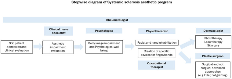
In the last decade, our Scleroderma Unit focused on aesthetic impairment in SSc patients and collaborated with plastic surgeons. Our clinical practice devised a specific aesthetic protocol involving rheumatologists, plastic surgeons, psychologists, physiotherapists and occupational therapists, as well as dermatologists (Figure 1). After being admitted to the Unit, each patient undergoes routine clinical investigations and imaging of hand and facial alterations. The rheumatologist provides an overall evaluation of the severity and impact that hand and facial alterations have on the quality of life, body image, and psychological well-being of each patients through validated questionnaires, in particular, the FACE-Q tools, 33 the Rosenberg Self-Esteem Scale, 34 the adapted Satisfaction with Appearance Scale, 14 and the Hospital Anxiety and Depression Scale. 35 Moreover, the perception of disability was measured by the Health Assessment Questionnaire 36 and by the Mouth Handicap in Systemic Sclerosis scale. 37 The following step consists in a psychology interview with the patient to improve awareness and acceptance of the disease. Each patient is also evaluated by a specialized physiotherapist for planning hand and face rehabilitation so as to safeguard and enhance the function of the affected areas. In the case of severe hand involvement, a meeting with an occupational therapist is scheduled to evaluate hand functionality and program a protocol which entails the use of assistive devices. Finally, the results of the rheumatological examination, patient questionnaires, and photographic documentation collected are discussed in a multidisciplinary clinic with dermatologists and plastic surgeons for a more tailored therapeutic strategy.
Figure 1.
The clinical pathway of the patient for the aesthetic program is managed by seven healthcare professionals who are coordinated by the rheumatologist.
Each professional has strong experience in rheumatic diseases, in particular in the treatment of scleroderma patients. For the correct management and the best effectiveness of the treatment, the contribution of every specialist is necessary to tailor the optimal treatment.
In the next future, all scleroderma clinics should share a similar multidisciplinary program, making this an integral part of the disease management. This approach may foster patient’s compliance by benefiting mood and quality of life.
In conclusion, the importance of aesthetic impairment in SSc patients is a growing unmet need which should be taken into consideration by rheumatologists and other specialists, being aware that aesthetic medicine and plastic surgery may have an important role on the well-being and quality of life of SSc patients.
Footnotes
The author(s) declared no potential conflicts of interest with respect to the research, authorship, and/or publication of this article.
Funding: The author(s) disclosed receipt of the following financial support for the research, authorship, and/or publication of this article: This paper has received funding from MUR under PNRR M4C2I1.3 Heal Italia project PE00000019 CUP E93C22001860006 University of Modena and Reggio Emilia to Prof. Dilia Giuggioli.
ORCID iDs: Martina Orlandi  https://orcid.org/0000-0001-6784-2235
Marco De Pinto  https://orcid.org/0000-0002-0947-8663
Gilda Sandri  https://orcid.org/0000-0003-0454-1093
Dilia Giuggioli  https://orcid.org/0000-0002-0041-3695
References
- 1. Farhat MM, Guerreschi P, Morell-Dubois S, et al. Perception of aesthetic impairment in patients with systemic sclerosis determined using a semi-quantitative scale and its association with disease characteristics. J Scleroderma Relat Disord 2024; 9(2): 124–133. [DOI] [PMC free article] [PubMed] [Google Scholar]
- 2. Volkmann ER, Andréasson K, Smith V. Systemic sclerosis. Lancet 2023; 401(10373): 304–318. [DOI] [PMC free article] [PubMed] [Google Scholar]
- 3. Smirani R, Poursac N, Naveau A, et al. Orofacial consequences of systemic sclerosis: a systematic review. J Scleroderma Relat Disord 2018; 3(1): 81–90. [DOI] [PMC free article] [PubMed] [Google Scholar]
- 4. Starnoni M, Pappalardo M, Spinella A, et al. Systemic sclerosis cutaneous expression: management of skin fibrosis and digital ulcers. Ann Med Surg 2021; 71: 102984. [DOI] [PMC free article] [PubMed] [Google Scholar]
- 5. Giuggioli D, Manfredi A, Lumetti F, et al. Scleroderma skin ulcers definition, classification, and treatment strategies our experience and review of the literature. Autoimmun Rev 2018; 17(2): 155–164. [DOI] [PubMed] [Google Scholar]
- 6. Park EH, Strand V, Oh YJ, et al. Health-related quality of life in systemic sclerosis compared with other rheumatic diseases: a cross-sectional study. Arthritis Res Ther 2019; 21(1): 61. [DOI] [PMC free article] [PubMed] [Google Scholar]
- 7. Maddali-Bongi S, Del Rosso A, Mikhaylova S, et al. Impact of hand and face disabilities on global disability and quality of life in systemic sclerosis patients. Clin Exp Rheumatol 2014; 32(6 suppl 86): S15–S20. [PubMed] [Google Scholar]
- 8. Tedeschini E, Pingani L, Simoni E, et al. Correlation of articular involvement, skin disfigurement and unemployment with depressive symptoms in patients with systemic sclerosis: a hospital sample. Int J Rheum Dis 2014; 17(2): 186–194. [DOI] [PubMed] [Google Scholar]
- 9. van Lankveld WG, Vonk MC, Teunissen H, et al. Appearance self-esteem in systemic sclerosis: subjective experience of skin deformity and its relationship with physician-assessed skin involvement, disease status and psychological variables. Rheumatology 2007; 46(5): 872–876. [DOI] [PubMed] [Google Scholar]
- 10. Malcarne V-L, Hansdottir I, Greenbergs H-L, et al. Appearance self-esteem in systemic sclerosis. Cognitive Therapy and Research 1999; 23: 197–208. [Google Scholar]
- 11. Benrud-Larson LM, Heinberg LJ, Boling C, et al. Body image dissatisfaction among women with scleroderma: extent and relationship to psychosocial function. Health Psychol 2003; 22(2): 130–139. [PubMed] [Google Scholar]
- 12. Pruzinsky T, Cash T-F. Assessing body image and quality of life in medical settings. In: Cash TF, Pruzinsky T. (eds) Body image: a handbook of theory, research, and clinical practice. New York: Guilford Press, 2002, pp. 171–182. [Google Scholar]
- 13. Steen VD, Medsger TA., Jr. The value of the Health Assessment Questionnaire and special patient-generated scales to demonstrate change in systemic sclerosis patients over time. Arthritis Rheum 1997; 40(11): 1984–1991. [DOI] [PubMed] [Google Scholar]
- 14. Heinberg LJ, Kudel I, White B, et al. Assessing body image in patients with systemic sclerosis (scleroderma): validation of the adapted Satisfaction with Appearance Scale. Body Image 2007; 4(1): 79–86. [DOI] [PMC free article] [PubMed] [Google Scholar]
- 15. Creadore A, Watchmaker J, Maymone MBC, et al. Cosmetic treatment in patients with autoimmune connective tissue diseases: best practices for patients with morphea/systemic sclerosis. J Am Acad Dermatol 2020; 83(2): 315–341. [DOI] [PubMed] [Google Scholar]
- 16. Gonzalez C, Pamatmat J, Hutto J, et al. Review of the current medical and surgical treatment options for microstomia in patients with scleroderma. Dermatol Surg 2021; 47: 780–784. [DOI] [PubMed] [Google Scholar]
- 17. Pirrello R, Verro B, Grasso G, et al. Hyaluronic acid and platelet-rich plasma, a new therapeutic alternative for scleroderma patients: a prospective open-label study. Arthritis Res Ther 2019; 21(1): 286. [DOI] [PMC free article] [PubMed] [Google Scholar]
- 18. Cumsky HJL, Michael M, Hoss E. Use of botulinum toxin and hyal-uronic acid filler to treat oral Involvement in scleroderma. Dermatol Surg 2022; 48(6): 698–699. [DOI] [PubMed] [Google Scholar]
- 19. Dinsdale G, Murray A, Moore T, et al. A comparison of intense pulsed light and laser treatment of telangiectases in patients with systemic sclerosis: a within-subject randomized trial. Rheumatology 2014; 53(8): 1422–1430. [DOI] [PubMed] [Google Scholar]
- 20. Murray AK, Moore TL, Richards H, et al. Pilot study of intense pulsed light for the treatment of systemic sclerosis-related telangiectases. Br J Dermatol 2012; 167(3): 563–569. [DOI] [PubMed] [Google Scholar]
- 21. Magalon G, Daumas A, Sautereau N, et al. Regenerative approach to scleroderma with fat grafting. Clin Plast Surg 2015; 42(3): 353–364, viii. [DOI] [PubMed] [Google Scholar]
- 22. Gimble JM, Katz AJ, Bunnel BA. Adipose-derived stem cells for regenerative medicine. Circ Res 2007; 100(9): 1249–1260. [DOI] [PMC free article] [PubMed] [Google Scholar]
- 23. Scuderi N, Ceccarelli S, Onesti MG, et al. Human adipose-derived stromal cells for cell-based therapies in the treatment of systemic sclerosis. Cell Transplant 2013; 22(5): 779–795. [DOI] [PubMed] [Google Scholar]
- 24. Manetti M. Could autologous adipose-derived stromal vascular fraction turn out an unwanted source of profibrotic myofibroblasts in systemic sclerosis? Ann Rheum Dis 2020; 79(5): e55. [DOI] [PubMed] [Google Scholar]
- 25. Giuggioli D, Spinella A, Cocchiara E, et al. Autologous fat grafting in the treatment of a scleroderma stump-skin ulcer: a case report. Case Rep Plast Surg Hand Surg 2021; 8(1): 18–22. [DOI] [PMC free article] [PubMed] [Google Scholar]
- 26. Pignatti M, Spinella A, Cocchiara E, et al. Autologous fat grafting for the oral and digital complications of systemic sclerosis: results of a prospective study. Aesthetic Plast Surg 2020; 44(5): 1820–1832. [DOI] [PubMed] [Google Scholar]
- 27. Sautereau N, Daumas A, Truillet R, et al. Efficacy of autologous microfat graft on facial handicap in systemic sclerosis patients. Plast Reconstr Surg Glob Open 2016; 4(3): e660. [DOI] [PMC free article] [PubMed] [Google Scholar]
- 28. Del Papa N, Caviggioli F, Sambataro D, et al. Autologous fat grafting in the treatment of fibrotic perioral changes in patients with systemic sclerosis. Cell Transplant 2015; 24(1): 63–72. [DOI] [PubMed] [Google Scholar]
- 29. Lipobank®, dove l’innovazione e il benessere si incontrano, https://lipobank.it
- 30. Almadori A, Griffin M, Ryan CM, et al. Stem cell enriched lipotransfer reverses the effects of fibrosis in systemic sclerosis. PLoS ONE 2019; 14(7): e0218068. [DOI] [PMC free article] [PubMed] [Google Scholar]
- 31. Onesti MG, Fioramonti P, Carella S, et al. Improvement of mouth functional disability in systemic sclerosis patients over one year in a trial of fat transplantation versus adipose-derived stromal cells. Stem Cells Int 2016; 2016: 2416192. [DOI] [PMC free article] [PubMed] [Google Scholar]
- 32. Kølle SF, Fischer-Nielsen A, Mathiasen AB, et al. Enrichment of autologous fat grafts with ex-vivo expanded adipose tissue-derived stem cells for graft survival: a randomised placebo-controlled trial. Lancet 2013; 382(9898): 1113–1120. [DOI] [PubMed] [Google Scholar]
- 33. Cogliandro A, Barone M, Persichetti P. Italian linguistic validation of the FACE-Q instrument. JAMA Facial Plast Surg 2017; 19(4): 336–337. [DOI] [PMC free article] [PubMed] [Google Scholar]
- 34. Sinclair SJ, Blais MA, Gansler DA, et al. Psychometric properties of the Rosenberg Self-Esteem Scale: overall and across demographic groups living within the United States. Eval Health Prof 2010; 33(1): 56–80. [DOI] [PubMed] [Google Scholar]
- 35. Zigmond AS, Snaith RP. The Hospital Anxiety and Depression Scale. Acta Psychiatr Scand 1983; 67(6): 361–370. [DOI] [PubMed] [Google Scholar]
- 36. Steen VD, Medsger TA. The value of the Health Assessment Questionnaire and special patient-generated scales to demonstrate change in systemic sclerosis patients over time. Arthritis Rheum 1997; 40(11): 1984–1991. [DOI] [PubMed] [Google Scholar]
- 37. Mouthon L, Rannou F, Bérezné A, et al. Development and validation of a scale for mouth handicap in systemic sclerosis: the Mouth Handicap in Systemic Sclerosis scale. Ann Rheum Dis 2007; 66(12): 1651–1655. [DOI] [PMC free article] [PubMed] [Google Scholar]


