Abstract
BACKGROUND
The development of tumor is closely linked to inflammation. Therefore, targeting molecules involved in inflammation may be effective in predicting cancer prognosis. Transarterial chemoembolization (TACE) holds significant therapeutic significance in addressing hepatocellular carcinoma (HCC). At present, no studies have evaluated the predictive value of γ-glutamyl transferase to albumin ratio (GAR) on the prognosis of HCC undergoing TACE.
AIM
To explore the potential prognostic significance of the GAR in individuals undergoing TACE for HCC.
METHODS
A total of 1231 patients from seven hospitals in China were randomized into a training cohort (n = 862) and a validation cohort (n = 369). To establish independent prognostic factors for overall survival (OS), we utilized multivariate and univariate Cox regression models. The best cut-off value of the GAR was determined with the X-tile software, with OS as the basis. Validations were performed using dual therapy cohort and triple therapy cohort.
RESULTS
X-tile software revealed a GAR threshold of 4.75 as optimal. Both pre- and post-propensity score matching analyses demonstrated that the median OS in the low-GAR group (< 4.75) was notably longer compared to the high-GAR group (≥ 4.75), showing results of 26.9 vs 9.8 months (P < 0.001) initially, and 18.1 vs 11.3 months (P < 0.001) after match. Furthermore, multivariate analysis identified GAR ≥ 4.75 as an independent prognostic factor (P < 0.001). The receiver operating characteristic curves for the nomogram showed area under receiver operating characteristic curves of 0.741, 0.747, and 0.708 for predicting 1-, 2-, and 3-year survival, respectively. Consistent findings were reiterated in the two cohorts involving TACE in combination with targeted therapy and TACE in combination with targeted therapy and immunotherapy. Calibration curve and decision curve analyses substantiated the model’s relatively robust predictive capabilities.
CONCLUSION
Our study validates the effective prognostic capacity of the GAR-based nomogram for HCC patients undergoing TACE or TACE in combination with systemic therapy.
Keywords: Transarterial chemoembolization, Immunotherapy, Targeted therapy, Hepatocellular carcinoma, Prognosis
Core Tip: γ-glutamyl transferase to albumin ratio has been confirmed for the first time to be predictive in hepatocellular carcinoma undergoing transarterial chemoembolization and transarterial chemoembolization combined with systemic therapy in this large-sample multicenter study. A nomogram model for predicting postoperative survival was also established based on γ-glutamyl transferase to albumin ratio, which was empirically demonstrated to have strong predictive ability.
INTRODUCTION
Hepatocellular carcinoma (HCC) is prevalent worldwide and is a life-threatening malignancy, witnessing an escalating incidence that represents a large proportion of deaths related to cancer. Projections indicate that the annual HCC incidence will exceed one million individuals by the year 2025[1]. Sadly, patients devoid of effective therapeutic interventions succumb to an abysmal median overall survival (mOS) of merely four months[2].
The latest guidelines released by the National Comprehensive Cancer Network advocate for the combined employment of atezolizumab and bevacizumab as the prime systemic therapy option for individuals affected by advanced HCC. Additionally, in cases of unresectable or inoperable HCC exceeding 5 centimeters, transarterial chemoembolization (TACE) may merit consideration to extend patient survival. Following the established staging system of Barcelona Clinical Liver Cancer (BCLC), TACE has become the mainstay of therapy for intermediate-stage HCC. Specifically, this encompasses patients boasting asymptomatic, multi-nodular tumors devoid of vascular invasion or extrahepatic spread[3]. Indeed, TACE has also yielded surprising results for some early patients who were not suitable for surgery or ablation[4-6]. In some series of studies, the majority of patients treated with TACE were in the early stages of disease, with more than 40% being solitary nodules[7,8]. Importantly, TACE has also garnered favorable treatment responses in advanced-stage HCC cases. An extensive longitudinal cohort study across 14 countries discovered that TACE served as the primary treatment for nearly 50% of individuals afflicted with BCLC-C-stage HCC[9]. This assertion is further supported by the findings of two prospective non-randomized trials[10,11], which confirmed the superiority of conventional TACE over basic supportive care.
Scientific investigations have illuminated the intricate relationship between long-standing inflammatory factors and the malignant transformation of tumors, highlighting the fundamental role of chronic inflammation and its associated factors in the emergence of tumorigenesis[12]. Inflammatory factors are important players in the regulation of the tumor microenvironment. They possess the capacity to instigate tumor epithelial-mesenchymal transition, generate reactive oxygen species, and thereby facilitate tumor cell proliferation, angiogenesis, invasion, and metastasis[12,13]. γ-glutamyl transferase (GGT) is a kind of glycoprotein secreted by Kupffer cells and bile duct endothelial cells. Elevated GGT levels are a marker of liver dysfunction and alcohol intake, and can be used to measure the severity of liver inflammation and cellular damage[14]. GGT’s main role is to mediate the hydrolysis of extracellular glutathione, which produces large amounts of hydrogen peroxide and reactive oxygen species and other free radicals, leading to oxidative stress in tissues[15]. Many experimental studies have also shown that the expression of GGT makes cancer cells more invasive and even leads to the emergence of anticancer drug resistance[16].
Albumin (ALB) is the most important protein in humans, synthesized by liver cells. It has a wide range of important physiological functions, such as regulating immunity, stabilizing endothelium, and binding to various drugs and toxins[17]. In addition, ALB binds to a series of inflammatory mediators and interacts with nitric oxide, playing anti-inflammatory and antioxidant roles[18]. Serum ALB levels reflect liver reserve function and nutritional status of the body. In cases of malnutrition, hepatitis, and cirrhosis, the concentration of ALB in the human body will significantly decrease[19]. Therefore, ALB is frequently utilized in the assessment of liver function prior to hepatic resection[20]. Recent research has confirmed that ALB can also exert a protective influence after a variety of radical surgeries[21]. Although some studies have shown that GAR can predict the outcome of HCC[22,23], these studies only focused on hepatectomy, and their sample sizes were small. In this study, we developed a predictive model for HCC based on a convenient and inexpensive liquid biopsy technique, which can effectively predict not only survival after TACE alone, but also outcomes of dual therapy or triple therapy.
MATERIALS AND METHODS
Study design
TACE alone cohort: During the period spanning from January 2019 to June 2023, 1231 patients with HCC, who received either drug-eluting beads TACE or conventional TACE, were gathered from seven hospitals in China. The diagnosis of HCC was confirmed by histology, at least two typical radiological features, or one typical imaging manifestation with a serum alpha-fetoprotein (AFP) level > 400 ng/mL. The inclusion criteria consisted of the following prerequisites: (1) Patients who had not previously received any form of antitumour therapy prior to TACE; (2) Patients exhibiting measurable lesions that aligned with the response assessment criteria specifically tailored for solid tumours (RECIST 1.1); and (3) GGT and ALB were measured within 7 days before TACE. The exclusion criteria were: (1) Presence of other malignant tumors other than HCC; (2) Patients with a history of other anti-tumor therapies; and (3) Lack of follow-up data.
Dual therapy cohort: Patients with HCC who received TACE in combination with targeted therapy at the above hospitals were included in the two-combination therapy cohort. The clinical data including demographic characteristics, hematological parameters, imaging data and tumor staging were collected retrospectively. At the same time, GGT and ALB values at baseline were obtained.
Triple therapy cohort: Patients with HCC who received TACE in conjunction with immunotherapy and targeted therapy at the same hospitals were enrolled in this cohort. Complete baseline clinical and laboratory data were recorded for subsequent analysis. The study intentionally excluded those patients who had incomplete follow-up information.
Data assessment
The laboratory parameters included AFP, GGT, alanine aminotransferase, ALB, total bilirubin, red blood cells, lactate dehydrogenase, alkaline phosphatase, hemoglobin, white blood cells, lymphocyte count, hepatitis B virus infection, and hepatitis C virus infection. Tumor burden was assessed by magnetic resonance imaging. Enhanced computer tomography or single-organ contrast-enhanced ultrasound, including tumor diameter and number, portal vein invasion, lymph node metastases and extrahepatic metastases. Additional data encompassed variables such as sex, age, presence of alcohol consumption, hepatitis B virus, hepatitis C virus infection, hypertension and diabetes mellitus. Both the training and validation cohorts underwent bi-monthly computer tomography or magnetic resonance imaging examinations following the initial treatment. All patients were evaluated according to RECIST 1.1.
Statistical analysis
All of the statistical analyses were implemented with R 4.1.3 software and SPSS 26.0 software. A bilateral P value of < 0.05 was viewed as statistically significant. Tumor response data and baseline features were summed up with descriptive statistics. Comparisons of categorical variables were done with χ2 test. The best threshold value for serum GAR was identified from OS utilizing X-tile software. To ensure balanced baseline characteristics, propensity score matching (PSM) scale was exploited for determining the low and high GAR groups. All patients were randomized into a validation group (n = 369) and a training group (n = 862). With the Kaplan-Meier statistical approach, the log-rank test was employed to estimate and compare mOS between groups. Subsequently, all covariates exhibiting an impact on survival in univariate analysis with P < 0.05 were included in the multivariate Cox proportional hazards model. Multivariate cox analysis was carried out to identify independent influencing factors, which were utilized to establish a nomogram. The nomogram’s prognostic effect was evaluated using receiver operating characteristic, calibration curve and decision curve analysis.
Ethical statements
The study protocol was authorized by the Ethics Committee of the Affiliated Hospital of Southwest Medical University (No. KY2021063) and complied with the guidelines of the Helsinki Declaration. Written informed consent was waived because of the retrospective study.
RESULTS
Patients and tumor characteristics
This retrospective study enrolled a total of 1231 HCC patients. There were 1044 (85%) males and 187 (15%) females. There were 501 (41%) patients aged ≥ 60 years. There were 749 patients (61%) who were positive for hepatitis B. AFP ≥ 500 ng/mL was detected in 528 patients (43%). Multiple tumor masses were present in 890 patients (72%). Tumor diameter ≥ 10 cm was found in 352 patients (29%). Portal vein tumor thrombus (PVTT) was found in 484 patients (39%). According to BCLC tumor staging, 177 (14%) were stage A, 188 (15%) were stage B, 853 (69%) were stage C, and 13 (1%) were stage D. The remaining baseline characteristics are shown in Table 1.
Table 1.
Baseline characteristics of low and high γ-glutamyl transferase to albumin ratio groups before propensity score matching
|
Variables
|
Total
|
GAR-low
|
GAR-high
|
P value
|
| Patients | 1231 | 706 | 525 | |
| Gender | < 0.001 | |||
| Female, n (%) | 187 (0.15) | 129 (0.18) | 58 (0.11) | |
| Male, n (%) | 1044 (0.85) | 577 (0.82) | 467 (0.89) | |
| Age ≥ 60 years, n (%) | 501 (0.41) | 305 (0.43) | 196 (0.37) | 0.044 |
| HBV, n (%) | 749 (0.61) | 432 (0.61) | 317 (0.60) | 0.819 |
| HCV, n (%) | 38 (0.03) | 27 (0.04) | 11 (0.02) | 0.117 |
| Alcohol, n (%) | 482 (0.39) | 243 (0.34) | 239 (0.46) | < 0.001 |
| Diabetes, n (%) | 122 (0.10) | 69 (0.10) | 53 (0.10) | 0.928 |
| Hypertension, n (%) | 178 (0.14) | 106 (0.15) | 72 (0.14) | 0.576 |
| ALT levels ≥ 40, U/L, n (%) | 639 (0.52) | 274 (0.39) | 365 (0.70) | < 0.001 |
| ALP levels ≥ 125, U/L, n (%) | 714 (0.58) | 249 (0.35) | 465 (0.89) | < 0.001 |
| Serum AFP ≥ 500, ng/mL, n (%) | 528 (0.43) | 254 (0.36) | 274 (0.52) | < 0.001 |
| Child-Pugh class, n (%) | < 0.001 | |||
| A | 920 (0.75) | 585 (0.83) | 335 (0.64) | |
| B | 297 (0.24) | 114 (0.16) | 183 (0.35) | |
| C | 14 (0.01) | 7 (0.01) | 7 (0.01) | |
| Lymph node metastasis, n (%) | 617 (0.50) | 312 (0.44) | 305 (0.58) | < 0.001 |
| Extrahepatic metastasis, n (%) | 238 (0.19) | 116 (0.16) | 122 (0.23) | 0.004 |
| PVTT, n (%) | 484 (0.39) | 196 (0.28) | 288 (0.55) | < 0.001 |
| BCLC stage, n (%) | < 0.001 | |||
| A | 177 (0.14) | 137 (0.19) | 40 (0.08) | |
| B | 188 (0.15) | 129 (0.18) | 59 (0.11) | |
| C | 853 (0.69) | 433 (0.61) | 420 (0.80) | |
| D | 13 (0.01) | 7 (0.01) | 6 (0.01) | |
| Number of tumors ≥ 2, n (%) | 890 (0.72) | 469 (0.66) | 421 (0.80) | < 0.001 |
| Tumor diameter, cm, n (%) | < 0.001 | |||
| < 3 | 172 (0.14) | 133 (0.19) | 39 (0.07) | |
| ≥ 3, < 5 | 216 (0.18) | 155 (0.22) | 61 (0.12) | |
| ≥ 5, < 10 | 491 (0.40) | 302 (0.43) | 189 (0.36) | |
| ≥ 10 | 352 (0.29) | 116 (0.16) | 236 (0.45) |
GAR-low: γ-glutamyl transferase to albumin ratio < 4.75; GAR-high: γ-glutamyl transferase to albumin ratio ≥ 4.75; HBV: Hepatitis B virus; HCV: Hepatitis C virus; ALT: Alanine aminotransferase; ALP: Alkaline phosphatase; AFP: Alpha-fetoprotein; PVTT: Portal vein tumor thrombus; BCLC: Barcelona Clinic Liver Cancer.
GAR levels and OS before and after PSM
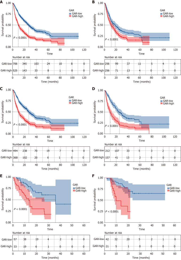
Both multivariate and univariate Cox analyses displayed that GAR value ≥ 4.75 was an independent prognostic risk factor for OS (Table 2). Based on X-tile software analysis, the best cut-off of GAR is 4.75. Based on the cutoff value, all 1231 patients were further divided into two groups, GAR ≥ 4.75 (n = 525) and GAR < 4.75 (n = 706). The high GAR group tended to exhibit higher tumor load before PSM (P < 0.05, Table 1). All patients in high and low GAR groups had a mOS of 9.8 months [95% confidence interval (CI): 8.9-11.5] and 26.9 months (95%CI: 24.0-30.2), respectively. Statistically significant differences existed between the two groups in mOS [hazard ratio (HR) = 2.079, 95%CI: 1.807-2.392, P < 0.001, Figure 1A]. There were no statistically significant differences between the two groups after PSM (Table 3). In the high GAR group, the mOS was 11.3 months (95%CI: 9.1-15.3), while in the low group, it was 18.1 months (95%CI: 13.4-23.3). The former was noticeably shorter as compared to the latter (HR = 1.461, 95%CI: 1.185-1.802, P < 0.001, Figure 1B).
Table 2.
Univariate and multivariate Cox regression analyses of overall survival before propensity score matching
|
Variables
|
HR (95%CI)
|
P value
|
HR (95%CI)
|
P value
|
| Gender (male/female) | 1.094 (0.898-1.334) | 0.372 | ||
| Age (≥ 60/< 60 years) | 0.888 (0.770-1.024) | 0.102 | ||
| HBV (positive/negative) | 1.087 (0.941-1.255) | 0.257 | ||
| HCV (positive/negative) | 0.657 (0.406-1.062) | 0.086 | ||
| Alcohol (yes/no) | 1.069 (0.927-1.233) | 0.360 | ||
| Diabetes (yes/no) | 0.913 (0.718-1.162) | 0.460 | ||
| Hypertension (yes/no) | 0.899 (0.732-1.104) | 0.310 | ||
| GAR-high | 2.075 (1.804-2.388) | < 0.001 | 1.533 (1.285-1.828) | < 0.001 |
| ALT levels ≥ 40, U/L | 1.313 (1.141-1.511) | < 0.001 | 1.003 (0.862-1.166) | 0.973 |
| ALP levels ≥ 125, U/L | 1.730 (1.495-2.001) | < 0.001 | 1.008 (0.840-1.209) | 0.932 |
| Serum AFP ≥ 500, ng/mL | 1.326 (1.152-1.525) | < 0.001 | 0.982 (0.846-1.140) | 0.814 |
| Child-Pugh A vs B/C | 1.812 (1.553-2.113) | < 0.001 | 1.389 (1.176-1.640) | < 0.001 |
| Lymph node metastasis | 1.435 (1.246-1.652) | < 0.001 | 0.940 (0.779-1.133) | 0.514 |
| Extrahepatic metastasis | 1.679 (1.424-1.979) | < 0.001 | 1.301 (1.091-1.550) | 0.003 |
| PVTT | 1.958 (1.701-2.253) | < 0.001 | 1.372 (1.150-1.637) | < 0.001 |
| BCLC stage A vs B vs C vs D | ||||
| A | 1 | < 0.001 | 1 | 0.106 |
| B | 1.409 (1.036-1.916) | 0.029 | 0.865 (0.603-1.241) | 0.432 |
| C | 2.447 (1.911-3.135) | < 0.001 | 1.142 (0.810-1.611) | 0.447 |
| D | 3.538 (1.823-6.868) | < 0.001 | 1.867 (0.929-3.752) | 0.079 |
| Number of tumors ≥ 2 | 1.614 (1.365-1.909) | < 0.001 | 1.381 (1.132-1.685) | 0.001 |
| Tumor diameter, cm | ||||
| < 3 | 1 | < 0.001 | 1 | < 0.001 |
| ≥ 3, < 5 | 1.692 (1.262-2.270) | < 0.001 | 1.684 (1.245-2.278) | 0.001 |
| ≥ 5, < 10 | 2.114 (1.633-2.737) | < 0.001 | 1.756 (1.337-2.306) | < 0.001 |
| ≥ 10 | 2.930 (2.250-3.816) | < 0.001 | 1.822 (1.364-2.435) | < 0.001 |
GAR-high: γ-glutamyl transferase to albumin ratio ≥ 4.75; HR: Hazard ratio; CI: Confidence interval; HBV: Hepatitis B virus; HCV: Hepatitis C virus; ALT: Alanine aminotransferase; ALP: Alkaline phosphatase; AFP: Alpha-fetoprotein; PVTT: Portal vein tumor thrombus; BCLC: Barcelona Clinic Liver Cancer.
Figure 1.
Kaplan-Meier survival curves stratified by γ-glutamyl transferase to albumin ratio. A: Overall survival of the transarterial chemoembolization alone cohort; B: The cohort after propensity score matching; C: Training set; D: Validation set; E: Dual therapy cohort; F: Triple therapy cohort. GAR: γ-glutamyl transferase to albumin ratio.
Table 3.
Baseline characteristics of high and low γ-glutamyl transferase to albumin ratio groups after propensity score matching, n (%)
|
Variables
|
Total
|
GAR-low
|
GAR-high
|
P value
|
| Patients | 512 | 256 | 256 | |
| Gender | 0.798 | |||
| Female | 71 (0.14) | 34 (0.13) | 37 (0.14) | |
| Male | 441 (0.86) | 222 (0.87) | 219 (0.86) | |
| Age ≥ 60 years | 215 (0.42) | 107 (0.42) | 108 (0.42) | 1 |
| HBV | 293 (0.57) | 148 (0.58) | 145 (0.57) | 0.858 |
| HCV | 17 (0.03) | 8 (0.03) | 9 (0.04) | 1 |
| Alcohol | 203 (0.40) | 99 (0.39) | 104 (0.41) | 0.718 |
| Diabetes | 47 (0.09) | 25 (0.1) | 22 (0.09) | 0.76 |
| Hypertension | 65 (0.13) | 29 (0.11) | 36 (0.14) | 0.426 |
| ALT levels ≥ 40, U/L | 304 (0.59) | 147 (0.57) | 157 (0.61) | 0.418 |
| ALP levels ≥ 125, U/L | 395 (0.77) | 196 (0.77) | 199 (0.78) | 0.833 |
| Serum AFP ≥ 500, ng/mL | 221 (0.43) | 108 (0.42) | 113 (0.44) | 0.721 |
| Child-Pugh class | 0.93 | |||
| A | 371 (0.72) | 186 (0.73) | 185 (0.72) | |
| B | 134 (0.26) | 67 (0.26) | 67 (0.26) | |
| C | 7 (0.01) | 3 (0.01) | 4 (0.02) | |
| Lymph node metastasis | 269 (0.53) | 139 (0.54) | 130 (0.51) | 0.479 |
| Extrahepatic metastasis | 99 (0.19) | 49 (0.19) | 50 (0.20) | 1 |
| PVTT | 232 (0.45) | 125 (0.49) | 107 (0.42) | 0.131 |
| BCLC stage | 0.347 | |||
| A | 58 (0.11) | 25 (0.10) | 33 (0.13) | |
| B | 67 (0.13) | 29 (0.11) | 38 (0.15) | |
| C | 380 (0.74) | 199 (0.78) | 181 (0.71) | |
| D | 7 (0.01) | 3 (0.01) | 4 (0.02) | |
| Number of tumors ≥ 2 | 385 (0.75) | 192 (0.75) | 193 (0.75) | 1 |
| Tumor diameter, cm | 0.797 | |||
| < 3 | 50 (0.10) | 22 (0.09) | 28 (0.11) | 0.797 |
| ≥ 3, < 5 | 79 (0.15) | 41 (0.16) | 38 (0.15) | |
| ≥ 5, < 10 | 234 (0.46) | 116 (0.45) | 118 (0.46) | |
| ≥ 10 | 149 (0.29) | 77 (0.30) | 72 (0.28) |
GAR-low: γ-glutamyl transferase to albumin ratio < 4.75; GAR-high: γ-glutamyl transferase to albumin ratio ≥ 4.75; HBV: Hepatitis B virus; HCV: Hepatitis C virus; ALT: Alanine aminotransferase; ALP: Alkaline phosphatase; AFP: Alpha-fetoprotein; PVTT: Portal vein tumor thrombus; BCLC: Barcelona Clinic Liver Cancer.
Prognostic factors influencing OS
Before PSM, we performed univariate and multivariate Cox analyses and identified six independent factors affecting OS prognosis, including number of tumors ≥ 2 (P = 0.001), GAR value ≥ 4.75 (P < 0.001), PVTT (P < 0.001), Child-Pugh B/C (P < 0.001), larger tumor diameter (P < 0.001), and extra-hepatic metastasis (P = 0.003) respectively (Table 2).
After PSM, each GAR group had 256 patients enrolled in the study cohort. Univariate and multivariate Cox regression analyses pinpointed several independent prognostic factors impacting OS: GAR value ≥ 4.75 (P < 0.001), PVTT (P = 0.001), higher Child-Pugh classification (P = 0.004), multiple tumors (P < 0.001), and larger tumor diameter (P < 0.001). Notably, the prognostic significance of high-GAR remained robust even after PSM (Supplementary Table 1).
GAR can predict OS in validation set and training set (TACE alone cohort)
A total of 1231 patients were grouped into the validation and training groups in a 3:7 ratio. There were no evident statistical differences in the baseline features of the patients in both groups (Supplementary Table 2). The mOS for patients in the low group in the training set was 27.1 months (95%CI: 24.2-32.2) vs 9.9 months (95%CI: 8.6-11.8) for patients in the high group (P < 0.001). Significantly shorter mOS was observed in the high GAR group, demonstrating statistical significance (HR = 2.061, 95%CI: 1.74-2.442, P < 0.001, Figure 1C). There were comparable results observed in the validation set, with the mOS for high GAR and low GAR groups being 25.4 (95%CI: 21.0-30.3) and 9.5 (95%CI: 7.6-12.5) months, respectively. The high group presented remarkably shorter mOS in contrast to the low group (HR = 2.119, 95%CI: 1.648-2.723, P < 0.001, Figure 1D).
Nomogram construction and validation
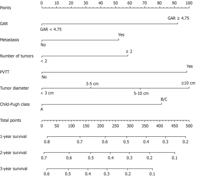
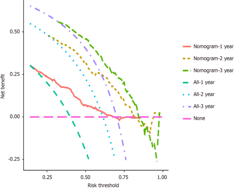
In the training cohort, we found six independent factors affecting OS prognosis, including GAR, number of tumors, PVTT, Child-Pugh classification, tumor diameter, and extra-hepatic metastasis respectively (Table 4). The nomogram was constructed based on independent factors that significantly affected OS (Figure 2). Time-varying receiver operating characteristic curves in the validation set displayed that the area under receiver operating characteristic curve values of this model in predicting 1-, 2-, and 3-year survival were 0.741, 0.747, and 0.708, respectively (Figure 3). Additionally, the calibration curves and decision curve analysis curves demonstrated good predictive potential of the model (Figures 4 and 5).
Table 4.
Univariate and multivariate Cox analyses for identifying clinical characteristics influencing overall survival in the training cohort
|
Variables
|
HR (95%CI)
|
P value
|
HR (95%CI)
|
P value
|
| Gender (male/female) | 1.129 (0.888-1.435) | 0.321 | ||
| Age (≥ 60/< 60 years) | 0.895 (0.754-1.063) | 0.206 | ||
| HBV (positive/negative) | 1.145 (0.962-1.362) | 0.128 | ||
| HCV (positive/negative) | 0.691 (0.406-1.175) | 0.172 | ||
| Alcohol (yes/no) | 1.017 (0.856-1.208) | 0.851 | ||
| Diabetes (yes/no) | 0.883 (0.662-1.179) | 0.399 | ||
| Hypertension (yes/no) | 0.856 (0.673-1.090) | 0.208 | ||
| GAR-high | 2.058 (1.737-2.438) | < 0.001 | 1.516 (1.218-1.885) | < 0.001 |
| ALT levels ≥ 40, U/L | 1.278 (1.079-1.514) | 0.005 | 0.904 (0.749-1.090) | 0.291 |
| ALP levels ≥ 125, U/L | 1.756 (1.475-2.091) | < 0.001 | 1.106 (0.882-1.388) | 0.382 |
| Serum AFP ≥ 500, ng/mL | 1.252 (1.057-1.484) | 0.009 | 0.904 (0.752-1.086) | 0.281 |
| Child-Pugh A vs B/C | 1.858 (1.544-2.234) | < 0.001 | 1.389 (1.133-1.703) | 0.002 |
| Lymph node metastasis | 1.354 (1.143-1.603) | < 0.001 | 0.951 (0.756-1.197) | 0.669 |
| Extrahepatic metastasis | 1.608 (1.311-1.973) | < 0.001 | 1.263 (1.013-1.575) | 0.038 |
| PVTT | 2.009 (1.696-2.380) | < 0.001 | 1.510 (1.214-1.879) | < 0.001 |
| BCLC stage A vs B vs C vs D | ||||
| A | 1 | < 0.001 | 1 | 0.146 |
| B | 1.363 (0.957-1.942) | 0.086 | 0.865 (0.571-1.311) | 0.494 |
| C | 2.183 (1.639-2.907) | < 0.001 | 0.999 (0.667-1.498) | 0.998 |
| D | 4.106 (1.949-8.648) | < 0.001 | 2.174 (0.989-4.775) | 0.053 |
| Number of tumors ≥ 2 | 1.511 (1.241-1.838) | < 0.001 | 1.335 (1.057-1.685) | 0.015 |
| Tumor diameter, cm | ||||
| < 3 | 1 | < 0.001 | 1 | 0.002 |
| ≥ 3, < 5 | 1.755 (1.226-2.513) | 0.002 | 1.752 (1.211-2.534) | 0.003 |
| ≥ 5, < 10 | 2.312 (1.691-3.162) | < 0.001 | 1.912 (1.372-2.665) | < 0.001 |
| ≥ 10 | 2.924 (2.117-4.039) | < 0.001 | 1.793 (1.256-2.559) | 0.001 |
GAR-high: γ-glutamyl transferase to albumin ratio ≥ 4.75; HR: Hazard ratio; CI: Confidence interval; HBV: Hepatitis B virus; HCV: Hepatitis C virus; ALT: Alanine aminotransferase; ALP: Alkaline phosphatase; AFP: Alpha-fetoprotein; PVTT: Portal vein tumor thrombus; BCLC: Barcelona Clinic Liver Cancer.
Figure 2.
Nomogram constructed based on independent risk factors for predicting 1-, 2-, and 3-year overall survival in patients with hepatocellular carcinoma. GAR: γ-glutamyl transferase to albumin ratio; PVTT: Portal vein tumor thrombus.
Figure 3.
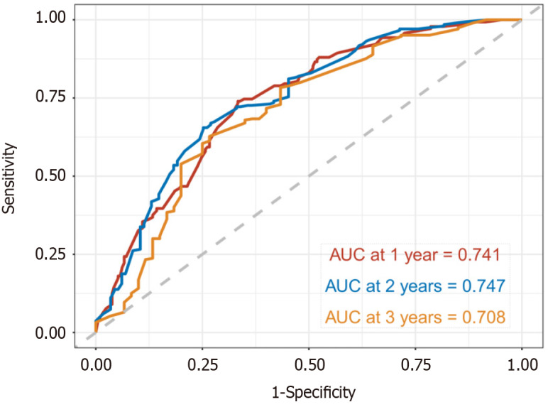
Time-dependent receiver operating characteristic curves of this predictive model. AUC: Area under receiver operating characteristic curve.
Figure 4.
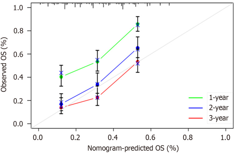
Calibration curve of validation cohort. OS: Overall survival.
Figure 5.
Decision curve analyses curve of validation cohort.
GAR can predict OS of cohort for dual therapy
In total, 90 patients who received the combination therapy of TACE along with targeted therapy were encompassed within the dual therapy cohort. The mOS was found to be 36.1 months (95%CI: 24.5 to not applicable) in the low group, whereas it reduced significantly to 13.6 months (95%CI: 9.2-26.8) in the high group. Comparative analysis revealed a noteworthy disparity in mOS between both GAR groups, and the survival time was obviously shorter in high GAR group (HR = 2.079, 95%CI: 1.807-2.392, P < 0.001; Figure 1E). Additionally, in this particular cohort, GAR demonstrated a substantial association with patient survival.
GAR can predict the OS of triple therapy cohort
There were 63 patients who received a combination therapy of TACE, targeted therapy, and immunotherapy were included in this analysis. The mOS for the low GAR group was not reached, while it amounted to 9.8 months (95%CI: 8.27 to not applicable) for the high GAR group. Upon comparing the two groups, it was evident that the high GAR group exhibited a significantly shorter mOS (HR = 4.615, 95%CI: 1.844-11.55, P < 0.001; Figure 1F). These findings strongly indicate a pronounced correlation between GAR and the survival rate of patients.
DISCUSSION
TACE has become an important treatment method in the era of tumor precision therapy[24]. Although some studies have shown that GAR can be applied for the prediction of HCC prognosis, the study sample is generally small, and the study objects are all surgical resection. At present, no study has confirmed the predictive value of GAR in TACE. Therefore, we initiated a large-sample, multicenter TACE cohort study and established a nomogram model of HCC patient survival based on GAR. In this study, the patients with GAR ≥ 4.75 had lower mOS than those with GAR < 4.75(P < 0.001). High predictive accuracy of GAR was also found in both the dual and triple therapy cohorts for the first time.
At present, TACE-based prognostic models have been gradually established. The neutrophil-lymphocyte ratio model and platelet-to-lymphocyte ratio have been demonstrated to predict survival prognosis and metastasis in HCC patients treated with TACE[25,26]. However, there is no prognostic marker for patients suffering from HCC after TACE in conjugation with immunotherapy or targeted therapy. TACE was once the first-line therapy for stage B BCLC patients, and recent prospective clinical studies have shown that TACE is also a novel ideal treatment option for patients with early-stage (solitary nodules) and some advanced-stage HCC[27]. More and more HCC patients are expected to benefit from TACE treatment, so it is of great clinical significance to search for an effective prognostic marker for TACE treatment.
The role of inflammation in cancer has long been the focus of researchers. Inflammation promotes tumor development through a multi-faceted process, including angiogenesis, extracellular matrix remodeling, immunosuppression, invasion, and distant infiltration[28,29]. Various inflammatory cells create a favorable environment for tumor growth, thus exacerbating the development of this pathological process[30]. High levels of serum GGT are often a marker of severe hepatitis, cirrhosis, and advanced cancer. There is evidence that it mediates the hydrolysis of glutathione, which can lead to the formation of free radicals, lipid peroxidation and induce malignant transformation of cells. GGT is also thought to promote rapid growth and turnover of tumor cells[16]. A number of published prospective studies have shown a positive correlation between the levels of GGT and a number of cancers, including liver, breast, and prostate cancer[31,32].
In contrast, ALB plays a vital role in counteracting oxidative stress, enhancing microcirculation, and inhibiting inflammatory responses. It has been demonstrated that ALB exerts DNA replication stabilization and immune response potentiation, thus impeding tumor progression[33]. Furthermore, hypoalbuminemia has been consistently linked to unfavorable prognoses in cancer patients[34,35]. McMillan et al[35] found that individuals with advanced lung or gastrointestinal malignancies exhibit a persistent systemic inflammatory response, which results in a continuous reduction in ALB concentration. To some extent, serum GGT levels can reflect tumor load, whereas liver metabolic ability and immune function are determined by ALB levels. Consequently, in comparison with other biomarkers, the GAR value more accurately reflects the relative tumor burden and liver function, thus affording a more effective prognostic tool for predicting the survival outcomes of patients diagnosed with HCC.
In preceding investigations, the prognostic outcome of HCC has been linked to factors such as GAR, PVTT, tumor number, tumor size, and Child grade[23,36]. The present study further corroborates this finding. Furthermore, we extended the nomogram model, demonstrating its commendable performance for prediction in the validation set. This has enhanced the GAR’s practical application in clinical settings. Particularly for patients with elevated GAR, vigilance and proactive management should be emphasized.
The strength of the current research is the large sample size, which improves the reliability of results. We performed a PSM analysis, which reduced the effect of bias and confounding variables in the study and could more truthfully reflect the association of GAR with OS. Post-PSM survival analysis reaffirmed the prognostic value of GAR, with the low-GAR group demonstrating superior mOS. Further statistical scrutiny of the matched cohort, including both univariate and multivariate models, consistently highlighted GAR as a pivotal, independent determinant of OS outcomes. GAR was not only internally validated by the test data set, but also validated TACE in conjunction with targeted cohorts and TACE in conjunction with immunotherapy and targeted therapy. Therefore, it further improves the clinical applicability in the future treatment of HCC.
This study does, however, have some limitations. In the first place, this was a retrospective study, so selection bias was inevitable. Second, GGT and ALB levels can be influenced by different laboratory hematologic techniques, which may lead to some deviations. Third, although the predictive performance of GAR in dual and triple therapy cohorts has been verified, the number of patients was small, which may compromise the reliability of the conclusion. Therefore, multicenter, a large population, and prospective studies are expected to confirm and update the findings of this study in the future.
CONCLUSION
Our study validates the effective prognostic capacity of the GAR-based nomogram for HCC patients undergoing TACE or TACE in combination with systemic therapy.
ACKNOWLEDGEMENTS
The authors would like to thank all the reviewers who participated in the review.
Footnotes
Institutional review board statement: The study protocol was authorized by the Ethics Committee of the Affiliated Hospital of Southwest Medical University (No. KY2021063) and complied with the Code of Ethics of the World Medical Association.
Informed consent statement: Written informed consent was waived because of the retrospective study.
Conflict-of-interest statement: All the authors report no relevant conflicts of interest for this article.
Provenance and peer review: Unsolicited article; Externally peer reviewed.
Peer-review model: Single blind
Specialty type: Oncology
Country of origin: China
Peer-review report’s classification
Scientific Quality: Grade C
Novelty: Grade C
Creativity or Innovation: Grade C
Scientific Significance: Grade B
P-Reviewer: Mirminachi S S-Editor: Wang JJ L-Editor: A P-Editor: Wang WB
Contributor Information
Zhen-Ying Wu, Department of Oncology, The Affiliated Hospital of Southwest Medical University, Luzhou 646000, Sichuan Province, China; Department of Oncology, Pangang Group General Hospital, Panzhihua 617000, Sichuan Province, China.
Han Li, Department of Oncology, The Affiliated Hospital of Southwest Medical University, Luzhou 646000, Sichuan Province, China.
Jia-Li Chen, Department of Oncology, The Affiliated Hospital of Southwest Medical University, Luzhou 646000, Sichuan Province, China.
Ke Su, Department of Oncology, National Cancer Center, Beijing 100000, China; Department of Oncology, National Clinical Research Center for Cancer, Beijing 100000, China; Department of Oncology, Cancer Hospital, Chinese Academy of Medical Sciences and Peking Union Medical College, Beijing 100000, China.
Mei-Ling Weng, Department of Oncology, The Affiliated Hospital of Southwest Medical University, Luzhou 646000, Sichuan Province, China.
Yun-Wei Han, Department of Oncology, The Affiliated Hospital of Southwest Medical University, Luzhou 646000, Sichuan Province, China. lanpaoxiansheng@126.com.
Data sharing statement
All data generated or analyzed during this study are included in this article and its Supplementary material files. Further inquiries can be directed to the corresponding author (lanpaoxiansheng@126.com).
References
- 1.Villanueva A. Hepatocellular Carcinoma. N Engl J Med. 2019;380:1450–1462. doi: 10.1056/NEJMra1713263. [DOI] [PubMed] [Google Scholar]
- 2.Schöniger-Hekele M, Müller C, Kutilek M, Oesterreicher C, Ferenci P, Gangl A. Hepatocellular carcinoma in Central Europe: prognostic features and survival. Gut. 2001;48:103–109. doi: 10.1136/gut.48.1.103. [DOI] [PMC free article] [PubMed] [Google Scholar]
- 3.European Association for the Study of the Liver. EASL Clinical Practice Guidelines: Management of hepatocellular carcinoma. J Hepatol. 2018;69:182–236. doi: 10.1016/j.jhep.2018.03.019. [DOI] [PubMed] [Google Scholar]
- 4.Burrel M, Reig M, Forner A, Barrufet M, de Lope CR, Tremosini S, Ayuso C, Llovet JM, Real MI, Bruix J. Survival of patients with hepatocellular carcinoma treated by transarterial chemoembolisation (TACE) using Drug Eluting Beads. Implications for clinical practice and trial design. J Hepatol. 2012;56:1330–1335. doi: 10.1016/j.jhep.2012.01.008. [DOI] [PubMed] [Google Scholar]
- 5.Malagari K, Pomoni M, Moschouris H, Bouma E, Koskinas J, Stefaniotou A, Marinis A, Kelekis A, Alexopoulou E, Chatziioannou A, Chatzimichael K, Dourakis S, Kelekis N, Rizos S, Kelekis D. Chemoembolization with doxorubicin-eluting beads for unresectable hepatocellular carcinoma: five-year survival analysis. Cardiovasc Intervent Radiol. 2012;35:1119–1128. doi: 10.1007/s00270-012-0394-0. [DOI] [PubMed] [Google Scholar]
- 6.Golfieri R, Cappelli A, Cucchetti A, Piscaglia F, Carpenzano M, Peri E, Ravaioli M, D'Errico-Grigioni A, Pinna AD, Bolondi L. Efficacy of selective transarterial chemoembolization in inducing tumor necrosis in small (<5 cm) hepatocellular carcinomas. Hepatology. 2011;53:1580–1589. doi: 10.1002/hep.24246. [DOI] [PubMed] [Google Scholar]
- 7.Terzi E, Golfieri R, Piscaglia F, Galassi M, Dazzi A, Leoni S, Giampalma E, Renzulli M, Bolondi L. Response rate and clinical outcome of HCC after first and repeated cTACE performed "on demand". J Hepatol. 2012;57:1258–1267. doi: 10.1016/j.jhep.2012.07.025. [DOI] [PubMed] [Google Scholar]
- 8.Takayasu K, Arii S, Kudo M, Ichida T, Matsui O, Izumi N, Matsuyama Y, Sakamoto M, Nakashima O, Ku Y, Kokudo N, Makuuchi M. Superselective transarterial chemoembolization for hepatocellular carcinoma. Validation of treatment algorithm proposed by Japanese guidelines. J Hepatol. 2012;56:886–892. doi: 10.1016/j.jhep.2011.10.021. [DOI] [PubMed] [Google Scholar]
- 9.Park JW, Chen M, Colombo M, Roberts LR, Schwartz M, Chen PJ, Kudo M, Johnson P, Wagner S, Orsini LS, Sherman M. Global patterns of hepatocellular carcinoma management from diagnosis to death: the BRIDGE Study. Liver Int. 2015;35:2155–2166. doi: 10.1111/liv.12818. [DOI] [PMC free article] [PubMed] [Google Scholar]
- 10.Niu ZJ, Ma YL, Kang P, Ou SQ, Meng ZB, Li ZK, Qi F, Zhao C. Transarterial chemoembolization compared with conservative treatment for advanced hepatocellular carcinoma with portal vein tumor thrombus: using a new classification. Med Oncol. 2012;29:2992–2997. doi: 10.1007/s12032-011-0145-0. [DOI] [PubMed] [Google Scholar]
- 11.Luo J, Guo RP, Lai EC, Zhang YJ, Lau WY, Chen MS, Shi M. Transarterial chemoembolization for unresectable hepatocellular carcinoma with portal vein tumor thrombosis: a prospective comparative study. Ann Surg Oncol. 2011;18:413–420. doi: 10.1245/s10434-010-1321-8. [DOI] [PubMed] [Google Scholar]
- 12.Coussens LM, Werb Z. Inflammation and cancer. Nature. 2002;420:860–867. doi: 10.1038/nature01322. [DOI] [PMC free article] [PubMed] [Google Scholar]
- 13.Comen EA, Bowman RL, Kleppe M. Underlying Causes and Therapeutic Targeting of the Inflammatory Tumor Microenvironment. Front Cell Dev Biol. 2018;6:56. doi: 10.3389/fcell.2018.00056. [DOI] [PMC free article] [PubMed] [Google Scholar]
- 14.Whitfield JB. Gamma glutamyl transferase. Crit Rev Clin Lab Sci. 2001;38:263–355. doi: 10.1080/20014091084227. [DOI] [PubMed] [Google Scholar]
- 15.Tate SS, Meister A. gamma-Glutamyl transpeptidase: catalytic, structural and functional aspects. Mol Cell Biochem. 1981;39:357–368. doi: 10.1007/BF00232585. [DOI] [PubMed] [Google Scholar]
- 16.Hanigan MH, Gallagher BC, Townsend DM, Gabarra V. Gamma-glutamyl transpeptidase accelerates tumor growth and increases the resistance of tumors to cisplatin in vivo. Carcinogenesis. 1999;20:553–559. doi: 10.1093/carcin/20.4.553. [DOI] [PMC free article] [PubMed] [Google Scholar]
- 17.Spinella R, Sawhney R, Jalan R. Albumin in chronic liver disease: structure, functions and therapeutic implications. Hepatol Int. 2016;10:124–132. doi: 10.1007/s12072-015-9665-6. [DOI] [PubMed] [Google Scholar]
- 18.Arroyo V, García-Martinez R, Salvatella X. Human serum albumin, systemic inflammation, and cirrhosis. J Hepatol. 2014;61:396–407. doi: 10.1016/j.jhep.2014.04.012. [DOI] [PubMed] [Google Scholar]
- 19.Domenicali M, Baldassarre M, Giannone FA, Naldi M, Mastroroberto M, Biselli M, Laggetta M, Patrono D, Bertucci C, Bernardi M, Caraceni P. Posttranscriptional changes of serum albumin: clinical and prognostic significance in hospitalized patients with cirrhosis. Hepatology. 2014;60:1851–1860. doi: 10.1002/hep.27322. [DOI] [PubMed] [Google Scholar]
- 20.Johnson PJ, Berhane S, Kagebayashi C, Satomura S, Teng M, Reeves HL, O'Beirne J, Fox R, Skowronska A, Palmer D, Yeo W, Mo F, Lai P, Iñarrairaegui M, Chan SL, Sangro B, Miksad R, Tada T, Kumada T, Toyoda H. Assessment of liver function in patients with hepatocellular carcinoma: a new evidence-based approach-the ALBI grade. J Clin Oncol. 2015;33:550–558. doi: 10.1200/JCO.2014.57.9151. [DOI] [PMC free article] [PubMed] [Google Scholar]
- 21.Gupta D, Lis CG. Pretreatment serum albumin as a predictor of cancer survival: a systematic review of the epidemiological literature. Nutr J. 2010;9:69. doi: 10.1186/1475-2891-9-69. [DOI] [PMC free article] [PubMed] [Google Scholar]
- 22.Liu KJ, Lv YX, Niu YM, Bu Y. Prognostic value of γ-glutamyl transpeptidase to albumin ratio combined with aspartate aminotransferase to lymphocyte ratio in patients with hepatocellular carcinoma after hepatectomy. Medicine (Baltimore) 2020;99:e23339. doi: 10.1097/MD.0000000000023339. [DOI] [PMC free article] [PubMed] [Google Scholar]
- 23.Pang S, Shi Y, Xu D, Sun Z, Chen Y, Yang Y, Zhao X, Si-Ma H, Yang N. Screening of Hepatocellular Carcinoma Patients with High Risk of Early Recurrence After Radical Hepatectomy Using a Nomogram Model Based on the γ-Glutamyl Transpeptidase-to-Albumin Ratio. J Gastrointest Surg. 2022;26:1–9. doi: 10.1007/s11605-022-05326-9. [DOI] [PubMed] [Google Scholar]
- 24.Pinato DJ, Howell J, Ramaswami R, Sharma R. Review article: delivering precision oncology in intermediate-stage liver cancer. Aliment Pharmacol Ther. 2017;45:1514–1523. doi: 10.1111/apt.14066. [DOI] [PubMed] [Google Scholar]
- 25.Liu C, Li L, Lu WS, Du H, Yan LN, Yang JY, Wen TF, Zeng GJ, Jiang L, Yang J. Neutrophil-lymphocyte Ratio Plus Prognostic Nutritional Index Predicts the Outcomes of Patients with Unresectable Hepatocellular Carcinoma After Transarterial Chemoembolization. Sci Rep. 2017;7:13873. doi: 10.1038/s41598-017-13239-w. [DOI] [PMC free article] [PubMed] [Google Scholar]
- 26.Fan W, Zhang Y, Wang Y, Yao X, Yang J, Li J. Neutrophil-to-lymphocyte and platelet-to-lymphocyte ratios as predictors of survival and metastasis for recurrent hepatocellular carcinoma after transarterial chemoembolization. PLoS One. 2015;10:e0119312. doi: 10.1371/journal.pone.0119312. [DOI] [PMC free article] [PubMed] [Google Scholar]
- 27.Raoul JL, Forner A, Bolondi L, Cheung TT, Kloeckner R, de Baere T. Updated use of TACE for hepatocellular carcinoma treatment: How and when to use it based on clinical evidence. Cancer Treat Rev. 2019;72:28–36. doi: 10.1016/j.ctrv.2018.11.002. [DOI] [PubMed] [Google Scholar]
- 28.Qian BZ. Inflammation fires up cancer metastasis. Semin Cancer Biol. 2017;47:170–176. doi: 10.1016/j.semcancer.2017.08.006. [DOI] [PubMed] [Google Scholar]
- 29.Chiba T, Marusawa H, Ushijima T. Inflammation-associated cancer development in digestive organs: mechanisms and roles for genetic and epigenetic modulation. Gastroenterology. 2012;143:550–563. doi: 10.1053/j.gastro.2012.07.009. [DOI] [PubMed] [Google Scholar]
- 30.Coffelt SB, de Visser KE. Cancer: Inflammation lights the way to metastasis. Nature. 2014;507:48–49. doi: 10.1038/nature13062. [DOI] [PubMed] [Google Scholar]
- 31.Van Hemelrijck M, Jassem W, Walldius G, Fentiman IS, Hammar N, Lambe M, Garmo H, Jungner I, Holmberg L. Gamma-glutamyltransferase and risk of cancer in a cohort of 545,460 persons - the Swedish AMORIS study. Eur J Cancer. 2011;47:2033–2041. doi: 10.1016/j.ejca.2011.03.010. [DOI] [PubMed] [Google Scholar]
- 32.Breitling LP, Claessen H, Drath C, Arndt V, Brenner H. Gamma-glutamyltransferase, general and cause-specific mortality in 19,000 construction workers followed over 20 years. J Hepatol. 2011;55:594–601. doi: 10.1016/j.jhep.2010.12.029. [DOI] [PubMed] [Google Scholar]
- 33.Bağırsakçı E, Şahin E, Atabey N, Erdal E, Guerra V, Carr BI. Role of Albumin in Growth Inhibition in Hepatocellular Carcinoma. Oncology. 2017;93:136–142. doi: 10.1159/000471807. [DOI] [PubMed] [Google Scholar]
- 34.McMillan DC. Systemic inflammation, nutritional status and survival in patients with cancer. Curr Opin Clin Nutr Metab Care. 2009;12:223–226. doi: 10.1097/MCO.0b013e32832a7902. [DOI] [PubMed] [Google Scholar]
- 35.McMillan DC, Watson WS, O'Gorman P, Preston T, Scott HR, McArdle CS. Albumin concentrations are primarily determined by the body cell mass and the systemic inflammatory response in cancer patients with weight loss. Nutr Cancer. 2001;39:210–213. doi: 10.1207/S15327914nc392_8. [DOI] [PubMed] [Google Scholar]
- 36.Fulgenzi CAM, Cheon J, D'Alessio A, Nishida N, Ang C, Marron TU, Wu L, Saeed A, Wietharn B, Cammarota A, Pressiani T, Personeni N, Pinter M, Scheiner B, Balcar L, Napolitano A, Huang YH, Phen S, Naqash AR, Vivaldi C, Salani F, Masi G, Bettinger D, Vogel A, Schönlein M, von Felden J, Schulze K, Wege H, Galle PR, Kudo M, Rimassa L, Singal AG, Sharma R, Cortellini A, Gaillard VE, Chon HJ, Pinato DJ. Reproducible safety and efficacy of atezolizumab plus bevacizumab for HCC in clinical practice: Results of the AB-real study. Eur J Cancer. 2022;175:204–213. doi: 10.1016/j.ejca.2022.08.024. [DOI] [PubMed] [Google Scholar]
Associated Data
This section collects any data citations, data availability statements, or supplementary materials included in this article.
Data Availability Statement
All data generated or analyzed during this study are included in this article and its Supplementary material files. Further inquiries can be directed to the corresponding author (lanpaoxiansheng@126.com).


