
FIGURE 5.
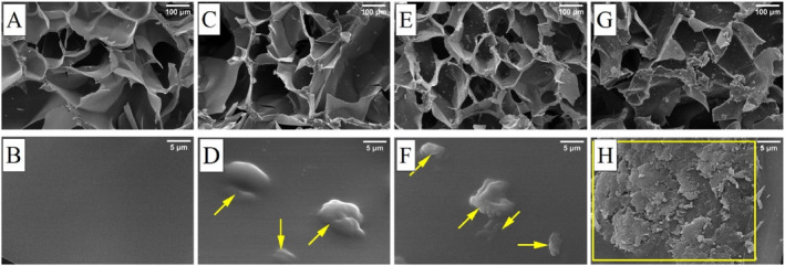
Morphological evaluation of cryogel samples with SEM images taken at 100× and 10,000× magnifications: (A, B) S0, (C, D) S25, (E, F) S50, and (G, H) S100. (Areas marked in yellow indicate the collection points of particles on polymer walls inside the pores.)