Abbreviations
ANC, Absolute neutrophil count
BP, Blood pressure
CT, Computer tomography
DNP, Dinitrophenol
ED, Emergency department
EF, Ejection friction
ICU, Intensive care unit
INTRODUCTION
Clenbuterol is a potent long-acting β2 adrenergic agonist which was reported to cause rhythm disturbance and cardiac ischemia.1 2,4 dinitrophenol (DNP) acts by uncoupling oxidative phosphorylation in mitochondria, leading to a hyper-metabolic state.2 Acute DNP intoxication carries a very high risk of mortality due to cardiorespiratory failure.3 Chronic exposure to DNP was reported to lead to cataracts, dermatitis, liver injury, myocardial injury, agranulocytosis and neutropenia.4
CASE
A 30-year-old gentleman with no past medical history presented to our emergency department (ED) with vertiginous giddiness, nausea, and vomiting for 1 day on a background of right ear pain and unsteady gait for 7 days. He was tachycardic and hypotensive with a heart rate of 105 beats/min and blood pressure (BP) of 67/26 mmHg respectively. O2 saturation was 98% on room air and tympanic temperature was 36.6 Celsius degree. Physical examination revealed right peri-auricular tenderness without ear discharge, no abdominal pain, no calf tenderness or peripheral oedema, no yellowing of skin or sclera. The cardiac and lung examinations were unremarkable. He denied taking any over-the-counter medications or supplements initially but subsequently admitted to using supplements for bodybuilding. He shared he was on both clenbuterol 40 mg and 2,4 DNP 50 mg daily for one month and only stopped taking them 5 days ago. He denied family history of sudden cardiac death. He is a social drinker and takes 1 vaping stick a day for 1-2 years. He was immediately transferred to the resuscitation room due to profound hypotension.
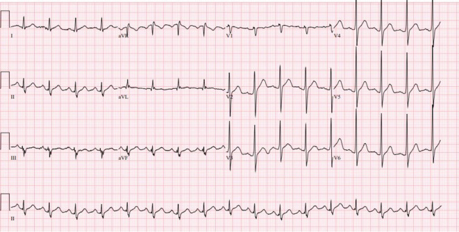
The initial electrocardiogram showed sinus tachycardia and early r-wave transition with small limb leads (Figure 1). Chest XR was unremarkable. The initial laboratory data revealed significant neutropenia with absolute neutrophil count (ANC) of 0.03 × 10⁹/L, white blood cell count of 0.4 × 10⁹/L, haematocrit 32%, and platelet count of 212 × 109/L. Inflammatory markers was elevated, C-reactive protein was 380 mg/L and procalcitonin was 5.84 g/L. Arterial blood gas showed acute respiratory alkalosis with pH 7.48, PCO2 29, PO2 97, bicarbonate 23 on room air. Lactate was normal at 1.04 mmol. Creatinine kinase was 29 U/L and methaemoglobin was normal. The troponin was 2 ng/L on arrival, then increased to 265 ng/L and peaked at 393 ng/L. Focused point of care ultrasound revealed global hypokinesia with severely reduced left ventricular ejection fraction of 30%. Right ventricular function was also moderately reduced, and both ventricular chambers appeared dilated. The cardiac valves appeared grossly intact. The serum toxicology screen was negative for acetaminophen, aspirin, and alcohol.
Figure 1.
Electrocardiogram showed sinus tachycardia with early R wave transition.
Fluid challenge and intravenous antibiotics were initiated immediately. The noradrenaline was started and titrated up to 0.3 mcg/kg/min for BP support. The cardiologist, intensivist, and toxicologist were consulted in the ED. In view of there being no specific antidotes for both clenbuterol and 2,4 DNP, the consensus was made to provide supportive care and treat the sepsis. The patient was transferred to the intensive care unit (ICU) for hemodynamic support. The haematologist was consulted for the severe neutropenia and suggested it was likely attributed to dinitrophenol exposure. One dose of subcutaneous filgrastim 300 mcg was given on admission with the ANC robustly increased to 2.17 × 10⁹/L on day 3 of hospitalization. Computer tomography (CT) of the neck, thorax, abdomen, and pelvis done on day 1 of hospitalisation demonstrated right parotitis with inflammatory peri-parotid fat stranding suggested early abscess formation. The otorhinolaryngologist was consulted, and bedside aspiration was attempted in the ICU but failed as a dry tap. The formal echo on day 2 of hospitalisation showed the ejection friction (EF) was 45% with mild global hypokinesia, both atria and ventricular were mildly dilated, and there was no pericardial effusion. Aspirin 100 mg daily and atorvastatin 20 mg daily were initiated to empirically treat type 2 myocardial infarction in view of the elevated troponin and cardiomyopathy noted. The patient’s condition improved and inotrope was weaned off on day 2 of hospitalisation, and he was transferred to general ward thereafter. Further source control with incision and drainage of the right auricular abscess was done due to enlarging size of the abscess noted from the repeated CT Neck on day 7 of hospitalisation. CT coronary angiogram on day 10 of hospitalisation revealed normal coronary arteries, suggestive of a non-ischemic aetiology of the cardiomyopathy. The patient recovered uneventfully and was discharged on day 12 of hospitalisation.
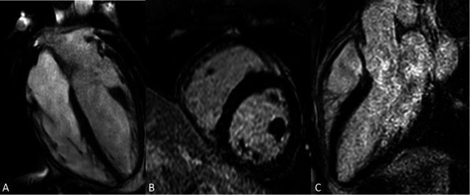
The patient was scheduled for outpatient otorhinolaryngologist follow-ups for the peri-auricular wound review, and the wound improved with the treatment of oral ciprofloxacin and wound dressing. The follow-up cardiac magnetic resonance imaging (Figure 2) of the three months after discharge revealed a mildly dilated ventricular and recovered EF of 59%, with no evidence of late gadolinium enhancement suggestive of either infarction, fibrosis, or infiltration. He resumed his usual life soon after discharge.
Figure 2.
(A) Cardiac MRI T1 sequence. Normal biventricular and atrial size. (B) No LGE detected on short axis phase sensitive inversion recovery sequence. (C) No LGE detected on apical 3 chamber phase sensitive inversion recovery sequence. LGE, late gadolinium enhancement; MRI, magnetic resonance imaging.
Our patient has clinical, biochemical, and radiological evidence of myocardial injury and cardiomyopathy, and we believe there are two culprits associated with adverse cardiovascular events in this case. The agranulocytosis led by 2,4 DNP predisposed him to fulminant infection, which triggered septic cardiomyopathy. To note, 2,4 DNP related agranulocytosis was reported in several cases.5,6 Meanwhile, both clenbuterol and 2,4 DNP can induce direct myocardial injury.
Clenbuterol is a beta-2 adrenergic agonist that is sometimes misused as a performance enhancer or for weight loss. Its primary health risks include tachycardia, hypertensive crisis, arrhythmia, muscle tremor, anxiety and hyperthermia.7 On the other hand, 2,4 DNP is a chemical that uncouples oxidative phosphorylation in mitochondria, leading to increased metabolism and heat production. Its misuse poses several severe health risks which encompass life-threatening hyperthermia, metabolic acidosis, dehydration and electrolyte imbalance.8
There are no antidotes for clenbuterol or 2,4 DNP. Hence, the mainstay of the management is supportive care and to treat reversible causes such as sepsis in this case.3 Questions about drug abuse and supplements should be an integral part of patient examination, particularly in young bodybuilders presenting with cardiomyopathy and hemodynamic instability.
The overlap of symptoms from cardiomyopathy and substance abuse can make diagnosis challenging. Clinicians must carefully differentiate between these conditions while managing their interplay. Identifying and managing withdrawal symptoms from clenbuterol and DNP is important. Continuous monitoring for adverse effects related to these substances is necessary to adjust treatment plans accordingly. Raising awareness about the risks associated with clenbuterol and DNP abuse can help prevent their misuse. Educating patients, especially those in sports or weight loss programs, about the potential for severe health consequences is vital.
LEARNING POINTS
• Importance of recognizing clenbuterol and 2,4 DNP abuse.
• Early identification is important in administration of appropriate life-saving therapy.
DECLARATION OF CONFLICT OF INTEREST
All the authors declare no conflict of interest.
Acknowledgments
Nil.
REFERENCES
- 1.Spiller HA, James KJ, Sholzen S, et al. A descriptive study of adverse events from clenbuterol misuse and abuse for weight loss and bodybuilding. Subst Abus. 2013:306–312. doi: 10.1080/08897077.2013.772083. [DOI] [PubMed] [Google Scholar]
- 2.Rognstad R, Katz J. The effect of 2,4-dinitrophenol on adipose-tissue metabolism. Biochem J. 1969:431–434. doi: 10.1042/bj1110431. [DOI] [PMC free article] [PubMed] [Google Scholar]
- 3.Bartlett J, Brunner M, Gough K. Deliberate poisoning with dinitrophenol (DNP): an unlicensed weight loss pill. Emerg Med J. 2010:159–160. doi: 10.1136/emj.2008.069401. [DOI] [PubMed] [Google Scholar]
- 4.Holb Grundlingh J, Dargan PI, El-Zanfaly M, et al. 2,4-dinitrophenol (DNP): a weight loss agent with significant acute toxicity and risk of death. J Med Toxicol. 2011:205–212. doi: 10.1007/s13181-011-0162-6. [DOI] [PMC free article] [PubMed] [Google Scholar]
- 5.Solomon S. A new danger in dinitrophenol therapy. Agranulocytosis with fatal outcome. J Am Med Assoc. 1934:1058. [Google Scholar]
- 6.Alexander GM. Acute complete granulopenia with death due to dinitrophenol poisoning. J Am Med Assoc. 1936:2115–2117. [Google Scholar]
- 7.Wong K, Boheler KR, Bishop J, et al. Clenbuterol induces cardiac hypertrophy with normal functional, morphological and molecular features. Cardiovasc Res. 1998:115–122. doi: 10.1016/s0008-6363(97)00190-9. [DOI] [PubMed] [Google Scholar]
- 8.Li YQ, Jiang JK, Huang WD. Clinical features and treatment in patients with acute 2,4 dinitrophenol poisoning. J Zhejiang Univ Sci B. 2011:189–192. doi: 10.1631/jzus.B1000265. [DOI] [PMC free article] [PubMed] [Google Scholar]


