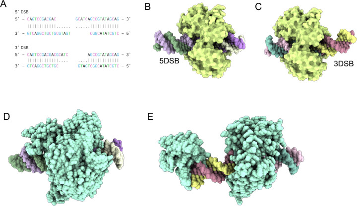
Figure 4. Conformational Dynamics of Human DNA Ligase I (LIG1) in Double-Strand Break Repair.
(A) 30mer DNA sequences with short 3’ (3DSB) and 5’ overhangs (5DSB) used for modeling LIG1 mediated ligation. (B) Conformational ensemble of LIG1 with 5DSB, illustrating suboptimal DNA positioning with a protruding end and reduced contact area with the enzymatic site. (C) Conformational ensemble of LIG1 with 3DSB, demonstrating optimal DNA positioning centrally within the catalytic site, facilitating efficient catalysis. (D) Structural representation of the LIG3b-5DSB complex, showing the DNA enveloped in the central cavity of LIG3b. (E) Structural representation of the LIG3b-3DSB complex, with a large protruding end stabilized by the N-terminal zinc finger domain of LIG3b.

