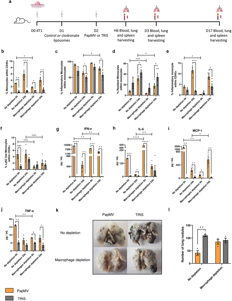
Figure 7.

Role of macrophages in the anti-metastatic properties of PapMV. (a) Injection schedule for the metastasis implantation model in Balb/c mice (created with BioRender.com). Balb/c mice were injected IV with 3×10⁵ 4T1 cells. After 24 hours, they received IP administration of control or clodronate containing liposomes. Subsequently, 24 hours later, they were injected IV with PapMV or TRIS. Mice were euthanized at 6 hours, 24 hours, or on day 17. At 6 hours and 24 hours (b-d) lungs and (e, f) blood were harvested for flow cytometric cell labeling. In the lungs, proportions of (b) monocytes and (c-d) monocyte subtypes were determined based the cell population indicated on the Y axis. In the blood, proportions of (e) inflammatory monocytes and (f) Ly6C low monocytes were determined based on total CD45+ immune cells. Serum was collected 6 hours and 24 hours following PapMV treatment and (g) ifn-α, (h) IL-6, (i) MCP-1, and (j) tnf-α concentrations were measured. (k) Lung metastases were visualized at D17 and (l) enumerated. Values are means ± SEM and complied from two independent experiments. (n = 2 for 6h, n = 4 for 24h).
