Contrast-enhanced harmonic endoscopic ultrasound (CH-EUS) has been reported to be useful in the diagnosis of pancreatobiliary disease. CH-EUS facilitates the differentiation of the cystic component from the parenchymal component by assessing the presence of blood flow 1 2 . Herein, we report a case of successful EUS-guided transluminal drainage (EUS-TD) for infected pancreatic fluid collection using CH-EUS.
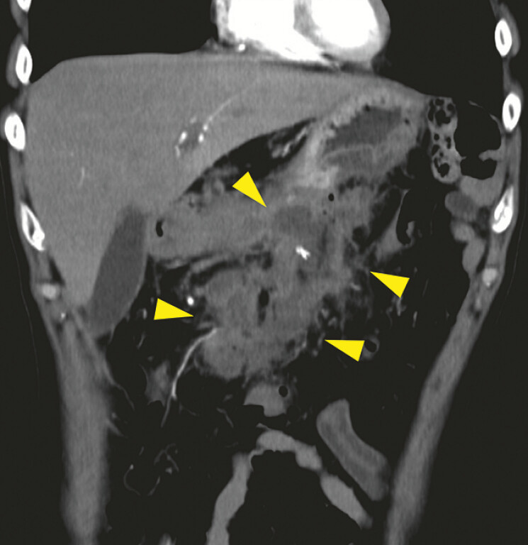
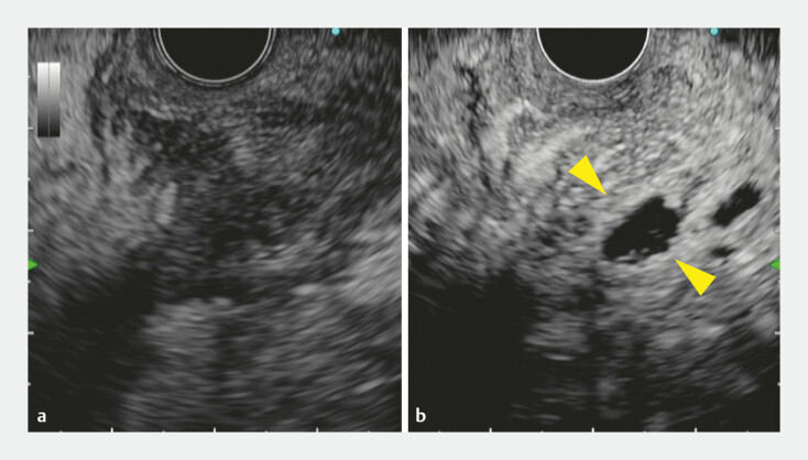
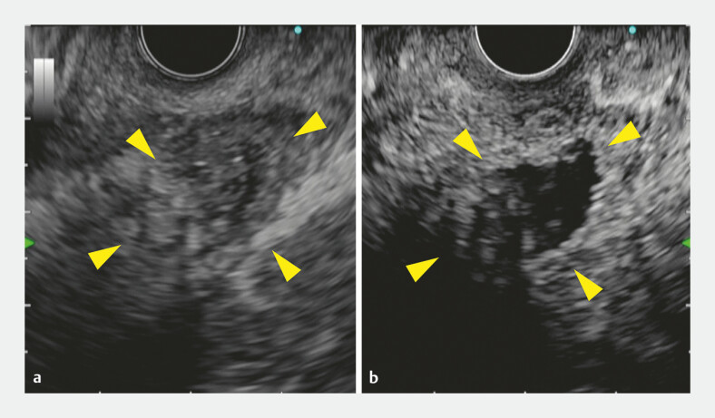
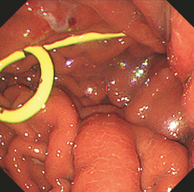
A 56-year-old man who had undergone distal pancreatectomy for pancreatic cancer two months ago was admitted to our hospital because of fever. Contrast-enhanced computed tomography revealed a postoperative pancreatic fistula (POPF) with fluid collection around the pancreas ( Fig. 1 ) and EUS-TD was attempted. Initially, we scanned the lesion with fundamental B-mode ultrasound, but the POPF was not well-recognized ( Fig. 2 a ). Consequently, CH-EUS was performed to identify the spread of the POPF cavity and its margins. The initially targeted location was recognized as only minimal avascular areas ( Fig. 2 b ). However, as a large avascular area was identified at another location ( Fig. 3 ), EUS-TD was successfully performed ( Fig. 4 , Fig. 5 ; Video 1 ). After the procedure, the patient’s symptoms resolved, and he was discharged five days later without any adverse events.
Fig. 1.
Contrast-enhanced computed tomography showed large post-operative pancreatic fluid collection (yellow arrowheads).
Fig. 2.
Endoscopic ultrasound images. a The initially targeted region. Despite the absence of an anechoic lesion, a mixed hypo- and hyperechoic area around the pancreas was observed under fundamental B-mode. b The initially targeted region was recognized as only minimal avascular areas (yellow arrowheads) on a contrast-enhanced harmonic image.
Fig. 3.
Another location with a large avascular area (yellow arrowheads) was identified.
Fig. 4.
Endoscopy image showing 7-Fr double pigtail plastic stent.
Fig. 5.
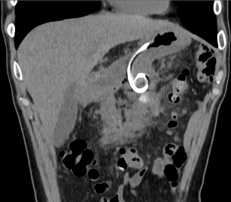
Computed tomography showed a successfully deployed 7-Fr double pigtail plastic stent.
Successful endoscopic ultrasound-guided drainage for infected pancreatic fluid collection using contrast-enhanced harmonic imaging.
Video 1
A POPF is usually well recognized in fundamental B-mode because of its predominantly liquid component. However, when it is composed mostly of solid components, such as necrosis, and has only a small liquid component, the boundary with the surrounding tissue is difficult to identify. In the present case, using CH-EUS the POPF cavity exhibited no enhancement owing to the absence of vascularity, whereas the surrounding tissue was enhanced. The application of CH-EUS may be useful in demarcating the boundary between the POPF cavity and its surrounding tissue in EUS-TD.
Endoscopy_UCTN_Code_TTT_1AS_2AJ
Footnotes
Conflict of Interest The authors declare that they have no conflict of interest.
Endoscopy E-Videos https://eref.thieme.de/e-videos .
E-Videos is an open access online section of the journal Endoscopy , reporting on interesting cases and new techniques in gastroenterological endoscopy. All papers include a high-quality video and are published with a Creative Commons CC-BY license. Endoscopy E-Videos qualify for HINARI discounts and waivers and eligibility is automatically checked during the submission process. We grant 100% waivers to articles whose corresponding authors are based in Group A countries and 50% waivers to those who are based in Group B countries as classified by Research4Life (see: https://www.research4life.org/access/eligibility/ ). This section has its own submission website at https://mc.manuscriptcentral.com/e-videos .
References
- 1.Minaga K, Kitano M, Yoshikawa T et al. Hepaticogastrostomy guided by real-time contrast-enhanced harmonic endoscopic ultrasonography: A novel technique. Endoscopy. 2016;48:E228–E229. doi: 10.1055/s-0042-109059. [DOI] [PubMed] [Google Scholar]
- 2.Minaga K, Takenaka M, Omoto S et al. A case of successful transluminal drainage of walled-off necrosis under contrast-enhanced harmonic endoscopic ultrasonography guidance. J Med Ultrason (2001) 2018;45:161–165. doi: 10.1007/s10396-017-0784-7. [DOI] [PubMed] [Google Scholar]


