Abstract
BACKGROUND
Non-invasive methods to diagnose non-alcoholic steatohepatitis (NASH), an inflammatory subtype of non-alcoholic fatty liver disease (NAFLD), are currently unavailable.
AIM
To develop an integrin αvβ3-targeted molecular imaging modality to differentiate NASH.
METHODS
Integrin αvβ3 expression was assessed in Human LO2 hepatocytes Scultured with palmitic and oleic acids (FFA). Hepatic integrin αvβ3 expression was analyzed in rabbits fed a high-fat diet (HFD) and in rats fed a high-fat, high-carbohydrate diet (HFCD). After synthesis, cyclic arginine-glycine-aspartic acid peptide (cRGD) was labeled with gadolinium (Gd) and used as a contrast agent in magnetic resonance imaging (MRI) performed on mice fed with HFCD.
RESULTS
Integrin αvβ3 was markedly expressed on FFA-cultured hepatocytes, unlike the control hepatocytes. Hepatic integrin αvβ3 expression significantly increased in both HFD-fed rabbits and HFCD-fed rats as simple fatty liver (FL) progressed to steatohepatitis. The distribution of integrin αvβ3 in the liver of NASH cases largely overlapped with albumin-positive staining areas. In comparison to mice with simple FL, the relative liver MRI-T1 signal value at 60 minutes post-injection of Gd-labeled cRGD was significantly increased in mice with steatohepatitis (P < 0.05), showing a positive correlation with the NAFLD activity score (r = 0.945; P < 0.01). Hepatic integrin αvβ3 expression was significantly upregulated during NASH development, with hepatocytes being the primary cells expressing integrin αvβ3.
CONCLUSION
After using Gd-labeled cRGD as a tracer, NASH was successfully distinguished by visualizing hepatic integrin αvβ3 expression with MRI.
Keywords: Non-alcoholic steatohepatitis, Cyclic peptides, Magnetic resonance imaging, Non-invasive diagnosis, Hepatic integrin αvβ3
Core Tip: Early identification of non-alcoholic steatohepatitis (NASH) patients and accurate assessment of non-alcoholic fatty liver disease severity are crucial for improving patient outcomes. Currently, no non-invasive method can replace liver biopsy to accurately discern NASH. Hepatic integrin αvβ3 expression significantly increased as simple fatty liver progressed to steatohepatitis. Inflammatory-injured hepatocytes, which might be the primary cells expressing integrin αvβ3 in steatohepatitis, were identified on the basis of steatosis. Utilizing gadolinium-labeled cyclic arginine-glycine-aspartic acid peptide as a contrast agent, steatohepatitis was successfully differentiated by visualizing hepatic integrin αvβ3 expression using a magnetic resonance imaging modality.
INTRODUCTION
Currently, non-alcoholic fatty liver disease (NAFLD) has become the most common cause of chronic liver disease worldwide and is estimated to affect about 25%-30% of the global population[1]. NAFLD encompasses a spectrum of liver disorders, ranging from simple fatty liver (FL) to non-alcoholic steatohepatitis (NASH), which can ultimately progress to cirrhosis[2]. NASH, an inflammatory subtype of NAFLD, is histologically characterized by hepatic steatosis accompanied by hepatocyte injury (ballooning) and intralobular inflammation, with or without fibrosis[3]. Due to the close association of NAFLD with metabolic disorders such as obesity, hyperlipidemia, and type 2 diabetes, and given the ongoing epidemic of these conditions, the prevalence of NAFLD, along with the proportion of NASH, is projected to continue rising in the coming decade[4]. Compared to the general population and patients with simple FL, a significantly higher proportion of NASH patients can progress to cirrhosis, hepatocellular carcinoma (HCC), and death[4-6]. Consequently, early identification of NASH and accurate assessment of NAFLD severity are crucial to improving patient outcomes.
Over the past two decades, numerous non-invasive methods have been explored to identify NASH among patients with NAFLD. These methods include conventional imaging examinations, imaging-based evaluation approaches such as magnetic resonance imaging-derived proton density fat fraction (MRI-PDFF), serum biomarkers (e.g., cytokeratin 18), and several complex scoring systems composed of clinical and laboratory values. The characteristics of various non-invasive diagnostic methods are summarized in Table 1. While the diagnosis of FL and advanced liver fibrosis, even cirrhosis, can be made using these methods, none can accurately distinguish NASH, and invasive liver biopsy remains the only accepted method for reliably diagnosing NASH[4,6]. However, performing a liver biopsy on every NAFLD patient is neither feasible nor necessary, as the procedure has limitations, including sampling errors that affect diagnostic accuracy and the risk of operative complications such as bleeding, infection, and, rarely, mortality[6,7]. Therefore, there is an urgent need to develop a novel non-invasive method to accurately distinguish NASH and monitor disease progression.
Table 1.
|
Methods
|
|
|
Advantages
|
Flaws
|
Patients (n)
|
AUROC
|
Sensitivity (%)
|
Specificity (%)
|
|
| Imaging modalities | Quantitative ultrasound biomarkers | Ultrasound attenuation coefficient[31] | Low cost and wide availability; High sensitivity and specificity in grading | The diagnostic performance is affected by the presence of fibrosis. | 125 (Excluding liver fibrosis) | S0 vs S1, S2, S3 | 0.951 | 82.1 | 95.5 |
| S0, S1 vs S2, S3 | 0.987 | 94.3 | 93.9 | ||||||
| S0, S1, S2 vs S3 | 0.931 | 94.1 | 85.5 | ||||||
| Ultrasound-derived fat fraction[32] | Low cost and wide availability; Real-time data collection | High interobserver variability | 46 | S0 vs S1, S2, S3 | 0.950 | 90.0 | 94.0 | ||
| S0, S1 vs S2, S3 | 0.980 | ||||||||
| CT | Contrast-enhanced CT[33] | Common in clinical practice; Quantify liver fat without additional radiation exposure | Exposes patients to ionizing radiation | 1204 | Steatosis≥ 5% | 0.669 | 34.0 | 94.2 | |
| Steatosis≥ 10% | 0.854 | ||||||||
| Steatosis ≥ 15% | 0.962 | 75.9 | 95.7 | ||||||
| Non-contrast dual-energy CT[34] | Reliable for detection of moderate steatosis | Influenced by varying contrast bolus and scan timing, cardiac output | 128 | Right lobe | 0.834 | 57.0 | 94.0 | ||
| Left lobe | 0.872 | 68.0 | 90.0 | ||||||
| MR-PDFF[35] | It is used as a reference for testing other clinical or biochemical markers | Expensive; availability-limited; unable to assess liver inflammation, ballooning | 77 | S0 vs S1, S2, S3 | 0.989 | 97 | 100 | ||
| S0, S1 vs S2, S3 | 0.825 | 61 | 90 | ||||||
| S0, S1, S2 vs S3 | 0.893 | 68 | 91 | ||||||
| Serum biomarkers | Laboratory parameter-based model | NAFLD bridge score[36] | Data availability and cost-effectiveness | Unable to differentiate NASH from simple steatosis with high sensitivity and specificity | 422 | With and without NAFLD | 0.880 | 95 | 87 |
MR-PDFF: Magnetic resonance imaging-estimated proton density fat fraction; AUROC: Area under the receiver operating characteristic curve; CT: Computed tomography; NAFLD: Non-alcoholic fatty liver disease; NASH: Non-alcoholic steatohepatitis.
Integrins are transmembrane glycoprotein heterodimeric receptors that primarily mediate adhesion between different cells or between cells and the extracellular matrix. They participate in signal transduction related to cell proliferation, activation, movement, and apoptosis, thereby playing a pivotal role in immune regulation, injury repair, tumor infiltration, and other processes[8-10]. A common feature of integrins is their ability to bind endogenous ligands by recognizing the amino acid binding motif arginine-glycine-aspartic acid (RGD)[9,11]. Among the 24 known integrin heterodimers, consisting of 18 α-subunit and 8 β-subunit variants, integrin αvβ3 has been the most studied over the past two decades. Numerous RGD-binding drugs targeting integrin αvβ3 have been developed for the treatment of integrin αvβ3-associated diseases[12-14].
In our previous studies, cyclic RGD pentapeptide was prepared and demonstrated to specifically bind to integrin αvβ3 on activated hepatic stellate cells both in vitro and in vivo[15,16]. Additionally, after the synthesized cyclic RGD peptide was directly radiolabeled or used to modify the dendrimer nanoprobe, molecular imaging approaches using single photon emission computed tomography or MRI were developed to non-invasively distinguish the severity of liver fibrosis[15,16]. In this study, hepatic expression of integrin αvβ3 was observed in different animal models of NAFLD. Furthermore, after synthesizing cyclic RGD peptides modified with gadolinium (Gd) and using them as an MRI contrast agent, we aimed to develop a molecular imaging modality to distinguish NASH by visualizing hepatic integrin αvβ3 expression.
MATERIALS AND METHODS
Animals
Eight-week-old male Sprague-Dawley rats, six-week-old male C57BL/6J mice, and four-month-old male New Zealand white rabbits were obtained from the Experimental Animals Department of Zhongshan Hospital, Fudan University (Shanghai, China). All experiments were conducted in accordance with governmental and international guidelines on animal experimentation. The study was reviewed and approved by the Zhongshan Hospital Institutional Review Board, and all experiments were performed following these guidelines.
Synthesis of cyclic RGD peptide and its derivative
Cyclic RGD (cRGD) peptide [cyclo (Arg-Gly-Asp-D-Tyr-Lys)] was synthesized and labeled with Gd as previously described[15-17]. Briefly, cRGD was initially synthesized using an Fmoc-protected solid-phase peptide synthesis method and subsequently labeled with Gd through 1,4,7,10-tetraazacyclododecane-1,4,7,10-tetraacetic acid (DOTA) to form cRGD-DOTA-Gd. The molecular weights of cRGD and cRGD-DOTA-Gd were determined using analytical reverse-phase high-performance liquid chromatography, and their purities were assessed by electrospray ionization mass spectrometry. The Gd3+ content of cRGD-DOTA-Gd was determined using a Hitachi P-4010 (Tokyo, Japan). Inductively Coupled Plasma Atomic Emission Spectroscopy system.
Expression of integrin αvβ3 in cultured hepatocytes
Human LO2 hepatocytes (from the Cell Bank of the Chinese Academy of Sciences, Shanghai, China) were cultured in a medium supplemented with 10% heat-inactivated fetal bovine serum and 1% penicillin/streptomycin in a 5% CO2 humidified atmosphere at 37 °C.
Firstly, LO2 cells were cultured in a medium containing 100 μmol/L palmitic acid and 200 μmol/L oleic acid (FFA) for 24 hours, after which they were stained with Oil-Red O. LO2 cells cultured in medium without FFA served as controls. Secondly, after being fixed with 4% paraformaldehyde and permeabilized with phosphate-buffered saline (PBS) containing 0.1% Triton X-100 and 0.1 mg/mL RNase A when appropriate, the control and FFA-cultured hepatocytes were incubated with primary antibodies against integrin αv subunit (1:1000, Abmart, T56887) or integrin β3 subunit (1:1000, Abmart, M015908), and albumin (1:1000, Servicebio, GB122080) at 4 °C overnight. Subsequently, these cells were incubated with secondary antibodies, including Alexa Fluor 594-conjugated immunoglobulin (Ig) G (1:200, Jackson, PA, United States) and Alexa Fluor 488-conjugated IgG (1:200, Jackson, PA, United States), counterstained with 6-diamidino-2-phenylindole at room temperature for 1 hour, and then photographed with a fluorescence microscope (Olympus, Japan). The message RNA (mRNA) levels and protein amounts of integrin αv and integrin β3 subunits were then determined in the control and FFA-cultured hepatocytes as described below.
Rabbit model of NAFLD induced by high-fat diet
Rabbits were fed a high-fat diet (HFD), which included an additional 20% corn oil in the regular diet containing 3.81% fat, 17.8% protein, and 46.5% carbohydrate (provided by the Experimental Animals Department of Zhongshan Hospital). Based on preliminary experimental results, rabbits fed with HFD for 2, 6, and 8 months were selected for further experiments [referred to as HFD-2 months (M), HFD-6M, and HFD-8M rabbits, respectively]. Rabbits fed with a regular diet for 8 months served as the control group (n = 10 for each group).
Murine models of NAFLD induced by high-fat high-carbohydrate diet
Sprague-Dawley rats and C57BL/6J mice were fed a high-fat high-carbohydrate diet (HFCD), which contained 60 kcal% fats, 20 kcal% proteins, and 20 kcal% carbohydrates (D12492, SHUYISHUER bio, Changzhou, China), along with high fructose/glucose (55% fructose and 45% glucose, Sigma, United States) in drinking water at a concentration of 42 g/L. Two months, four months, or six months after the treatment, treated rats and mice were used for further experiments (referred to as HFCD-2M, HFCD-4M, and HFCD-6M rats or mice, respectively). Rats and mice fed with a regular diet for 6 months served as the control group.
Histological analysis
After fixation in neutralized formalin for 48 hours, liver sections were stained with hematoxylin and eosin. Liver sections from rats and mice were stained with Masson, while those from rabbits were stained with Sirius red to assess the degree of liver fibrosis. Additionally, Oil-Red O staining was used to evaluate the degree of hepatic steatosis in frozen liver sections. The severity of NAFLD was semi-quantitatively scored according to the NAFLD activity score, which included steatosis (0-3), lobular inflammation (0-3), and ballooning (0-2)[18]. A score greater than 4 was defined as NASH.
Immunohistochemistry analysis
Hepatic expression of integrin αvβ3 was initially assessed in control and HFD-fed rabbits using immunohistochemistry analysis as previously described[17]. Briefly, mouse monoclonal anti-integrin β3 antibody (1:400 in blocking solution, Millipore, Massachusetts, United States, MAB1974) and biotin-conjugated goat polyclonal anti-mouse IgG (1:100 in blocking solution, Abcam, ab6788) were used as the primary and secondary antibodies, respectively. Ten fields (400 × magnification) from each liver section were randomly selected and recorded, and the integrin β3 subunit positive-staining areas were quantified using NIN Image 1.62 software. The percentage of positive-staining areas in each section was then calculated.
Quantitative real-time polymerase chain reaction analysis
Hepatic mRNA levels of integrin αv and integrin β3 subunits in control and HFCD-fed rats were quantified using quantitative real-time polymerase chain reaction analysis analysis as described previously[15]. The forward primers for integrin αv and integrin β3 subunits were CGAAGCCTTAGCAAGACTGTCCTG and GACTCGGACTGGACTGGCTACTAC, respectively. The reverse primers were CAGTTGAGTTCCAGCCTTCATCGG and ACTTCTCGCAGGTGTCTCCATAGG, respectively. GAPDH was used as the reference, with forward and reverse primers TCCCTCAAGATTGTCAGCAA and AGATCCACAACGGATACATT, respectively. The relative expression of integrin αv and integrin β3 subunits mRNA was analyzed using the comparative cycle threshold method. Additionally, total RNA was extracted from control and FFA-cultured LO2 hepatocytes, and the mRNA expression levels of integrin αv and integrin β3 subunits in these cells were also determined.
Western blot assay
The protein amounts of integrin αv and integrin β3 subunits in the livers of control and HFCD-fed rats were determined by western blot analysis. Additionally, protein was extracted from control and FFA-cultured LO2 hepatocytes, and the protein expression levels of integrin αv and integrin β3 subunits in these cells were analyzed. β-tubulin was used as the reference. After chemiluminescent signals were generated and detected on radiographic film, the relative expression of integrin αv and integrin β3 subunits was quantified by scanning densitometry analysis.
Immunofluorescent co-localization of integrin αvβ3 in various hepatic cells
Immunofluorescent staining was performed to reveal the co-localization of integrin β3 subunits with markers of various hepatic cells, including albumin (hepatocytes), α-smooth muscle actin (α-SMA) (activated hepatic stellate cell), cluster of differentiation (CD) 31 (hepatic sinusoidal endothelial cells), CD68 (macrophages), and CD163 (Kupffer cells), in liver sections of control and HFCD-fed rats as previously described[15]. Primary antibodies against integrin β3 subunit (1:500; Abmart), monoclonal anti-albumin (1:500; Servicebio), monoclonal anti-SMA (1:500, Servicebio, GB111364), monoclonal anti-CD31 (1:500, Servicebio, GB11063-1), monoclonal anti-CD68 (1:500, Servicebio, GB11067), and monoclonal anti-CD163 (1:500, Servicebio, GB11340-1) were used. Secondary antibodies included Alexa Fluor 594-conjugated IgG (1:200, Jackson) and Alexa Fluor 488-conjugated IgG (1:200, Jackson). Multicolored fluorescent staining of liver sections was analyzed, and 10 randomly selected amplifying fields (400 ×) in each section were assessed using Image-Pro Plus version 6.0.0.260. The percentage of integrin β3 subunit-stained green area in each section and the ratio of the merged yellow color area to the total integrin β3 subunit-stained area in each section were calculated.
In vivo MRI studies
MRI was performed in mice using a Biospec 70/20 MRI scanner (7.0 T, Bruker, Germany) to assess the accumulation of cRGD-DOTA-Gd in livers after cRGD-DOTA-Gd was injected intravenously at a dose of 0.05 mmol/kg [Gd3+] in a total of 0.25 mL PBS solution in the control and HFCD-fed mice, as described previously[16]. Briefly, dynamic T1-weighted MRIs of the liver were collected before and at 30, 60, 90, and 120 minutes after cRGD-DOTA-Gd injection. Cross-sectional images of the liver were acquired using a fast low-angle shot sequence and processed with Weasis software version 3.8.1. The signal intensity of the hepatic region [T1 (liver)] was measured, and the muscle signal intensity [T1 (muscle)] from the region of interest in the same section was used to normalize the hepatic signal intensity. The relative hepatic signal intensity was denoted as T1 (liver)/T1 (muscle). The relative hepatic signal intensity before (T0) and at different time points (Tt) after the injection of cRGD-DOTA-Gd were calculated, and the relative liver MRI-T1 signal value at a specific time point post-injection was determined by subtracting T0 from Tt.
Statistical analysis
Data were presented as the mean ± SD and analyzed by a one-way analysis of variance followed by the least significant difference test. Statistical product and service solutions 24.0 statistical software (Chicago, United States) was used, and a P value of < 0.05 was considered statistically significant.
RESULTS
Properties of cRGD and its derivative
Cyclic peptides (cRGD) were prepared and labeled with Gd (cRGD-DOTA-Gd). The molecular weights of cRGD and cRGD-DOTA-Gd were 623 and 1362 Da, respectively, and their purities exceeded 95%. The Gd3+ content in cRGD-DOTA-Gd was 8.1%.
Expression of integrin αvβ3 in hepatocytes in vitro
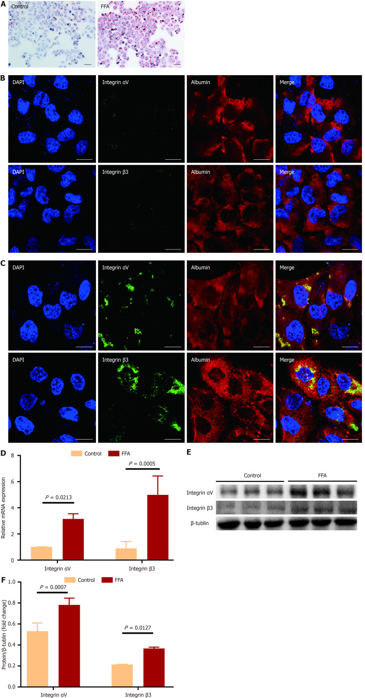
After LO2 hepatocytes were cultured with FFA for 24 hours, the accumulation of fat droplets was observed in most cells through Oil-Red O staining (Figure 1A), indicating the development of hepatocyte steatosis. Integrin αvβ3 was expressed on FFA-cultured hepatocytes as shown by immunofluorescent staining, but not on the control hepatocytes (Figure 1B and C). Compared to the control cells, the mRNA expression levels and protein amounts of integrin αv and integrin β3 subunits were significantly increased in FFA-cultured hepatocytes (Figure 1B-F, P < 0.05 for all comparisons).
Figure 1.
Expression of integrin αvβ3 in cultured hepatocytes. Human LO2 hepatocytes were cultured with the medium containing 100 μmol/L palmitate acid and 200 μmol/L oleic acid (FFA) for 24 hours, and the cells cultured in the medium without FFA served as the control. A: Representative micrographs of the control and FFA-cultured hepatocytes after stained with Oil-Red O. Images were taken at original magnification (200 ×), scale bars = 20 μm; B and C: Representative fluorescent images of the control (B) and FFA-cultured (C) hepatocytes after separately stained with integrin αv and β3 subunits antibody (green color) and counterstained with albumin antibody (red color). 6-diamidino-2-phenylindole was used for nuclei staining. The merged images show the yellow color area by overlaying images of the counterstaining. Images were taken at original magnification (400 ×), scale bars = 100 μm; D: Comparison of the message RNA levels of integrin αv and β3 subunits in the control and FFA-cultured hepatocytes. The message RNA levels of integrin αv and β3 subunit were determined by quantitative real-time polymerase chain reaction analysis; E and F: Comparison of the protein amounts of integrin αv and β3 subunits in the control and FFA-cultured hepatocytes. The protein amounts of integrin αv and β3 subunits were analyzed by western-blot assay, and β-Tublin was used as the reference. All experiments were undertaken in triplicates. In all panels, data are expressed in means ± SD. FFA: Oleic acid; DAPI: 6-diamidino-2-phenylindole.
Expression of integrin αvβ3 in livers of rabbits with NAFLD
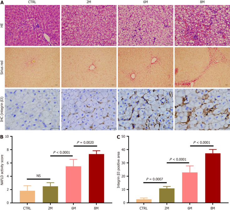
The expression of integrin αvβ3 in NAFLD livers was initially observed in HFD-fed rabbits. After 2 months of HFD feeding, lipid accumulation in hepatocytes was observed, indicating the development of hepatic steatosis. With prolonged HFD feeding for 6 and 8 months, hepatic steatosis worsened, and hepatocyte ballooning became prominent, accompanied by inflammatory cell infiltration in hepatic lobules and the formation of perisinusoidal fibrosis, even bridge fibrosis (Figure 2A). The NAFLD activity score was significantly higher in HFD-6M and HFD-8M rabbits compared to control and HFD-2M rabbits (Figure 2B, P < 0.05 for all comparisons). After immunohistochemical staining of hepatic slices for integrin β3 subunits, abundant positive-staining areas were observed in HFD-6M and HFD-8M rabbits (Figure 2A). The percentage of integrin β3 subunit positive-staining areas in the livers of HFD-6M and HFD-8M rabbits (22.83 ± 4.93 and 37.22 ± 2.88, respectively) was significantly higher than in control (2.68 ± 0.97) and HFD-2M rabbits (10.82 ± 1.48) (Figure 2C, P < 0.05 for all comparisons). These results indicated that hepatic expression of integrin αvβ3 increased consistently with the progression of NAFLD, with a significant increase observed when the condition progressed to NASH.
Figure 2.
Expression of integrin αvβ3 in livers of rabbits with non-alcoholic fatty liver disease. Non-alcoholic fatty liver disease was induced in rabbits by high-fat diet (HFD) for 2, 6, and 8 months (referred to as HFD-2M, 6M and 8M), and rabbits fed with regular diet for 8 months served as the control group (n = 6 per group). A: Representative micrographs of hepatic histology stained with hematoxylin-eosin staining (200 ×), Sirius red (100 ×), and immunohistochemistry for integrin β3 subunit (400 ×). In immunohistochemistry staining images, the brown areas indicated integrin β3 subunit positive staining, scale bars = 50 μm; B: Comparison of non-alcoholic fatty liver disease activity score in the control and HFD-fed rabbits; C: Comparison of the percentage of integrin β3 subunit positive-staining area in liver sections of the control and HFD-fed rabbits. For semi-quantitative analysis of hepatic integrin αvβ3 expression level, 10 fields were randomly selected and recorded from each section stained with immunohistochemistry for integrin β3 subunit. Then integrin β3 subunit positive-staining area was measured, and the percentages of the positive-staining area in liver sections were compared. In all panels, data are expressed in means ± SD. IHC: Immunohistochemistry; HE: Hematoxylin-eosin staining; CTRL: Normal control group; M: Month; NS: Not significant; NAFLD: Non-alcoholic fatty liver disease.
Expression of integrin αvβ3 in livers of rats with NAFLD
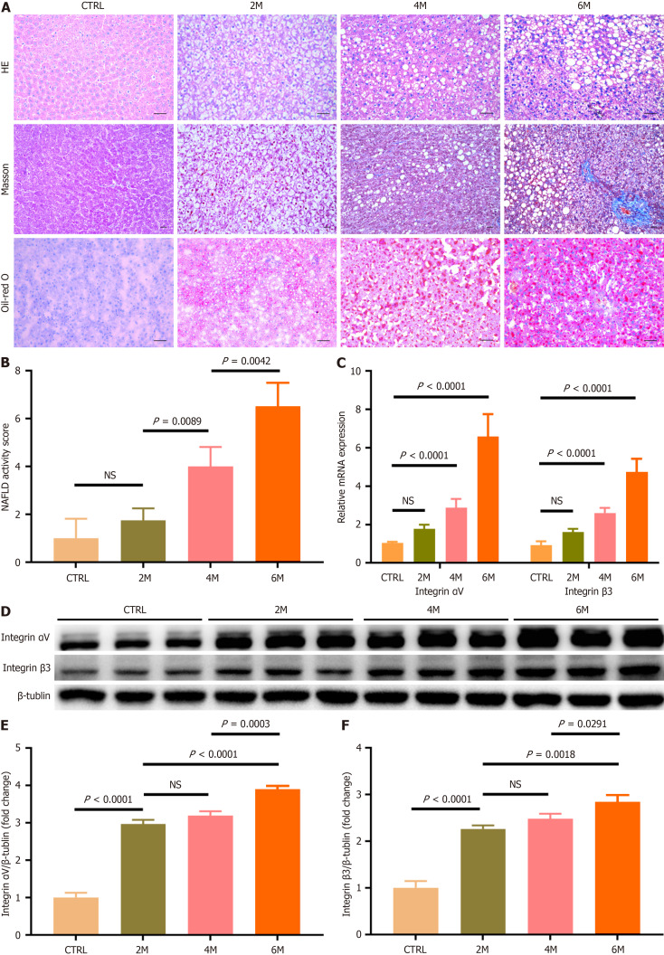
As shown in Figure 3A, after rats were fed with HFCD, massive fat droplets accumulated in hepatocytes, which was most prominent in HFCD-6M rats. As hepatic steatosis worsened, hepatocyte ballooning and inflammatory cell infiltration became more pronounced in hepatic lobules after 4 months of HFCD feeding. When the HFCD feeding was prolonged to 6 months, perisinusoidal fibrosis was observed in hepatic lobules. Compared to control rats (1.17 ± 0.41) and HFCD-2M rats (1.83 ± 0.75), the NAFLD activity score of hepatic tissue was significantly increased in HFCD-4M and HFCD-6M rats (4.00 ± 0.89 and 6.83 ± 0.75, respectively), with the highest score in HFCD-6M rats (Figure 3B, P < 0.05 for all comparisons). These results indicated that simple FL developed in rats after 2 months of HFCD feeding, progressed to steatohepatitis after 4 months, and further advanced to NASH with liver fibrosis after 6 months.
Figure 3.
Expression of integrin αvβ3 in livers of rats with non-alcoholic fatty liver disease. Non-alcoholic fatty liver disease was induced in rats by high-fat high-carbohydrate diet (HFCD) for 2, 4, and 6 months (referred to as HFCD-2M, 4M and 6M), and rats fed with regular diet for 6 months served as the control group (n= 6 per group). A: Representative micrographs of hepatic histology stained with H&E (200 ×), Masson (100 ×), and Oil-red O (200 ×), scale bars = 50 μm; B: Comparison of non-alcoholic fatty liver disease activity score in the control and HFCD-fed rats; C: Comparison of hepatic integrin αvβ3 message RNA level in the control and HFCD-fed rats. Hepatic message RNA levels of integrin αv and β3 subunits were respectively determined by quantitative real-time polymerase chain reaction analysis; D-F: Comparison of the protein amounts of integrin αvβ3 in livers of the control and HFCD-fed rats. The protein amounts of integrin αv and β3 subunits were analyzed by western-blot assay, and β-Tublin was used as the reference. In all panels, data are expressed in means ± SD. HE: Hematoxylin-eosin staining; CTRL: Normal control group; M: Month; NS: Not significant; NAFLD: Non-alcoholic fatty liver disease.
Hepatic integrin αvβ3 expression during the development and progression of NAFLD was further assessed in HFCD-fed rats. Compared to control and HFCD-2M rats, hepatic mRNA expression levels of integrin αv and integrin β3 subunits were significantly increased after 4 months of HFCD feeding, reaching the highest levels in HFCD-6M rats (Figure 3C, P < 0.05 for all comparisons). Additionally, the protein amounts of integrin αv and integrin β3 subunits in hepatic tissue gradually increased in HFCD-fed rats, with levels significantly higher than those in control rats (Figure 3D-F, P < 0.05 for all comparisons). These results indicated that hepatic integrin αvβ3 expression progressively increased with the development and progression of NAFLD.
Localization of integrin αvβ3 in livers with NAFLD
To identify the principal cells expressing integrin αvβ3 in livers with NAFLD, double immunofluorescent staining was performed to visualize the co-localization of the integrin β3 subunit with albumin, α-SMA, CD31, CD68, and CD163 in control and HFCD-fed rats. Integrin αvβ3 expression was negligible in the livers of control rats, as no integrin β3 subunit positive-staining (green) areas were found in the liver slices (Figure 4A). In contrast, increasing integrin β3 subunit positive-staining areas were observed in the liver slices of HFCD-fed rats, particularly in HFCD-6M rats (Figure 4B-D). The percentage of integrin β3 subunit positive-staining areas in the liver slices of HFCD-4M and HFCD-6M rats was significantly higher than in control and HFCD-2M rats (Figure 4E, P < 0.05 for all comparisons).
Figure 4.
Immunofluorescent co-localization of integrin αvβ3 and the markers of various hepatic cells including α-smooth muscle actin, cluster of differentiation 31, cluster of differentiation 68 or cluster of differentiation 163 in livers of the control and high-fat high-carbohydrate diet-fed rats. Representative fluorescent images of integrin β3 subunit (green color) and albumin, α-smooth muscle actin, cluster of differentiation (CD) 31, CD68 and CD163 (red color) in liver sections, which were separately stained with specific first antibodies and visualized by second antibodies, and counterstained with 6-diamidino-2-phenylindole for nuclei staining. The merged images show the yellow color area by overlaying images of the counterstaining, and amplified images corresponding to the indicated areas in boxes are present. Images were recorded at original magnification (400 ×), scale bars = 100 μm. A: Control; B: High-fat high-carbohydrate diet (HFCD)-2M; C: HFCD-4M; D: HFCD-6M rats; E: Comparison of the percentage of integrin β3 subunit positive-staining area in liver sections of the control and HFCD-fed rats. Ten fields were randomly selected and recorded from each section. Then integrin β3 positive-staining area was measured, and the percentages of the positive-staining area in liver sections were compared; F: Comparison of the ratio of the overlapped yellow area to integrin β3 subunit positive-staining green area in liver sections of HFCD-6M rats. Ten fields were randomly selected and recorded from each section. Then integrin β3 subunit positive-staining area and the area of integrin β3 subunit positive-staining overlapped with hepatic cellular markers positive-staining were respectively measured, and the ratios were compared. In all panels, data are expressed in means ± SD. DAPI: 6-diamidino-2-phenylindole; CTRL: Normal control group; M: Month; CD: Cluster of differentiation; αSMA: α-smooth muscle actin.
Additionally, in the livers of HFCD-6M rats, the positive staining of the integrin β3 subunit (green) extensively overlapped (yellow) with albumin staining (red) and showed minimal overlap with α-SMA, CD31, CD68, or CD163 staining (Figure 4D). The ratio of the overlapped area of integrin β3 subunit staining with albumin positive-staining was significantly higher than the overlapped staining with other cellular markers (Figure 4F, P < 0.05 for all comparisons).
These findings suggest that hepatic expression of integrin αvβ3 markedly increased as simple FL progressed to steatohepatitis, with hepatocytes being the primary cells expressing integrin αvβ3 in livers with NASH.
Imaging the progression of NAFLD by MRI in vivo
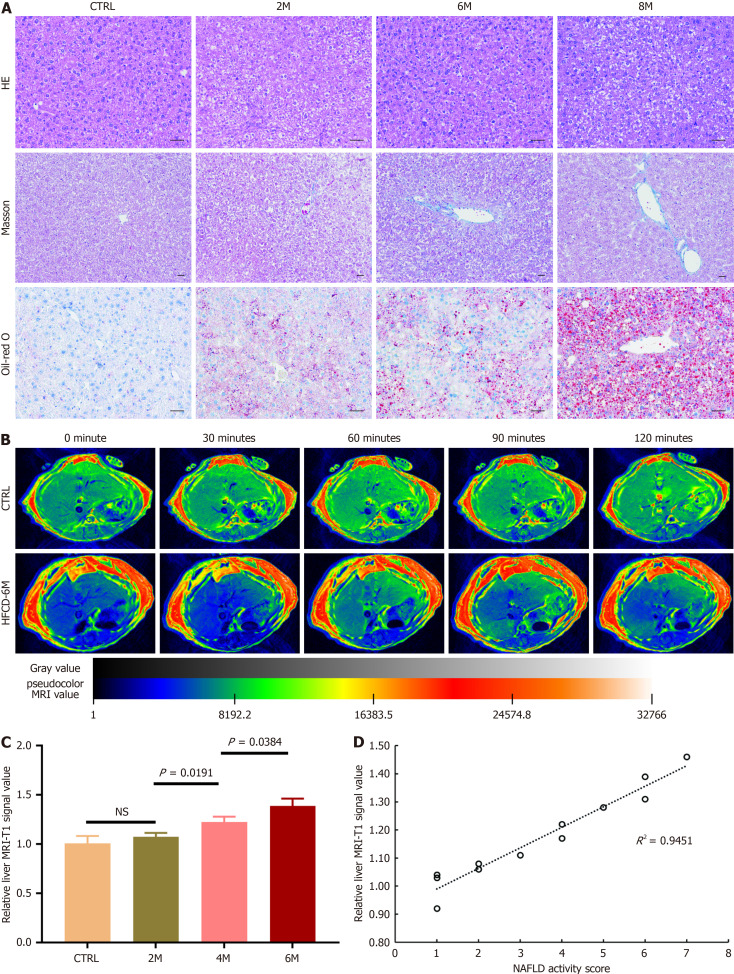
After being fed with HFCD for 6 months, the rats became so obese that no suitable coil was available for MRI examination. Therefore, mice were used for in vivo MRI experiments. After the mice were fed with HFCD, similar histopathological findings were observed in their livers: Simple FL developed in HFCD-2M mice and progressed to steatohepatitis in HFCD-4M and HFCD-6M mice (Figure 5A).
Figure 5.
Imaging non-alcoholic fatty liver disease using a magnetic resonance imaging modality with cyclic arginine-glycine-aspartic acid peptides labeled with gadolinium through 1,4,7,10-tetraazacyclododecane-1,4,7,10-tetraacetic acid as a tracer in mice. Non-alcoholic fatty liver disease was induced in mice by high-fat high-carbohydrate diet (HFCD) for 2, 4, and 6 months (referred to as HFCD-2M, 4M and 6M), and mice fed with regular diet for 6 months served as the control group (n= 3 per group). A: Representative micrographs of hepatic histology stained with hematoxylin-eosin staining (200 ×), Masson (100 ×), and Oil-red O (200 ×), scale bars = 50 μm; B: Representative hepatic T1-weighed magnetic resonance imaging (MRI) of the control and HFCD-6M mice prior to and at 30, 60, 90 and 120 minutes after cyclic arginine-glycine-aspartic acid (cRGD)-1,4,7,10-tetraazacyclododecane-1,4,7,10-tetraacetic acid (DOTA)-gadolinium (Gd) injection; C: Comparison of the relative liver MRI-T1 signal value in the control and HFCD-fed mice 60 minutes after cRGD-DOTA-Gd injection. Data are expressed in means ± SD; D: Correlation of the relative liver MRI-T1 signal value at 60 minutes post-injection of cRGD-DOTA-Gd with non-alcoholic fatty liver disease activity score was assessed. HE: Hematoxylin-eosin staining; CTRL: Normal control group; M: Month; NS: Not significant; NAFLD: Non-alcoholic fatty liver disease; HFCD: High-fat high-carbohydrate diet; MRI: Magnetic resonance imaging.
A dynamic T1-weighted MRI approach was employed to assess the deposition of cRGD-DOTA-Gd in the livers after the radiotracers were intravenously injected in control and HFCD-fed mice. As shown in Figure 5B, there was no significant change in hepatic signal intensity in control mice before and up to 120 minutes after cRGD-DOTA-Gd injection. In contrast, in HFCD-6M mice, compared to pre-injection levels, the hepatic signal was markedly intensified at 60 minutes post-injection of cRGD-DOTA-Gd and then gradually decreased. Compared to control mice, the relative liver MRI-T1 signal value at 60 minutes post-injection of cRGD-DOTA-Gd was significantly increased in mice fed with HFCD for 4 months, with the highest values observed in HFCD-6M mice (Figure 5C, P < 0.05 for all comparisons). Furthermore, the relative hepatic MRI-T1 signal value at 60 minutes post-injection of cRGD-DOTA-Gd showed a strong positive correlation with the NAFLD activity score in HFCD-fed mice (r = 0.945, P < 0.01, Figure 5D).
DISCUSSION
To date, the development of a non-invasive method that can accurately diagnose NASH and determine the disease severity of NAFLD, thereby replacing the need for liver biopsy, remains one of the urgent unmet needs in the NASH field[4]. Compared to other conventional imaging modalities, such as ultrasound and computed tomography, MRI offers the highest spatial resolution for soft tissues and is considered the most sensitive modality for evaluating hepatic steatosis[19]. While these imaging modalities can also detect advanced liver fibrosis and cirrhosis, none can accurately distinguish NASH. Recently, several MRI-based imaging modalities, such as MRI-PDFF and magnetic resonance elastography (MRE), have been developed for NAFLD diagnosis. MRI-PDFF is currently the gold standard for diagnosing and quantifying hepatic steatosis, showing excellent accuracy in detecting dynamic changes in steatosis, but it fails to provide information on steatohepatitis and fibrosis[20,21]. Conversely, MRE is recognized as the most accurate imaging modality for diagnosing liver fibrosis in NAFLD, primarily by measuring elastic shear wave propagation through liver parenchyma[22,23]. However, MRE cannot accurately discriminate NASH without significant fibrosis, and more importantly, the presence of inflammation in the liver with NASH negatively impacts its accuracy in diagnosing fibrosis. Therefore, no current non-invasive imaging method can accurately discern NASH.
Many animal models are currently available for basic and translational research on NAFLD, primarily grouped into two categories: specific gene knockout models and special diet-induced models. Compared to gene knockout NAFLD models, the animal models induced by special diets, particularly high-fat (± high-carbohydrate) diets, more comprehensively mimic the spectrum of metabolic and histological features of human NAFLD[24]. In the present study, the HFD-induced NAFLD rabbit model was initially used to observe the expression of integrin αvβ3 in the liver with NAFLD. However, significant individual variability in hepatic histopathological manifestations was found in HFD-fed rabbits. Therefore, HFCD-induced NAFLD murine models were used to further assess hepatic integrin αvβ3 expression and conduct in vivo MRI examinations. With prolonged HFCD feeding, hepatic steatosis developed and progressed to steatohepatitis in most rats and mice, as demonstrated histologically.
In our previous study, it was found that integrin αvβ3 in fibrotic livers was primarily expressed on activated hepatic stellate cells, and its expression level strongly correlated with the degree of liver fibrosis, while it was scarcely expressed on hepatocytes[15,25]. Additionally, Rokugawa et al[26] recently reported quantitatively evaluating hepatic integrin αvβ3 expressed on activated hepatic stellate cells using positron emission tomography imaging in rats with NASH[26,27]. However, in the present study, hepatocytes - not activated hepatic stellate cells-were found to be the primary cells expressing integrin αvβ3 in NASH livers. In the in vivo study, hepatic expression of integrin αvβ3 was significantly increased in NASH livers, with no expression in normal livers and mild expression in livers with simple FL. More importantly, most integrin αvβ3 was found to be expressed on hepatocytes in NASH livers, as demonstrated by double immunofluorescent staining for integrin αvβ3 and markers of various hepatic cells. In the in vitro study, integrin αvβ3 was markedly expressed on hepatocytes cultured with palmitic acid and FFA, but not on normal control hepatocytes. It has been demonstrated that hepatocytes develop not only steatosis, but also inflammatory injury and apoptosis after being cultured with palmitic acid and FFA[28]. These results indicate that on the basis of steatosis, hepatocytes injured by inflammation express abundant integrin αvβ3 in NASH livers. Additionally, based on these results, we speculate that integrin αvβ3 might play a role in the pathogenesis of NASH, which needs to be further elucidated in future studies.
In this study, hepatic integrin αvβ3 expression significantly increased as simple FL progressed to steatohepatitis, with or without fibrosis, which correlated with the increased NAFLD activity score. Therefore, we aimed to visualize hepatic integrin αvβ3 expression during the development and progression of NAFLD using an MRI-based molecular imaging modality. After Gd-labeled cRGD, targeted to integrin αvβ3, was injected, the hepatic signal markedly intensified at 60 minutes post-injection compared to pre-injection levels, and the relative liver MRI-T1 signal value at 60 minutes post-injection was significantly increased in mice with NASH compared to control mice and those with simple FL, showing a linear correlation with the NAFLD activity score. These results indicated that the binding amount of Gd-labeled cRGD in the liver significantly increased secondary to elevated hepatic integrin αvβ3 expression during the development of NASH. Thus, NASH was successfully differentiated from simple FL in NAFLD by visualizing hepatic integrin αvβ3 expression with the targeted molecular imaging modality.
Recently, it has been found that long-term outcomes, including overall mortality in patients with NAFLD, are strongly associated with the severity of liver fibrosis but not with the NAFLD activity score[29]. However, non-NASH NAFLD seldom progresses to significant fibrosis, indicating that steatohepatitis is a cornerstone in the development and progression of liver fibrosis in NAFLD[4-6]. More importantly, HCC has been found to develop in NASH patients without liver cirrhosis[30]. Consequently, it is essential to discern NASH early and regularly monitor the progression of NAFLD, but invasive liver biopsy, the only currently accepted method for diagnosing NASH, fails to achieve this goal. In contrast, non-invasive molecular imaging modalities could potentially be utilized for routine screening and regular follow-up of NASH in the NAFLD population. Over the past decades, numerous molecular imaging modalities have been developed for diagnosing tumors and their metastases, especially those with integrin αvβ3-positive expression[14]. Furthermore, in our previous studies, two molecular imaging modalities were explored to diagnose liver fibrosis by visualizing hepatic αvβ3 expression in the development and progression of fibrosis[15,16]. All these developments will aid in promoting the clinical application of integrin αvβ3-targeted molecular imaging modalities in NASH diagnosis.
However, the availability and cost-effectiveness of MRI are still worth considering. There is no doubt that MRI is more expensive than ultrasound or computed tomography-based imaging modalities and tends to be more time-consuming. Nevertheless, the reliability of MRI is widely accepted, and other non-invasive methods are often compared with MRI-based methods like MRI-PDFF, which are limited in assessing inflammation, to evaluate their potential in clinical applications. In this study, we developed an RGD-based MRI contrast agent to differentiate steatohepatitis by visualizing hepatic integrin αvβ3 expression, which could counteract the interference of inflammation in MRI detection. This approach holds promise for future development and use.
CONCLUSION
In conclusion, this study demonstrated that hepatic integrin αvβ3 expression significantly increased as simple FL progressed to NASH. Hepatocytes, injured by inflammation on the basis of hepatic steatosis, were the primary cells expressing integrin αvβ3 in NASH livers. After cyclic RGD peptides were labeled with Gd and used as a contrast agent, NASH was successfully distinguished by visualizing hepatic integrin αvβ3 expression with an MRI modality.
ACKNOWLEDGEMENTS
The authors would like to acknowledge Pang ZY (Shanghai East Hospital) for valuable assistance with fluorescence microscopy and MRI analysis.
Footnotes
Institutional review board statement: This study does not involve human subject research.
Institutional animal care and use committee statement: This study was performed in line with the Declaration of Helsinki and the ethical approval was obtained from the Institutional Ethical Committee of Animal Experimentation, Zhongshan Hospital affiliated to Fudan University.
Conflict-of-interest statement: The authors declare that they have no conflict of interest.
ARRIVE guidelines statement: The authors have read the ARRIVE guidelines, and the manuscript was prepared and revised according to the ARRIVE guidelines.
Provenance and peer review: Unsolicited article; Externally peer reviewed.
Peer-review model: Single blind
Specialty type: Gastroenterology and hepatology
Country of origin: China
Peer-review report’s classification
Scientific Quality: Grade C
Novelty: Grade B
Creativity or Innovation: Grade B
Scientific Significance: Grade B
P-Reviewer: Liu XY S-Editor: Fan M L-Editor: A P-Editor: Xu ZH
Contributor Information
Xiao-Quan Huang, Department of Gastroenterology and Hepatology, Zhongshan Hospital, Fudan University, Shanghai 200032, China.
Ling Wu, Department of Gastroenterology and Hepatology, Zhongshan Hospital, Fudan University, Shanghai 200032, China.
Chun-Yan Xue, Department of Gastroenterology and Hepatology, Zhongshan Hospital, Fudan University, Shanghai 200032, China.
Chen-Yi Rao, Department of Gastroenterology and Hepatology, Zhongshan Hospital, Fudan University, Shanghai 200032, China.
Qing-Qing Fang, Department of Gastroenterology, Minhang Hospital, Fudan University, Shanghai 201100, China.
Ying Chen, Department of Gastroenterology, Minhang Hospital, Fudan University, Shanghai 201100, China.
Cao Xie, Department of Pharmacy, Fudan University, Shanghai 200032, China.
Sheng-Xiang Rao, Department of Radiology, Zhongshan Hospital, Fudan University, Shanghai 200032, China.
Shi-Yao Chen, Department of Gastroenterology and Hepatology, Zhongshan Hospital, Fudan University, Shanghai 200032, China; Department of Gastroenterology, Minhang Hospital, Fudan University, Shanghai 201100, China.
Feng Li, Department of Gastroenterology and Hepatology, Zhongshan Hospital, Fudan University, Shanghai 200032, China; Department of Gastroenterology, Minhang Hospital, Fudan University, Shanghai 201100, China. li.feng2@zs-hospital.sh.cn.
Data sharing statement
Data will be made available upon reasonable request.
References
- 1.Younossi Z, Tacke F, Arrese M, Chander Sharma B, Mostafa I, Bugianesi E, Wai-Sun Wong V, Yilmaz Y, George J, Fan J, Vos MB. Global Perspectives on Nonalcoholic Fatty Liver Disease and Nonalcoholic Steatohepatitis. Hepatology. 2019;69:2672–2682. doi: 10.1002/hep.30251. [DOI] [PubMed] [Google Scholar]
- 2.Pouwels S, Sakran N, Graham Y, Leal A, Pintar T, Yang W, Kassir R, Singhal R, Mahawar K, Ramnarain D. Non-alcoholic fatty liver disease (NAFLD): a review of pathophysiology, clinical management and effects of weight loss. BMC Endocr Disord. 2022;22:63. doi: 10.1186/s12902-022-00980-1. [DOI] [PMC free article] [PubMed] [Google Scholar]
- 3.Chalasani N, Younossi Z, Lavine JE, Charlton M, Cusi K, Rinella M, Harrison SA, Brunt EM, Sanyal AJ. The diagnosis and management of nonalcoholic fatty liver disease: Practice guidance from the American Association for the Study of Liver Diseases. Hepatology. 2018;67:328–357. doi: 10.1002/hep.29367. [DOI] [PubMed] [Google Scholar]
- 4.Cotter TG, Rinella M. Nonalcoholic Fatty Liver Disease 2020: The State of the Disease. Gastroenterology. 2020;158:1851–1864. doi: 10.1053/j.gastro.2020.01.052. [DOI] [PubMed] [Google Scholar]
- 5.Rinella ME, Neuschwander-Tetri BA, Siddiqui MS, Abdelmalek MF, Caldwell S, Barb D, Kleiner DE, Loomba R. AASLD Practice Guidance on the clinical assessment and management of nonalcoholic fatty liver disease. Hepatology. 2023;77:1797–1835. doi: 10.1097/HEP.0000000000000323. [DOI] [PMC free article] [PubMed] [Google Scholar]
- 6.Sheka AC, Adeyi O, Thompson J, Hameed B, Crawford PA, Ikramuddin S. Nonalcoholic Steatohepatitis: A Review. JAMA. 2020;323:1175–1183. doi: 10.1001/jama.2020.2298. [DOI] [PubMed] [Google Scholar]
- 7.Ajmera V, Loomba R. Imaging biomarkers of NAFLD, NASH, and fibrosis. Mol Metab. 2021;50:101167. doi: 10.1016/j.molmet.2021.101167. [DOI] [PMC free article] [PubMed] [Google Scholar]
- 8.Xu S, Zhang T, Cao Z, Zhong W, Zhang C, Li H, Song J. Integrin-α9β1 as a Novel Therapeutic Target for Refractory Diseases: Recent Progress and Insights. Front Immunol. 2021;12:638400. doi: 10.3389/fimmu.2021.638400. [DOI] [PMC free article] [PubMed] [Google Scholar]
- 9.Slack RJ, Macdonald SJF, Roper JA, Jenkins RG, Hatley RJD. Emerging therapeutic opportunities for integrin inhibitors. Nat Rev Drug Discov. 2022;21:60–78. doi: 10.1038/s41573-021-00284-4. [DOI] [PMC free article] [PubMed] [Google Scholar]
- 10.De Marco R, Tolomelli A, Juaristi E, Gentilucci L. Integrin Ligands with α/β-Hybrid Peptide Structure: Design, Bioactivity, and Conformational Aspects. Med Res Rev. 2016;36:389–424. doi: 10.1002/med.21383. [DOI] [PubMed] [Google Scholar]
- 11.Ruoslahti E. My scientific journey to and through extracellular matrix. Matrix Biol. 2024;133:57–63. doi: 10.1016/j.matbio.2024.08.003. [DOI] [PubMed] [Google Scholar]
- 12.Sani S, Messe M, Fuchs Q, Pierrevelcin M, Laquerriere P, Entz-Werle N, Reita D, Etienne-Selloum N, Bruban V, Choulier L, Martin S, Dontenwill M. Biological Relevance of RGD-Integrin Subtype-Specific Ligands in Cancer. Chembiochem. 2021;22:1151–1160. doi: 10.1002/cbic.202000626. [DOI] [PubMed] [Google Scholar]
- 13.Bhatwadekar AD, Kansara V, Luo Q, Ciulla T. Anti-integrin therapy for retinovascular diseases. Expert Opin Investig Drugs. 2020;29:935–945. doi: 10.1080/13543784.2020.1795639. [DOI] [PMC free article] [PubMed] [Google Scholar]
- 14.Li ZH, Zhou Y, Ding YX, Guo QL, Zhao L. Roles of integrin in tumor development and the target inhibitors. Chin J Nat Med. 2019;17:241–251. doi: 10.1016/S1875-5364(19)30028-7. [DOI] [PubMed] [Google Scholar]
- 15.Li F, Song Z, Li Q, Wu J, Wang J, Xie C, Tu C, Wang J, Huang X, Lu W. Molecular imaging of hepatic stellate cell activity by visualization of hepatic integrin αvβ3 expression with SPECT in rat. Hepatology. 2011;54:1020–1030. doi: 10.1002/hep.24467. [DOI] [PubMed] [Google Scholar]
- 16.Li F, Yan H, Wang J, Li C, Wu J, Wu S, Rao S, Gao X, Jin Q. Non-invasively differentiating extent of liver fibrosis by visualizing hepatic integrin αvβ3 expression with an MRI modality in mice. Biomaterials. 2016;102:162–174. doi: 10.1016/j.biomaterials.2016.06.026. [DOI] [PubMed] [Google Scholar]
- 17.Wu L, Huang XQ, Li N, Xie C, Rao SX, Chen SY, Li F. A magnetic resonance imaging modality for non-invasively distinguishing progression of liver fibrosis by visualizing hepatic platelet-derived growth factor receptor-beta expression in mice. J Gastroenterol Hepatol. 2021;36:3448–3456. doi: 10.1111/jgh.15628. [DOI] [PubMed] [Google Scholar]
- 18.Kleiner DE, Brunt EM, Van Natta M, Behling C, Contos MJ, Cummings OW, Ferrell LD, Liu YC, Torbenson MS, Unalp-Arida A, Yeh M, McCullough AJ, Sanyal AJ Nonalcoholic Steatohepatitis Clinical Research Network. Design and validation of a histological scoring system for nonalcoholic fatty liver disease. Hepatology. 2005;41:1313–1321. doi: 10.1002/hep.20701. [DOI] [PubMed] [Google Scholar]
- 19.Selvaraj EA, Mózes FE, Jayaswal ANA, Zafarmand MH, Vali Y, Lee JA, Levick CK, Young LAJ, Palaniyappan N, Liu CH, Aithal GP, Romero-Gómez M, Brosnan MJ, Tuthill TA, Anstee QM, Neubauer S, Harrison SA, Bossuyt PM, Pavlides M LITMUS Investigators. Diagnostic accuracy of elastography and magnetic resonance imaging in patients with NAFLD: A systematic review and meta-analysis. J Hepatol. 2021;75:770–785. doi: 10.1016/j.jhep.2021.04.044. [DOI] [PubMed] [Google Scholar]
- 20.Caussy C, Reeder SB, Sirlin CB, Loomba R. Noninvasive, Quantitative Assessment of Liver Fat by MRI-PDFF as an Endpoint in NASH Trials. Hepatology. 2018;68:763–772. doi: 10.1002/hep.29797. [DOI] [PMC free article] [PubMed] [Google Scholar]
- 21.Xia T, Du M, Li H, Wang Y, Zha J, Wu T, Ju S. Association between Liver MRI Proton Density Fat Fraction and Liver Disease Risk. Radiology. 2023;309:e231007. doi: 10.1148/radiol.231007. [DOI] [PubMed] [Google Scholar]
- 22.Castera L, Friedrich-Rust M, Loomba R. Noninvasive Assessment of Liver Disease in Patients With Nonalcoholic Fatty Liver Disease. Gastroenterology. 2019;156:1264–1281.e4. doi: 10.1053/j.gastro.2018.12.036. [DOI] [PMC free article] [PubMed] [Google Scholar]
- 23.Ozturk A, Olson MC, Samir AE, Venkatesh SK. Liver fibrosis assessment: MR and US elastography. Abdom Radiol (NY) 2022;47:3037–3050. doi: 10.1007/s00261-021-03269-4. [DOI] [PMC free article] [PubMed] [Google Scholar]
- 24.Jahn D, Kircher S, Hermanns HM, Geier A. Animal models of NAFLD from a hepatologist's point of view. Biochim Biophys Acta Mol Basis Dis. 2019;1865:943–953. doi: 10.1016/j.bbadis.2018.06.023. [DOI] [PubMed] [Google Scholar]
- 25.Zhong L, Zhao J, Huang L, Liu Y, Pang X, Zhan K, Li S, Xue Q, Pan X, Deng L. Runx2 activates hepatic stellate cells to promote liver fibrosis via transcriptionally regulating Itgav expression. Clin Transl Med. 2023;13:e1316. doi: 10.1002/ctm2.1316. [DOI] [PMC free article] [PubMed] [Google Scholar]
- 26.Rokugawa T, Konishi H, Ito M, Iimori H, Nagai R, Shimosegawa E, Hatazawa J, Abe K. Evaluation of hepatic integrin αvβ3 expression in non-alcoholic steatohepatitis (NASH) model mouse by (18)F-FPP-RGD(2) PET. EJNMMI Res. 2018;8:40. doi: 10.1186/s13550-018-0394-4. [DOI] [PMC free article] [PubMed] [Google Scholar]
- 27.Hiroyama S, Rokugawa T, Ito M, Iimori H, Morita I, Maeda H, Fujisawa K, Matsunaga K, Shimosegawa E, Abe K. Quantitative evaluation of hepatic integrin α(v)β(3) expression by positron emission tomography imaging using (18)F-FPP-RGD(2) in rats with non-alcoholic steatohepatitis. EJNMMI Res. 2020;10:118. doi: 10.1186/s13550-020-00704-3. [DOI] [PMC free article] [PubMed] [Google Scholar]
- 28.Zhang M, Bai X, Du Q, Xu J, Wang D, Chen L, Dong K, Chen Z, Yang J. The Different Mechanisms of Lipid Accumulation in Hepatocytes Induced by Oleic Acid/Palmitic Acid and High-Fat Diet. Molecules. 2023;28 doi: 10.3390/molecules28186714. [DOI] [PMC free article] [PubMed] [Google Scholar]
- 29.Ekstedt M, Hagström H, Nasr P, Fredrikson M, Stål P, Kechagias S, Hultcrantz R. Fibrosis stage is the strongest predictor for disease-specific mortality in NAFLD after up to 33 years of follow-up. Hepatology. 2015;61:1547–1554. doi: 10.1002/hep.27368. [DOI] [PubMed] [Google Scholar]
- 30.Shah PA, Patil R, Harrison SA. NAFLD-related hepatocellular carcinoma: The growing challenge. Hepatology. 2023;77:323–338. doi: 10.1002/hep.32542. [DOI] [PMC free article] [PubMed] [Google Scholar]
- 31.Gao J, Zapata I, Chen J, Erpelding TN, Adamson C, Park D. Quantitative Ultrasound Biomarkers to Assess Nonalcoholic Fatty Liver Disease. J Ultrasound Med. 2023;42:1675–1688. doi: 10.1002/jum.16185. [DOI] [PubMed] [Google Scholar]
- 32.Zalcman M, Barth RA, Rubesova E. Real-time ultrasound-derived fat fraction in pediatric population: feasibility validation with MR-PDFF. Pediatr Radiol. 2023;53:2466–2475. doi: 10.1007/s00247-023-05752-0. [DOI] [PubMed] [Google Scholar]
- 33.Pickhardt PJ, Blake GM, Graffy PM, Sandfort V, Elton DC, Perez AA, Summers RM. Liver Steatosis Categorization on Contrast-Enhanced CT Using a Fully Automated Deep Learning Volumetric Segmentation Tool: Evaluation in 1204 Healthy Adults Using Unenhanced CT as a Reference Standard. AJR Am J Roentgenol. 2021;217:359–367. doi: 10.2214/AJR.20.24415. [DOI] [PubMed] [Google Scholar]
- 34.Zhang PP, Choi HH, Ohliger MA. Detection of fatty liver using virtual non-contrast dual-energy CT. Abdom Radiol (NY) 2022;47:2046–2056. doi: 10.1007/s00261-022-03482-9. [DOI] [PMC free article] [PubMed] [Google Scholar]
- 35.Tang A, Tan J, Sun M, Hamilton G, Bydder M, Wolfson T, Gamst AC, Middleton M, Brunt EM, Loomba R, Lavine JE, Schwimmer JB, Sirlin CB. Nonalcoholic fatty liver disease: MR imaging of liver proton density fat fraction to assess hepatic steatosis. Radiology. 2013;267:422–431. doi: 10.1148/radiol.12120896. [DOI] [PMC free article] [PubMed] [Google Scholar]
- 36.Yip TC, Ma AJ, Wong VW, Tse YK, Chan HL, Yuen PC, Wong GL. Laboratory parameter-based machine learning model for excluding non-alcoholic fatty liver disease (NAFLD) in the general population. Aliment Pharmacol Ther. 2017;46:447–456. doi: 10.1111/apt.14172. [DOI] [PubMed] [Google Scholar]
Associated Data
This section collects any data citations, data availability statements, or supplementary materials included in this article.
Data Availability Statement
Data will be made available upon reasonable request.


