

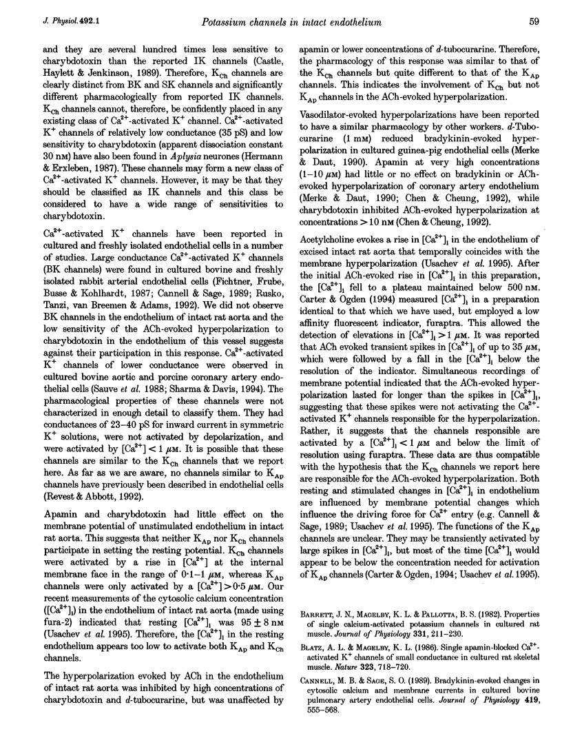
Abstract
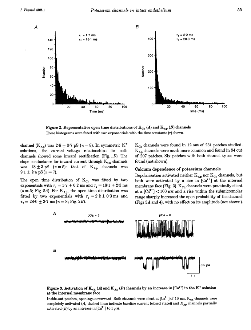
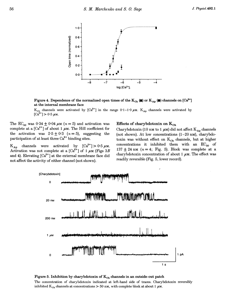
1. Single K+ channel currents and membrane potential were recorded in the endothelium of excised intact rat aorta. 2. Two types of K+ channel were found in excised patches, KCh and KAp. With Na+ and K+ as the main external and internal cations, outward conductances were 6.7 pS (KCh) and 2.8 pS (KAp). In symmetric 150 mM K+, the inward conductances were 18 and 9.1 pS. 3. Activation by Ca2+ was concentration dependent. KCh channels were activated by [Ca2+] > 0.1 microM and KAp by [Ca2+] > 0.5 microM. 4. Apamin at concentrations > 1 nM inhibited KAp Channels. Block was complete at 10 nM. KAp channels were insensitive to charybdotoxin. KCh channels were inhibited by charybdotoxin at concentrations > 50 nM, but were insensitive to apamin. 5. d-Tubocurarine (dTC) evoked flickering activity of KAp channels at concentrations > 5 microM and complete block at 100 microM. At these doses, dTC did not affect KCh channels, but at concentrations > 1 mM it decreased the single channel amplitude. 6. Hyperpolarization evoked by acetylcholine was unaffected by apamin or dTC at low concentrations ( < or = 100 microM), but inhibited by high concentrations of charybdotoxin ( > 50 nM) or dTC ( > 1 mM). 7. These data suggest that KCh channels are novel Ca(2+)-activated K+ channels responsible for the ACh-evoked hyperpolarization in the endothelium of rat aorta.
Full text
PDF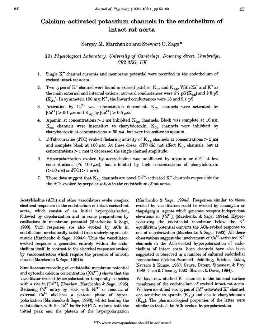
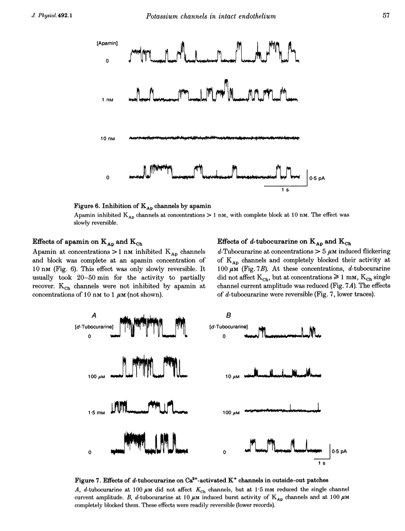
Selected References
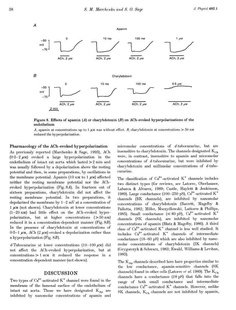
These references are in PubMed. This may not be the complete list of references from this article.
- Blatz A. L., Magleby K. L. Single apamin-blocked Ca-activated K+ channels of small conductance in cultured rat skeletal muscle. Nature. 1986 Oct 23;323(6090):718–720. doi: 10.1038/323718a0. [DOI] [PubMed] [Google Scholar]
- Colden-Stanfield M., Schilling W. P., Ritchie A. K., Eskin S. G., Navarro L. T., Kunze D. L. Bradykinin-induced increases in cytosolic calcium and ionic currents in cultured bovine aortic endothelial cells. Circ Res. 1987 Nov;61(5):632–640. doi: 10.1161/01.res.61.5.632. [DOI] [PubMed] [Google Scholar]
- Fichtner H., Fröbe U., Busse R., Kohlhardt M. Single nonselective cation channels and Ca2+-activated K+ channels in aortic endothelial cells. J Membr Biol. 1987;98(2):125–133. doi: 10.1007/BF01872125. [DOI] [PubMed] [Google Scholar]
- Hermann A., Erxleben C. Charybdotoxin selectively blocks small Ca-activated K channels in Aplysia neurons. J Gen Physiol. 1987 Jul;90(1):27–47. doi: 10.1085/jgp.90.1.27. [DOI] [PMC free article] [PubMed] [Google Scholar]
- Marchenko S. M., Sage S. O. Electrical properties of resting and acetylcholine-stimulated endothelium in intact rat aorta. J Physiol. 1993 Mar;462:735–751. doi: 10.1113/jphysiol.1993.sp019579. [DOI] [PMC free article] [PubMed] [Google Scholar]