Abstract
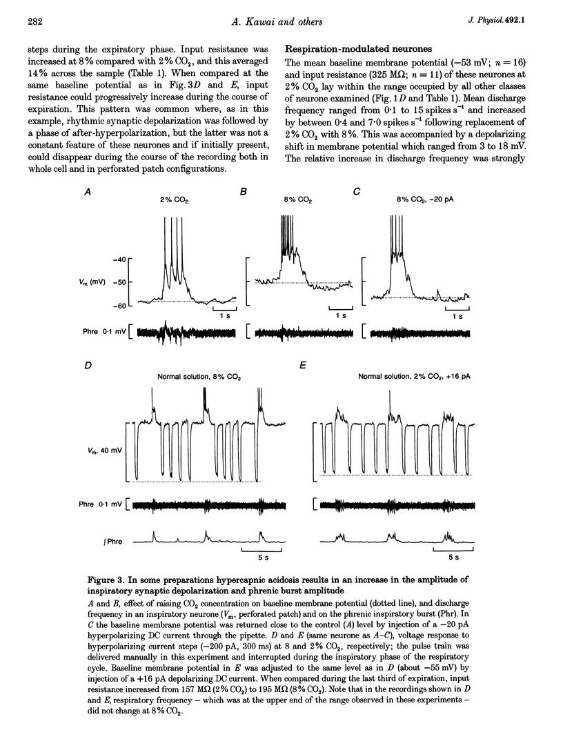
1. Using the isolated medulla and spinal cord of the neonatal rat, the response to CO2-induced changes in superfusate pH was examined in whole cell and perforated patch recordings from ventral medullary neurones which were identified by injection of Lucifer Yellow. The respiratory response to changing the CO2 concentration (from 2 to 8%) consisted of an increase in phrenic burst frequency, which could be accompanied by an increase, decrease or no change in burst amplitude. 2. Five classes of neurone - inspiratory, post-inspiratory, expiratory, respiration-modulated and ionic - were distinguished on the basis of their membrane potential and discharge patterns. Almost all (112 of 123) responded rapidly to 8% CO2 with a sustained change in membrane potential. Depolarizing responses (3-18 mV) occurred in inspiratory, respiration-modulated and 45% of tonic neurones. Hyperpolarizing responses (2-19 mV) occurred in expiratory and post-inspiratory neurones. The remaining tonic neurones were inhibited or showed no response. 3. In representatives of each class of neurone, membrane potential responses to 8% CO2 were retained when tested in the presence of tetrodotoxin (n = 7), low (0.2 mM) Ca(2+)-high (5 mM) Mg2+ (n = 23) or Cd2+ (0.2 mM) (n = 3)-containing superfusate, implying that they are mediated by intrinsic membrane or cellular mechanisms. 4. Neurones were distributed between 1200 microns rostral and 400 microns caudal to obex, and their cell bodies were located between 50 and 700 microns below the ventral surface (n = 104). Almost all responsive neurones (n = 78) showed dendritic projections to within 50 microns of the surface. 6. These experiments indicate that significant numbers of ventral medullary neurones, including respiratory neurones, are intrinsically chemosensitive. The consistency with which these neurones show surface dendritic projections suggests that this sensitivity may arise in part at this level.
Full text
PDF


Selected References
These references are in PubMed. This may not be the complete list of references from this article.
- Arata A., Onimaru H., Homma I. Respiration-related neurons in the ventral medulla of newborn rats in vitro. Brain Res Bull. 1990 Apr;24(4):599–604. doi: 10.1016/0361-9230(90)90165-v. [DOI] [PubMed] [Google Scholar]
- Bainton C. R., Kirkwood P. A. The effect of carbon dioxide on the tonic and the rhythmic discharges of expiratory bulbospinal neurones. J Physiol. 1979 Nov;296:291–314. doi: 10.1113/jphysiol.1979.sp013006. [DOI] [PMC free article] [PubMed] [Google Scholar]
- Bradley G. W., von Euler C., Marttila I., Roos B. Transient and steady state effects of CO-2 on mechanisms determining rate and depth of breathing. Acta Physiol Scand. 1974 Nov;92(3):341–350. doi: 10.1111/j.1748-1716.1974.tb05752.x. [DOI] [PubMed] [Google Scholar]
- Brockhaus J., Ballanyi K., Smith J. C., Richter D. W. Microenvironment of respiratory neurons in the in vitro brainstem-spinal cord of neonatal rats. J Physiol. 1993 Mar;462:421–445. doi: 10.1113/jphysiol.1993.sp019562. [DOI] [PMC free article] [PubMed] [Google Scholar]
- Dean J. B., Bayliss D. A., Erickson J. T., Lawing W. L., Millhorn D. E. Depolarization and stimulation of neurons in nucleus tractus solitarii by carbon dioxide does not require chemical synaptic input. Neuroscience. 1990;36(1):207–216. doi: 10.1016/0306-4522(90)90363-9. [DOI] [PubMed] [Google Scholar]
- Dean J. B., Lawing W. L., Millhorn D. E. CO2 decreases membrane conductance and depolarizes neurons in the nucleus tractus solitarii. Exp Brain Res. 1989;76(3):656–661. doi: 10.1007/BF00248922. [DOI] [PubMed] [Google Scholar]
- Harada Y., Kuno M., Wang Y. Z. Differential effects of carbon dioxide and pH on central chemoreceptors in the rat in vitro. J Physiol. 1985 Nov;368:679–693. doi: 10.1113/jphysiol.1985.sp015883. [DOI] [PMC free article] [PubMed] [Google Scholar]
- Issa F. G., Remmers J. E. Identification of a subsurface area in the ventral medulla sensitive to local changes in PCO2. J Appl Physiol (1985) 1992 Feb;72(2):439–446. doi: 10.1152/jappl.1992.72.2.439. [DOI] [PubMed] [Google Scholar]
- Jarolimek W., Misgeld U., Lux H. D. Neurons sensitive to pH in slices of the rat ventral medulla oblongata. Pflugers Arch. 1990 May;416(3):247–253. doi: 10.1007/BF00392060. [DOI] [PubMed] [Google Scholar]
- Loeschcke H. H. Central chemosensitivity and the reaction theory. J Physiol. 1982 Nov;332:1–24. doi: 10.1113/jphysiol.1982.sp014397. [DOI] [PMC free article] [PubMed] [Google Scholar]
- Marino P. L., Lamb T. W. Effects of CO-2 and extracellular H+ iontophoresis on single cell activity in the cat brainstem. J Appl Physiol. 1975 Apr;38(4):688–695. doi: 10.1152/jappl.1975.38.4.688. [DOI] [PubMed] [Google Scholar]
- McLean H. A., Remmers J. E. Respiratory motor output of the sectioned medulla of the neonatal rat. Respir Physiol. 1994 Apr;96(1):49–60. doi: 10.1016/0034-5687(94)90105-8. [DOI] [PubMed] [Google Scholar]
- Morin-Surun M. P., Boudinot E., Schäfer T., Denavit-Saubié M. Localization of chemosensitive structures in the isolated brainstem of adult guinea-pig. J Physiol. 1995 May 15;485(Pt 1):203–212. doi: 10.1113/jphysiol.1995.sp020724. [DOI] [PMC free article] [PubMed] [Google Scholar]
- Neubauer J. A., Gonsalves S. F., Chou W., Geller H. M., Edelman N. H. Chemosensitivity of medullary neurons in explant tissue cultures. Neuroscience. 1991;45(3):701–708. doi: 10.1016/0306-4522(91)90282-s. [DOI] [PubMed] [Google Scholar]
- Okada Y., Mückenhoff K., Holtermann G., Acker H., Scheid P. Depth profiles of pH and PO2 in the isolated brain stem-spinal cord of the neonatal rat. Respir Physiol. 1993 Sep;93(3):315–326. doi: 10.1016/0034-5687(93)90077-n. [DOI] [PubMed] [Google Scholar]
- Okada Y., Mückenhoff K., Scheid P. Hypercapnia and medullary neurons in the isolated brain stem-spinal cord of the rat. Respir Physiol. 1993 Sep;93(3):327–336. doi: 10.1016/0034-5687(93)90078-o. [DOI] [PubMed] [Google Scholar]
- Onimaru H., Arata A., Homma I. Firing properties of respiratory rhythm generating neurons in the absence of synaptic transmission in rat medulla in vitro. Exp Brain Res. 1989;76(3):530–536. doi: 10.1007/BF00248909. [DOI] [PubMed] [Google Scholar]
- Onimaru H., Homma I. Whole cell recordings from respiratory neurons in the medulla of brainstem-spinal cord preparations isolated from newborn rats. Pflugers Arch. 1992 Mar;420(3-4):399–406. doi: 10.1007/BF00374476. [DOI] [PubMed] [Google Scholar]
- Peers C., Buckler K. J. Transduction of chemostimuli by the type I carotid body cell. J Membr Biol. 1995 Mar;144(1):1–9. doi: 10.1007/BF00238411. [DOI] [PubMed] [Google Scholar]
- Pilowsky P. M., Jiang C., Lipski J. An intracellular study of respiratory neurons in the rostral ventrolateral medulla of the rat and their relationship to catecholamine-containing neurons. J Comp Neurol. 1990 Nov 22;301(4):604–617. doi: 10.1002/cne.903010409. [DOI] [PubMed] [Google Scholar]
- Rae J., Cooper K., Gates P., Watsky M. Low access resistance perforated patch recordings using amphotericin B. J Neurosci Methods. 1991 Mar;37(1):15–26. doi: 10.1016/0165-0270(91)90017-t. [DOI] [PubMed] [Google Scholar]
- Richerson G. B. Response to CO2 of neurons in the rostral ventral medulla in vitro. J Neurophysiol. 1995 Mar;73(3):933–944. doi: 10.1152/jn.1995.73.3.933. [DOI] [PubMed] [Google Scholar]
- Richter D. W., Ballanyi K., Schwarzacher S. Mechanisms of respiratory rhythm generation. Curr Opin Neurobiol. 1992 Dec;2(6):788–793. doi: 10.1016/0959-4388(92)90135-8. [DOI] [PubMed] [Google Scholar]
- Richter D. W. Generation and maintenance of the respiratory rhythm. J Exp Biol. 1982 Oct;100:93–107. doi: 10.1242/jeb.100.1.93. [DOI] [PubMed] [Google Scholar]
- Rigatto H., Fitzgerald S. C., Willis M. A., Yu C. In search of the central respiratory neurons: II. Electrophysiologic studies of medullary fetal cells inherently sensitive to CO2 and low pH. J Neurosci Res. 1992 Dec;33(4):590–597. doi: 10.1002/jnr.490330411. [DOI] [PubMed] [Google Scholar]
- Schwarzacher S. W., Wilhelm Z., Anders K., Richter D. W. The medullary respiratory network in the rat. J Physiol. 1991 Apr;435:631–644. doi: 10.1113/jphysiol.1991.sp018529. [DOI] [PMC free article] [PubMed] [Google Scholar]
- Shams H. Differential effects of CO2 and H+ as central stimuli of respiration in the cat. J Appl Physiol (1985) 1985 Feb;58(2):357–364. doi: 10.1152/jappl.1985.58.2.357. [DOI] [PubMed] [Google Scholar]
- Smith J. C., Ballanyi K., Richter D. W. Whole-cell patch-clamp recordings from respiratory neurons in neonatal rat brainstem in vitro. Neurosci Lett. 1992 Jan 6;134(2):153–156. doi: 10.1016/0304-3940(92)90504-z. [DOI] [PubMed] [Google Scholar]
- Smith J. C., Ellenberger H. H., Ballanyi K., Richter D. W., Feldman J. L. Pre-Bötzinger complex: a brainstem region that may generate respiratory rhythm in mammals. Science. 1991 Nov 1;254(5032):726–729. doi: 10.1126/science.1683005. [DOI] [PMC free article] [PubMed] [Google Scholar]
- Smith J. C., Greer J. J., Liu G. S., Feldman J. L. Neural mechanisms generating respiratory pattern in mammalian brain stem-spinal cord in vitro. I. Spatiotemporal patterns of motor and medullary neuron activity. J Neurophysiol. 1990 Oct;64(4):1149–1169. doi: 10.1152/jn.1990.64.4.1149. [DOI] [PubMed] [Google Scholar]
- Suzue T. Respiratory rhythm generation in the in vitro brain stem-spinal cord preparation of the neonatal rat. J Physiol. 1984 Sep;354:173–183. doi: 10.1113/jphysiol.1984.sp015370. [DOI] [PMC free article] [PubMed] [Google Scholar]
- Völker A., Ballanyi K., Richter D. W. Anoxic disturbance of the isolated respiratory network of neonatal rats. Exp Brain Res. 1995;103(1):9–19. doi: 10.1007/BF00241960. [DOI] [PubMed] [Google Scholar]


