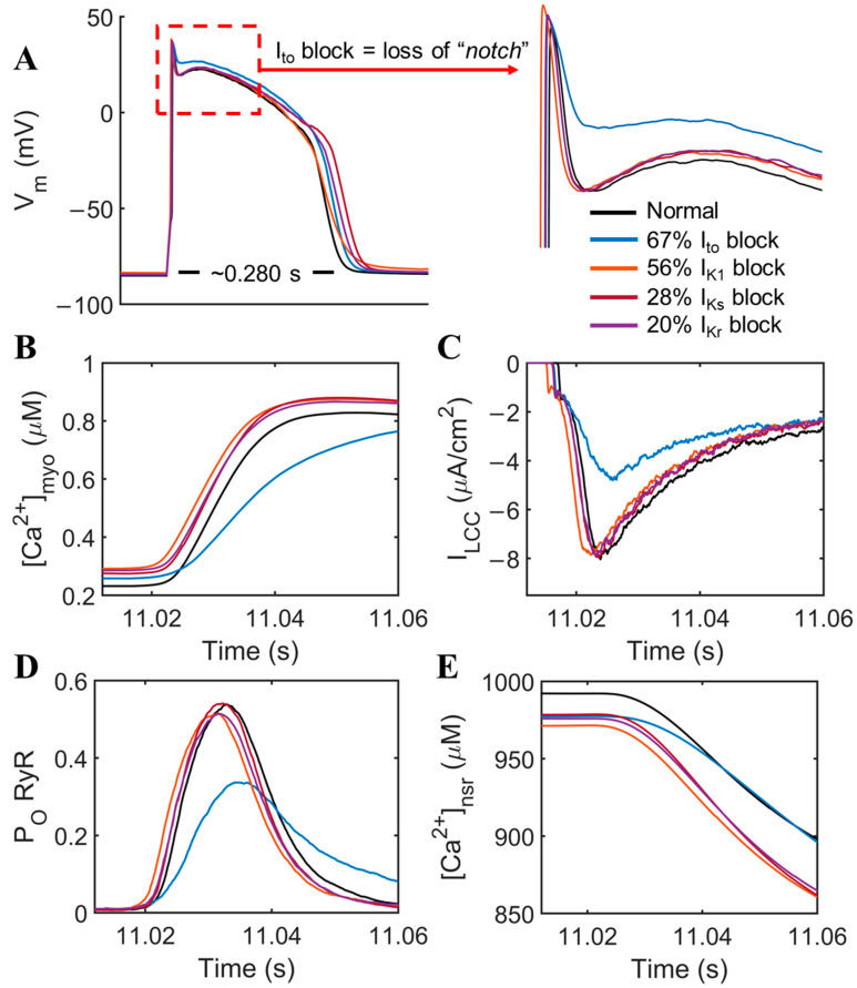
Figure 2.
Prominent effects of Ito among other K+ currents. (A) APD prolongation from the downregulated repolarizing K+ currents present in failing human cardiomyocytes with decreased Ito (blue) showing a loss of the distinctive “notch” in the action potential shape. (B) Roughly two-fold increase in time-to-peak Ca2+ transient from 0.02 to 0.04 s duration was observed. (C) ILCC peak density also decreased roughly 30–35%, respectively, by the effects of Ito alone. (D) RyR open probability is reduced ~20–25% and (E) delayed kinetics of Ca2+ release from the SR by Ito.

