Abstract
Background: The Fibrosis-4 (FIB-4) index is widely recommended as a first-tier method for screening advanced hepatic fibrosis; however, its diagnostic performance is known to be suboptimal in patients with Type 2 diabetes mellitus (T2DM). We aim to propose a modified FIB-4, using the parameters of the existing FIB-4, tailored specifically for diabetic patients with metabolic dysfunction-associated steatotic liver disease (MASLD). Methods: A total of 1503 patients who underwent liver biopsy were divided into T2DM (n = 517) and non-T2DM (n = 986) groups. The model was developed using multiple regression analysis in the derivation cohort and validated in the validation cohort. Diagnostic accuracy was evaluated using the area under the receiver operating characteristic (AUC) curves. Results: Among the 1503 individuals, those with T2DM were older, more likely to be male, and had a higher prevalence of advanced hepatic fibrosis (≥F3) compared to non-T2DM individuals. Independent risk factors for advanced fibrosis in T2DM included age, AST, AST/ALT ratio, albumin, triglycerides, and platelet count. The optimized FIB-4 model for T2DM with MASLD (Diabetes Fibrosis Index) demonstrated superior diagnostic accuracy (AUC 0.771) compared to the FIB-4 (AUC 0.735, p = 0.012). The model showed a higher negative predictive value than the original FIB-4 across all age groups in the diabetic group. Conclusions: The newly optimized FIB-4 model for T2DM with MASLD (Diabetes Fibrosis Index), incorporating a non-linear predictive model, improves diagnostic performance (AUC) and the negative predictive value in MASLD with T2DM.
Keywords: hepatic fibrosis, diabetes, FIB-4
1. Introduction
Metabolic dysfunction-associated steatotic liver disease (MASLD) is the most common cause of chronic liver disease, affecting approximately one in four individuals in most developed countries [1]. The prevalence of significant hepatic fibrosis in MASLD patients is reported to be around 5.9–8.8% [2,3]. The most important risk factor for the development of hepatic fibrosis is T2DM [4]. The rate of significant hepatic fibrosis in T2DM is higher than in MASLD (5.9~8.8%), estimated to be between 12.5% and 20% [5,6,7]. Early detection and active lifestyle modification for significant or advanced hepatic fibrosis in MASLD is cost-effective and can reduce liver-related events and overall mortality associated with the disease [8,9]. Moreover, with the recent FDA approval of the first treatment for MASLD, early identification and management of significant or advanced hepatic fibrosis in MASLD patients can help reduce the social and economic burden of the disease [9,10].
In relation to the assessment of advanced hepatic fibrosis in T2DM, the American Association for the Study of Liver Diseases (AASLD), the American Diabetes Association (ADA), and the American Gastroenterological Association (AGA) issued recommendations in 2023 for evaluating liver disease in all patients with Type 2 diabetes [11]. Similarly, in 2022, the American Association of Clinical Endocrinology (AACE) released comparable guidelines [12]. Given the high prevalence of MASLD in T2DM, these organizations recommend regular screening for advanced fibrosis in this patient group, even if hepatic steatosis is not clinically evident. This recommendation reflects recent research highlighting the high prevalence of advanced fibrosis in T2DM. The recommended screening methods include an initial risk assessment using the Fibrosis-4 (FIB-4) index and, for at-risk patients, a secondary risk assessment with vibration-controlled transient elastography (VCTE) or the enhanced liver fibrosis (ELF) test [13]. However, the practicality of universal fibrosis screening in T2DM patients remains debatable [14,15,16,17]. Initial screening with the FIB-4 index has limitations, with sensitivity and specificity reported as 73% and 62%, respectively, which may result in missed diagnoses [18]. So, the AGA recommends a second-tier non-invasive test (NIT), such as VCTE, for all T2DM patients, regardless of FIB-4 results, differing from other organizations [19]. Therefore, the current guidelines for screening advanced hepatic fibrosis in T2DM patients have significant unmet needs in both accuracy and practical feasibility [20].
Therefore, there is a need for a new NIT or algorithm to effectively screen for liver fibrosis in patients with T2DM accompanied by hepatic steatosis [21]. To date, many non-invasive serologic tests have been proposed for diagnosing hepatic fibrosis in MASLD patients [22,23]. However, for widespread use in primary care settings, these tests must be cost-effective and free from patent restrictions. For this reason, many clinical guidelines still recommend FIB-4 as the first-tier NIT [24].
This study aims to develop a non-invasive test to identify advanced liver fibrosis in patients with MASLD accompanied by T2DM, confirmed through liver biopsy. We aim to propose a modified FIB-4, using the parameters of the existing FIB-4, tailored specifically for diabetic patients with MASLD.
2. Patients and Methods
2.1. Study Design
This study was conducted using retrospective liver biopsy data collected from several centers across multiple countries. The study received approval from the Hanyang University Institutional Review Board (HYIRB- 2023-07-063-005). Given the retrospective nature of the research, the requirement for informed consent was waived.
2.2. Inclusion and Exclusion Criteria
The study included adults aged 19 years or older who had been diagnosed with T2DM and hepatic steatosis. The diagnosis of hepatic steatosis was confirmed through liver biopsy. Individuals were excluded from the study if their blood test results were insufficient or if their history of alcohol consumption was unclear. We diagnosed NAFLD based on the AASLD guidelines. The inclusion criteria are as follows: (1) the presence of hepatic steatosis was observed through liver biopsy with at least one cardiometabolic risk factor (obesity, impaired fasting glucose, hypertension, hypertriglyceridemia, low HDL cholesterolemia), (2) there must be no excessive alcohol consumption (ethanol intake less than 210 g per week for men and less than 140 g per week for women), and (3) other causes of fatty liver, such as medications, must be excluded. The exclusion criteria are as follows: (1) insufficient data variables, (2) use of medications that can induce fatty liver for more than two weeks, and (3) the presence of chronic liver disease causes such as viral infections (hepatitis C or B virus), primary biliary cholangitis, or autoimmune hepatitis must be excluded.
2.3. Definition
MASLD was defined as the absence of significant alcohol consumption (defined as >140 g per week for women and >210 g per week for men) in the past two years. Patients with chronic viral hepatitis, such as hepatitis B or C, were excluded from the study. Additionally, those who were taking medications known to induce hepatic steatosis were not included. Diabetes was defined by the use of diabetes-related medications, a fasting blood glucose level of 126 mg/dL or higher, or a hemoglobin A1c (HbA1C) level greater than 6.5%.
2.4. Liver Histology
Liver biopsy samples were scored using the non-alcoholic fatty liver disease activity score (NAS) system, which assigns separate scores for steatosis (0–3), hepatocellular ballooning (0–2), and lobular inflammation (0–3). The fibrosis stage was classified using Brunt’s pathological grading system, ranging from stage 0 to stage 4, with stage 4 being defined as cirrhosis.
2.5. In-Depth Analysis of FIB-4 Variables
We investigated the impact of the clinical parameters that constitute FIB-4 on advanced hepatic fibrosis in MASLD patients with T2DM. The interactions between the variables that make up FIB-4—age, platelet count, alanine transaminase (ALT), and aspartate aminotransferase (AST)—were analyzed in the presence of advanced hepatic fibrosis.
2.6. Statistical Analysis
To identify independent risk factors for advanced hepatic fibrosis in patients with T2DM, a logistic regression analysis was performed along with T2DM patients’ data. A scoring system based on Polynomial Logistic Regression was created using the factors identified as risk factors (p < 0.05) in the logistic regression analysis. The variables used in the model include age, platelets, AST, ALT, and BMI. To determine the coefficients of the model, which consists of the quadratic terms of the main factors, a Genetic Algorithm optimization was employed. The coefficients that yielded the highest AUROC for detecting advanced hepatic fibrosis in T2DM patients were obtained: (Diabetes Fibrosis Index) = 1.4013 × Age − 2.9859 × Plt × (1 − 0.00159 × Plt) + 5.8155 × AST × (1 − 0.00365 × AST) − 1.2014 × ALT + 56.7468 × BMI × (1 − 0.01467 × BMI). Using the formula, a cut-off point was selected from the ROC curve, and the diagnostic performance using this cut-off point was compared with FIB-4. All statistical analyses were conducted using R version 4.4.0.
3. Results
3.1. Baseline Characteristics of Study Population
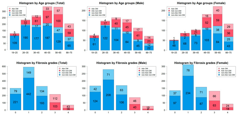
Among the 1503 individuals who underwent full liver biopsy, 517 were diagnosed with T2DM. Compared to the non-T2DM group, the T2DM group within the total cohort was older, had a higher proportion of males, and exhibited a greater prevalence of advanced hepatic fibrosis (≥F3) (Table 1). The prevalence of advanced hepatic fibrosis increased with age, and the proportion of patients with diabetes rose as the stage of fibrosis advanced (Figure 1). In the T2DM group, the proportion of individuals with advanced hepatic fibrosis (≥F3) was 30.0% (155/517) (Table 2). Patients with T2DM who had advanced hepatic fibrosis (≥F3) were older, had a higher male proportion, and had a higher BMI compared to those in the F0~2 fibrosis group.
Table 1.
Baseline characteristics of total population (T2DM and non-T2DM).
| Total (n = 1503) |
Non-T2DM (n = 986) |
T2DM (n = 517) |
p | |
|---|---|---|---|---|
| Age (years) † | 48.10 ± 14.85 | 45.11 ± 14.95 | 53.81 ± 12.88 | <0.001 a |
| Male | 48.4% | 49.3% | 46.6% | 0.352 b |
| BMI (kg/m2) † | 29.39 ± 6.59 | 29.41 ± 7.23 | 29.355 ± 5.16 | 0.854 a |
| Waist circumference (cm) † | 95.83 ± 12.46 | 95.41 ± 11.75 | 96.50 ± 13.54 | 0.469 a |
| Hypertension | 42.4% | 33.9% | 58.6% | <0.001 b |
| Diabetes | 34.4% | |||
| Triglyceride (mg/dL) † | 159.46 ± 100.72 | 156.62 ± 102.69 | 164.89 ± 96.71 | 0.129 a |
| HDL-CROL (mg/dL) † | 48.06 ± 14.77 | 49.00 ± 14.56 | 46.27 ± 15.00 | 0.001 a |
| Cholesterol (mg/dL) | 124.39 ± 50.58 | 127.54 ± 47.84 | 118.12 ± 55.16 | 0.004 a |
| Albumin (mg/dL) | 4.33 ± 0.44 | 4.35 ± 0.42 | 4.29 ± 0.46 | 0.007 a |
| Glucose (mg/dL) † | 113.90 ± 35.59 | 99.45 ± 16.10 | 141.41 ± 45.07 | <0.001 a |
| AST (IU/L) † | 51.54 ± 35.39 | 47.74 ± 33.87 | 58.79 ± 37.09 | <0.001 a |
| ALT (IU/L) † | 73.63 ± 58.89 | 71.19 ± 60.20 | 78.29 ± 56.07 | 0.023 a |
| AST/ALT † | 0.8697 ± 0.4423 | 0.8650 ± 0.4634 | 0.8786 ± 0.3993 | 0.555 a |
| Platelets (×109/L) † | 236.65 ± 72.67 | 246.55 ± 70.32 | 217.78 ± 73.41 | <0.001 a |
| FIB-4 index † | 1.58 ± 1.47 | 1.32 ± 1.24 | 2.07 ± 1.73 | <0.001 a |
| NAFLD fibrosis score † | −1.82 ± 1.67 | −2.46 ± 1.39 | −0.58 ± 1.45 | <0.001 a |
| Steatosis | 1.39 ± 1.01 | 1.24 ± 1.03 | 1.74 ± 0.86 | <0.001 a |
| Inflammation | 1.11 ± 0.99 | 0.91 ± 0.96 | 1.56 ± 0.90 | <0.001 a |
| Fibrosis | 285 (19.0%) | 130 (13.2%) | 155 (30.0%) | <0.001 b |
| Prevalence of ≥F2 | 1218 | 856 | 362 | |
| Prevalence of ≥F3 | 285 | 130 | 155 |
Data are expressed as number (percent). † Data are shown as mean ± standard deviation. Abbreviations: AST, aspartate transaminase; ALT, alanine transaminase; BMI, body mass index; HDL, high-density lipoprotein; NAFLD, non-alcoholic fatty liver disease; a Student’s t-test; b Chi-square test.
Figure 1.
Prevalence of Type 2 diabetes according to fibrosis stage and age.
Table 2.
Baseline characteristics of Type 2 diabetes.
| Total (n = 517) |
≥F3 (n = 155) |
F0~2 (n = 362) |
p | |
|---|---|---|---|---|
| Age (years) † | 53.8 ± 12.9 | 57.5 ± 12.2 | 52.2 ± 12.9 | <0.001 a |
| Male | 46.6% | 41.9% | 48.6% | 0.194 b |
| BMI (kg/m2) † | 29.36 ± 5.16 | 29.41 ± 4.48 | 29.33 ± 5.43 | 0.865 a |
| Waist circumference (cm) † | 96.50 ± 13.54 | 98.28 ± 7.75 | 95.90 ± 14.99 | 0.266 a |
| Hypertension | 58.6% | 65.8% | 55.4% | 0.036 b |
| Triglyceride (mg/dL) † | 164.89 ± 96.71 | 141.09 ± 64.46 | 175.49 ± 106.39 | <0.001 a |
| HDL-CROL (mg/dL) † | 46.27 ± 15.00 | 47.60 ± 17.15 | 45.68 ± 13.92 | 0.226 a |
| Cholesterol (mg/dL) | 188.12 ± 55.16 | 122.30 ± 82.12 | 116.31 ± 38.01 | 0.444 a |
| Albumin (mg/dL) | 4.29 ± 0.46 | 4.20 ± 0.47 | 4.32 ± 0.45 | 0.005 a |
| Glucose (mg/dL) † | 141.41 ± 45.07 | 142.56 ± 46.87 | 140.91 ± 44.34 | 0.711 a |
| AST (IU/L) † | 58.79 ± 37.09 | 65.35 ± 33.84 | 55.99 ± 38.10 | 0.006 a |
| ALT (IU/L) † | 78.29 ± 56.08 | 75.90 ± 47.52 | 79.31 ± 59.39 | 0.490 a |
| AST/ALT † | 0.8786 ± 0.3993 | 0.9893 ± 0.4348 | 0.8312 ± 0.3738 | <0.001 a |
| Platelets (×109/L) † | 217.78 ± 73.41 | 183.91 ± 73.51 | 232.279 ± 68.52 | <0.001 a |
| FIB-4 index † | 2.07 ± 1.73 | 3.02 ± 2.20 | 1.67 ± 1.29 | <0.001 a |
| NAFLD fibrosis score † | −0.58 ± 1.45 | 0.16 ± 1.56 | −0.91 ± 1.27 | <0.001 a |
| Steatosis (mean ± SD) | 1.74 ± 0.86 | 1.77 ± 0.68 | 1.73 ± 0.92 | 0.805 a |
| Inflammation (mean ± SD) | 1.56 ± 0.90 | 1.86 ± 0.80 | 1.44 ± 0.91 | 0.006 a |
| Hepatic fibrosis (%) | 30.0% | 155 | 362 | |
| F0/1 | 228 (63.0%) | |||
| F2 | 134 (37.0%) | |||
| F3 | 112 (72.3%) | |||
| F4 | 43 (27.7%) |
Data are expressed as number (percent). † Data are shown as mean ± standard deviation. Abbreviations: AST, aspartate transaminase; ALT, alanine transaminase; BMI, body mass index; HDL, high-density lipoprotein; NAFLD, non-alcoholic fatty liver disease; a Student’s t-test; b Chi-square test.
3.2. Major Clinical Risk Factors and Their Interactions in Advanced Hepatic Fibrosis
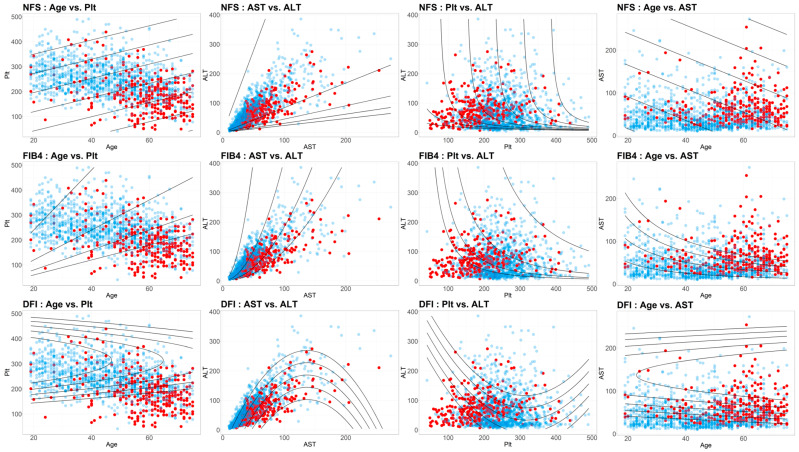
To identify the independent risk factors for advanced hepatic fibrosis in patients with T2DM, a logistic regression analysis was performed (Table 3). The results indicated that age, AST, AST/ALT ratio, albumin, triglycerides, and platelet count were independent risk factors for advanced hepatic fibrosis in the T2DM group. In a separate logistic regression analysis conducted on the non-T2DM group, additional factors such as diabetes status, fasting blood glucose, HbA1c, and gender were identified as independent risk factors for advanced hepatic fibrosis. Interactions among the independent risk factors for advanced hepatic fibrosis in T2DM were also assessed. Notably, age and platelet count, AST and ALT, and platelet count and ALT, as well as age and AST, exhibited non-linear two-dimensional interactions concerning advanced hepatic fibrosis (Figure 2).
Table 3.
Logistic regression for major clinical risk factors of advanced hepatic fibrosis.
| Total | T2DM | |||||||
|---|---|---|---|---|---|---|---|---|
| Odds Ratio | 95% CI Lower |
95% CI Upper |
p | Odds Ratio | 95% CI Lower |
95% CI Upper |
p | |
| T2DM | 2.799 | 2.145 | 3.657 | <0.001 | ||||
| Age (years) † | 1.064 | 1.053 | 1.076 | <0.001 | 1.063 | 1.044 | 1.084 | <0.001 |
| Gender (M) | 1.518 | 1.166 | 1.983 | <0.001 | 1.540 | 1.020 | 2.342 | 0.041 |
| BMI (kg/m2) † | 0.991 | 0.970 | 1.010 | 0.355 | 0.991 | 0.953 | 1.028 | 0.636 |
| AST (IU/L) † | 1.011 | 1.008 | 1.014 | <0.001 | 1.007 | 1.002 | 1.013 | 0.008 |
| ALT (IU/L) † | 1.001 | 0.998 | 1.003 | 0.532 | 0.998 | 0.994 | 1.002 | 0.285 |
| AST/ALT † | 2.031 | 1.539 | 2.680 | <0.001 | 3.099 | 1.927 | 5.071 | <0.001 |
| Albumin (mg/dL) | 0.381 | 0.270 | 0.534 | <0.001 | 0.476 | 0.274 | 0.820 | 0.008 |
| Glucose (mg/dL) † | 1.009 | 1.006 | 1.012 | <0.001 | 1.006 | 1.002 | 1.011 | 0.004 |
| HBA1c (%) | 1.195 | 1.069 | 1.340 | 0.002 | 1.130 | 0.979 | 1.317 | 0.105 |
| Triglyceride (mg/dL) † | 0.999 | 0.997 | 1.000 | 0.111 | 0.997 | 0.994 | 0.999 | 0.040 |
| HDL-CROL (mg/dL) † | 0.997 | 0.988 | 1.006 | 0.532 | 1.004 | 0.990 | 1.016 | 0.563 |
| LDL-CROL (mg/dL) † | 1.000 | 0.996 | 1.002 | 0.770 | 0.998 | 0.991 | 1.004 | 0.516 |
| Platelets | 0.987 | 0.985 | 0.989 | <0.001 | 0.987 | 0.984 | 0.991 | <0.001 |
Data are expressed as number (percent). † Data are shown as mean ± standard deviation. Abbreviations: AST, aspartate transaminase; ALT, alanine transaminase; BMI, body mass index; HDL, high-density lipoprotein.
Figure 2.
Interaction analysis of major clinical risk factors for advanced hepatic fibrosis.
3.3. Optimized FIB-4 Model for T2DM with MASLD for Advanced Hepatic Fibrosis in T2DM
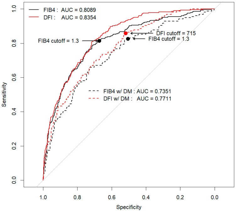
A non-linear predictive model was constructed to account for the interactions among independent risk factors for advanced hepatic fibrosis in patients with T2DM. The model incorporated five variables: age, platelet count, AST, ALT, and BMI. The diagnostic accuracy of this optimized FIB-4 model for T2DM with MASLD, as measured by the area under the curve (AUC), was 0.771, which was significantly higher than the AUC of 0.735 for FIB-4 (p = 0.012) (Figure 3). The optimal cut-off value for the optimized FIB-4 in T2DM, termed the Diabetes Fibrosis Index (calculated as 1.4013 × Age − 2.9859 × Plt × (1 − 0.00159 × Plt) + 5.8155 × AST × (1 − 0.00365 × AST) − 1.2014 × ALT + 56.7468 × BMI × (1 − 0.01467 × BMI)), was 715, yielding a sensitivity of 0.85 and a specificity of 0.52. This performance was superior to the FIB-4 model, which had a sensitivity of 0.82 and a specificity of 0.50. Notably, the diagnostic performance of the FIB-4 model significantly declined with age, particularly in diabetic patients aged 65 years and older, where the AUC dropped to 0.68. In contrast, the optimized FIB-4 model for T2DM with MASLD demonstrated improved diagnostic performance in this older age group, with an AUC of 0.72, surpassing that of the FIB-4 (Table 4).
Figure 3.
Diagnostic performance of new NIT for T2DM.
Table 4.
Diagnostic performance according to age.
| FIB4 | Total | T2DM | ||||||||||||
| Cut-Off | Age Group | AUROC | Acc | Sens | Spec | PPV | NPV | Age Group | AUROC | Acc | Sens | Spec | PPV | NPV |
| 1.3 | Total | 0.8089 | 0.6993 | 0.8134 | 0.6727 | 0.3667 | 0.9393 | Total | 0.7351 | 0.6035 | 0.8258 | 0.5083 | 0.4183 | 0.872 |
| ~25 | 0.6325 | 0.9531 | 0.1667 | 0.9918 | 0.5000 | 0.9603 | ~25 | 0.5846 | 0.7778 | 0.2000 | 1.0000 | 1.0000 | 0.7647 | |
| 26~35 | 0.6642 | 0.9231 | 0.1667 | 0.9640 | 0.2000 | 0.9554 | 26~35 | 0.6094 | 0.7027 | 0.0000 | 0.8125 | 0.0000 | 0.8387 | |
| 36~45 | 0.6366 | 0.8427 | 0.4483 | 0.8908 | 0.3333 | 0.4483 | 36~45 | 0.6093 | 0.7808 | 0.5000 | 0.8361 | 0.3750 | 0.8947 | |
| 46~55 | 0.7384 | 0.6469 | 0.6604 | 0.6444 | 0.2574 | 0.9104 | 46~55 | 0.6982 | 0.6077 | 0.6970 | 0.5773 | 0.3594 | 0.8485 | |
| 56~65 | 0.7804 | 0.5285 | 0.9615 | 0.3319 | 0.3953 | 0.9500 | 56~65 | 0.7754 | 0.5669 | 0.9649 | 0.3400 | 0.4545 | 0.9444 | |
| 66~75 | 0.7031 | 0.4608 | 1.0000 | 0.1129 | 0.4211 | 1.0000 | 66~75 | 0.6878 | 0.4608 | 1.0000 | 0.0678 | 0.4388 | 1.0000 | |
| New Model | Total | T2DM | ||||||||||||
| Cut-Off | Age Group | AUROC | Acc | Sens | Spec | PPV | NPV | Age Group | AUROC | Acc | Sens | Spec | PPV | NPV |
| 715 | Total | 0.8354 | 0.7279 | 0.8134 | 0.708 | 0.3935 | 0.9421 | Total | 0.7710 | 0.6228 | 0.8581 | 0.5221 | 0.4346 | 0.8957 |
| ~25 | 0.8757 | 0.9531 | 0.3333 | 0.9836 | 0.5000 | 0.9677 | ~25 | 0.7538 | 0.7222 | 0.4000 | 0.8462 | 0.5000 | 0.7857 | |
| 26~35 | 0.7911 | 0.8590 | 0.4167 | 0.8829 | 0.1613 | 0.9655 | 26~35 | 0.6656 | 0.7027 | 0.6000 | 0.7188 | 0.2500 | 0.9200 | |
| 36~45 | 0.7867 | 0.7603 | 0.5517 | 0.7857 | 0.2388 | 0.9350 | 36~45 | 0.6995 | 0.6438 | 0.5833 | 0.6557 | 0.2500 | 0.8889 | |
| 46~55 | 0.7902 | 0.7211 | 0.7170 | 0.7218 | 0.3248 | 0.9318 | 46~55 | 0.7423 | 0.6538 | 0.7879 | 0.6082 | 0.4062 | 0.8939 | |
| 56~65 | 0.7748 | 0.6096 | 0.8942 | 0.4803 | 0.4387 | 0.9091 | 56~65 | 0.7912 | 0.5987 | 0.9474 | 0.4000 | 0.4737 | 0.9302 | |
| 66~75 | 0.7345 | 0.5980 | 0.9625 | 0.3629 | 0.4936 | 0.9375 | 66~75 | 0.7217 | 0.5588 | 0.9535 | 0.2712 | 0.4881 | 0.8889 | |
4. Discussion
This study proposes a novel Diabetes Fibrosis Index for identifying patients with advanced hepatic fibrosis in those MASLD patients with T2DM. The optimized FIB-4 model for T2DM with MASLD utilizes clinical parameters similar to those used in the widely adopted FIB-4 index, commonly employed in primary care settings. However, it enhances sensitivity and specificity compared to the traditional FIB-4, particularly addressing the issue of decreased sensitivity associated with age, which is a significant limitation of the FIB-4 [25,26,27]. Our study introduces a Diabetes Fibrosis Index for identifying patients with advanced hepatic fibrosis in those with T2DM based on a large cohort of 1503 biopsy-confirmed patients. The original FIB-4 index has a good ability to rule out advanced fibrosis when a cut-off of 1.3 is used [28]. The novel marker in this study is useful because it shows a higher negative predictive value (NPV) than the original FIB-4 across all age groups in the diabetic group. However, the issue is that the positive predictive value (PPV) is low when using the current cut-off point, suggesting that other NITs, including elastography, may be necessary to confirm advanced fibrosis.
FIB-4 is generally recommended as a first-tier screening tool for high-risk groups in patients with MASLD. It has shown relatively good diagnostic performance for advanced hepatic fibrosis, with reported AUCs ranging from 0.75 to 0.83. However, FIB-4′s diagnostic performance is known to be suboptimal in patients under 35 years of age and those over 65, as well as in those with T2DM, where its sensitivity is notably reduced [29]. Studies such as those by Qadri et al. have reported a diagnostic performance of FIB-4 in predicting advanced hepatic fibrosis in MASLD patients with T2DM with a sensitivity of 73% and a specificity of 62% using the 1.30 cut-off [30]. This suggests that using FIB-4 as a first-tier test in this population could result in missing up to 27% of patients with advanced hepatic fibrosis [31]. Consequently, current guidelines acknowledge the lack of robust evidence for the diagnostic performance of FIB-4 in MASLD patients with T2DM and highlight the need for further research [30].
In this study, we focused on using the same biochemical parameters as the original FIB-4, excluding BMI, to identify advanced hepatic fibrosis in T2DM patients. While various NITs have been proposed for hepatic fibrosis across different at-risk groups, including MASLD and T2DM, their application in primary care settings remains limited. This is largely due to the absence of a clear screening algorithm and the significant societal burden associated with additional costly tests or blood work, particularly when effective treatments are lacking. For this reason, despite its limitations, most clinical guidelines continue to recommend FIB-4 as a first-tier test [32,33,34]. Therefore, our research group sought to improve the performance of the FIB-4 test by utilizing a non-linear equation that reflects the non-linear relationship of the biochemical parameters with advanced hepatic fibrosis (Figure 2).
Despite recommendations in all guidelines to screen for advanced hepatic fibrosis in patients with diabetes due to its high prevalence, this practice is not widely implemented in clinical settings [35]. Simulation studies in the U.S. have suggested that applying FIB-4 as a screening test could result in 40% of all diabetic patients requiring annual vibration-controlled transient elastography (VCTE) tests, and 19% needing referral to a hepatologist each year [30].
The strength of this novel score is that it can exclude cases of advanced fibrosis progression in T2DM-associated SLD using only data obtained from physical examinations. While serum NITs unaffected by T2DM, such as ELF and type 4 collagen 7s, exist, they cannot be used in general health examinations. Therefore, this novel marker will be particularly useful in primary care settings. If additional analysis is conducted, it may be beneficial to compare ROC curves by BMI or degree of T2DM, provided there is sufficient data on HbA1c, not just age.
The limitations of our study are as follows: First, these data are based on a cross-sectional study focused on identifying at-risk groups in T2DM. However, it is also important to verify whether the newly proposed modified FIB-4 score provides superior predictive ability for long-term clinical outcomes in T2DM compared to the conventional FIB-4. Moreover, our use of retrospective data in the study has inherent biases in data collection, such as selection bias or incomplete data, which may still exist and could impact the reliability of the study’s findings. Prospective validation is indeed necessary to confirm the model’s clinical utility. Unfortunately, due to the nature of the database, we were unable to compare the predictive ability for long-term clinical outcomes with the conventional FIB-4. Second, although the new model’s NPV has improved, the improvement in PPV is relatively low. Sensitivity, specificity, NPV, and PPV are highly sensitive to changes in the cut-off point. The authors prioritized maintaining sensitivity and NPV above 90% as the primary goal of a screening test, rather than focusing on increasing PPV. In screening tests, sensitivity and NPV are generally prioritized over specificity and PPV, particularly when used as a first-tier screening tool, where minimizing missed high-risk cases and clearly identifying non-urgent cases are essential. For these reasons, the authors selected an optimal cut-off to achieve sensitivity and NPV close to 90%. Third, the presence of T2DM and insulin resistance are known to be significant independent risk factors for the degree of hepatic fibrosis in MASLD. Additionally, the duration of T2DM and the specific antidiabetic medications used are also reported to play important roles in hepatic fibrosis. However, in this study, data on the duration of T2DM and a detailed history of drug usage were not collected.
In conclusion, this study presents a novel, non-linear predictive model for diagnosing advanced hepatic fibrosis in patients with T2DM, demonstrating superior accuracy compared to the traditional FIB-4 index. By utilizing commonly available clinical parameters, the optimized FIB-4 model for T2DM with MASLD addresses the limitations of FIB-4, particularly its reduced sensitivity in older patients. This model has the potential to improve early detection of advanced fibrosis in T2DM, thereby enhancing patient outcomes. Further validation in clinical settings is warranted to confirm its utility and effectiveness.
Author Contributions
K.C. performed the statistical analyses. J.K. was responsible for preparing the first draft and revising the manuscript. T.A., M.A., M.K., H.T., T.H., M.-L.Y. and M.H.N. prepared data. E.L.Y. and T.I. reviewed and revised the manuscript. D.W.J. was responsible for the conceptual work and design, and critically reviewed and supervised the overall study. All authors have read and agreed to the published version of the manuscript.
Institutional Review Board Statement
This study was approved by the Hanyang University Institutional Review Board (HYUH 2022-11-006-003). This protocol approved on 31 July 2023.
Informed Consent Statement
This study was conducted based on data previously collected through routine practice, and informed consent was waived by the Institutional Review Board.
Data Availability Statement
Data in this study include patients’ clinical information, which may be shared upon request to the authors following Institutional Review Board approval.
Conflicts of Interest
The authors declare no conflicts of interest.
Funding Statement
This work was supported by the National Research Foundation of Korea (NRF) grant funded by the Korea government (MSIT) (RS-2024-00440477, and RS-2024-00347603). This research was supported by the Basic Science Research Program through the National Research Foundation of Korea (NRF) funded by the Ministry of Education (2020R1A6A1A06046728).
Footnotes
Disclaimer/Publisher’s Note: The statements, opinions and data contained in all publications are solely those of the individual author(s) and contributor(s) and not of MDPI and/or the editor(s). MDPI and/or the editor(s) disclaim responsibility for any injury to people or property resulting from any ideas, methods, instructions or products referred to in the content.
References
- 1.European Association for the Study of the Liver (EASL) European Association for the Study of Diabetes (EASD) European Association for the Study of Obesity (EASO) EASL-EASD-EASO Clinical Practice Guidelines on the management of metabolic dysfunction-associated steatotic liver disease (MASLD) J. Hepatol. 2024;81:492–542. doi: 10.1016/j.jhep.2024.04.031. [DOI] [PubMed] [Google Scholar]
- 2.Lee C.M., Yoon E.L., Kim M., Kang B.K., Cho S., Nah E.H., Jun D.W. Prevalence, distribution, and hepatic fibrosis burden of the different subtypes of steatotic liver disease in primary care settings. Hepatology. 2024;79:1393–1400. doi: 10.1097/HEP.0000000000000664. [DOI] [PubMed] [Google Scholar]
- 3.Kim H.Y., Yu J.H., Chon Y.E., Kim S.U., Kim M.N., Han J.W., Lee H.A., Jin Y.J., An J., Choi M., et al. Prevalence of clinically significant liver fibrosis in the general population: A systematic review and meta-analysis. Clin. Mol. Hepatol. 2024;30:S199–S213. doi: 10.3350/cmh.2024.0351. [DOI] [PMC free article] [PubMed] [Google Scholar]
- 4.Qi X., Li J., Caussy C., Teng G.J., Loomba R. Epidemiology, screening, and co-management of type 2 diabetes mellitus and metabolic dysfunction-associated steatotic liver disease. Hepatology. 2024 doi: 10.1097/HEP.0000000000000913. [DOI] [PubMed] [Google Scholar]
- 5.Ajmera V., Cepin S., Tesfai K., Hofflich H., Cadman K., Lopez S., Madamba E., Bettencourt R., Richards L., Behling C., et al. A prospective study on the prevalence of NAFLD, advanced fibrosis, cirrhosis and hepatocellular carcinoma in people with type 2 diabetes. J. Hepatol. 2023;78:471–478. doi: 10.1016/j.jhep.2022.11.010. [DOI] [PMC free article] [PubMed] [Google Scholar]
- 6.Mertens J., Weyler J., Dirinck E., Vonghia L., Kwanten W.J., Mortelmans L., Peleman C., Chotkoe S., Spinhoven M., Vanhevel F., et al. Prevalence, risk factors and diagnostic accuracy of non-invasive tests for NAFLD in people with type 1 diabetes. JHEP Rep. 2023;5:100753. doi: 10.1016/j.jhepr.2023.100753. [DOI] [PMC free article] [PubMed] [Google Scholar]
- 7.Ortiz-Lopez C., Lomonaco R., Orsak B., Finch J., Chang Z., Kochunov V.G., Hardies J., Cusi K. Prevalence of prediabetes and diabetes and metabolic profile of patients with nonalcoholic fatty liver disease (NAFLD) Diabetes Care. 2012;35:873–878. doi: 10.2337/dc11-1849. [DOI] [PMC free article] [PubMed] [Google Scholar]
- 8.Park H., Yoon E.L., Kim M., Kwon S.H., Kim D., Cheung R., Kim H.L., Jun D.W. Cost-effectiveness study of FIB-4 followed by transient elastography screening strategy for advanced hepatic fibrosis in a NAFLD at-risk population. Liver Int. 2024;44:944–954. doi: 10.1111/liv.15838. [DOI] [PubMed] [Google Scholar]
- 9.Noureddin M., Jones C., Alkhouri N., Gomez E.V., Dieterich D.T., Rinella M.E., Therapondos G., Girgrah N., Mantry P.S., Sussman N.L., et al. Screening for Nonalcoholic Fatty Liver Disease in Persons with Type 2 Diabetes in the United States Is Cost-effective: A Comprehensive Cost-Utility Analysis. Gastroenterology. 2020;159:1985–1987.e4. doi: 10.1053/j.gastro.2020.07.050. [DOI] [PubMed] [Google Scholar]
- 10.Harrison S.A., Bedossa P., Guy C.D., Schattenberg J.M., Loomba R., Taub R., Labriola D., Moussa S.E., Neff G.W., Rinella M.E., et al. A Phase 3, Randomized, Controlled Trial of Resmetirom in NASH with Liver Fibrosis. N. Engl. J. Med. 2024;390:497–509. doi: 10.1056/NEJMoa2309000. [DOI] [PubMed] [Google Scholar]
- 11.Rinella M.E., Neuschwander-Tetri B.A., Siddiqui M.S., Abdelmalek M.F., Caldwell S., Barb D., Kleiner D.E., Loomba R. AASLD Practice Guidance on the clinical assessment and management of nonalcoholic fatty liver disease. Hepatology. 2023;77:1797–1835. doi: 10.1097/HEP.0000000000000323. [DOI] [PMC free article] [PubMed] [Google Scholar]
- 12.Cusi K., Isaacs S., Barb D., Basu R., Caprio S., Garvey W.T., Kashyap S., Mechanick J.I., Mouzaki M., Nadolsky K., et al. American Association of Clinical Endocrinology Clinical Practice Guideline for the Diagnosis and Management of Nonalcoholic Fatty Liver Disease in Primary Care and Endocrinology Clinical Settings: Co-Sponsored by the American Association for the Study of Liver Diseases (AASLD) Endocr. Pract. 2022;28:528–562. doi: 10.1016/j.eprac.2022.03.010. [DOI] [PubMed] [Google Scholar]
- 13.Chan W.K., Wong V.W., Adams L.A., Nguyen M.H. MAFLD in adults: Non-invasive tests for diagnosis and monitoring of MAFLD. Hepatol. Int. 2024;18((Suppl. 2)):909–921. doi: 10.1007/s12072-024-10661-x. [DOI] [PubMed] [Google Scholar]
- 14.Vieira Barbosa J., Lai M. Nonalcoholic Fatty Liver Disease Screening in Type 2 Diabetes Mellitus Patients in the Primary Care Setting. Hepatol. Commun. 2021;5:158–167. doi: 10.1002/hep4.1618. [DOI] [PMC free article] [PubMed] [Google Scholar]
- 15.Kim R.G., Deng J., Reaso J.N., Grenert J.P., Khalili M. Noninvasive Fibrosis Screening in Fatty Liver Disease Among Vulnerable Populations: Impact of Diabetes and Obesity on FIB-4 Score Accuracy. Diabetes Care. 2022;45:2449–2451. doi: 10.2337/dc22-0556. [DOI] [PMC free article] [PubMed] [Google Scholar]
- 16.Meritsi A., Latsou D., Manesis E., Gatos I., Theotokas I., Zoumpoulis P., Rapti S., Tsitsopoulos E., Moshoyianni H., Manolakopoulos S., et al. Noninvasive, Blood-Based Biomarkers as Screening Tools for Hepatic Fibrosis in People with Type 2 Diabetes. Clin. Diabetes. 2022;40:327–338. doi: 10.2337/cd21-0104. [DOI] [PMC free article] [PubMed] [Google Scholar]
- 17.Sporea I., Mare R., Popescu A., Nistorescu S., Baldea V., Sirli R., Braha A., Sima A., Timar R., Lupusoru R. Screening for Liver Fibrosis and Steatosis in a Large Cohort of Patients with Type 2 Diabetes Using Vibration Controlled Transient Elastography and Controlled Attenuation Parameter in a Single-Center Real-Life Experience. J. Clin. Med. 2020;9:1032. doi: 10.3390/jcm9041032. [DOI] [PMC free article] [PubMed] [Google Scholar]
- 18.Park H., Yoon E.L., Kim M., Lee J., Kim H.L., Cho S., Nah E.H., Jun D.W. Diagnostic performance of the fibrosis-4 index and the NAFLD fibrosis score for screening at-risk individuals in a health check-up setting. Hepatol. Commun. 2023;7:e0249. doi: 10.1097/HC9.0000000000000249. [DOI] [PMC free article] [PubMed] [Google Scholar]
- 19.Patel K., Sebastiani G. Limitations of non-invasive tests for assessment of liver fibrosis. JHEP Rep. 2020;2:100067. doi: 10.1016/j.jhepr.2020.100067. [DOI] [PMC free article] [PubMed] [Google Scholar]
- 20.Younossi Z.M., Henry L. Understanding the Burden of Nonalcoholic Fatty Liver Disease: Time for Action. Diabetes Spectr. 2024;37:9–19. doi: 10.2337/dsi23-0010. [DOI] [PMC free article] [PubMed] [Google Scholar]
- 21.Chan W.K., Petta S., Noureddin M., Goh G.B.B., Wong V.W. Diagnosis and non-invasive assessment of MASLD in type 2 diabetes and obesity. Aliment. Pharmacol. Ther. 2024;59((Suppl. 1)):S23–S40. doi: 10.1111/apt.17866. [DOI] [PubMed] [Google Scholar]
- 22.De A., Mehta M., Duseja A., ICOM-D Study Group Substantial overlap between NAFLD and MASLD with comparable disease severity and non-invasive test performance: An analysis of the Indian Consortium on MASLD (ICOM-D) cohort. J. Hepatol. 2024;81:e162–e164. doi: 10.1016/j.jhep.2024.05.027. [DOI] [PubMed] [Google Scholar]
- 23.Vali Y., Lee J., Boursier J., Spijker R., Loffler J., Verheij J., Brosnan M.J., Bocskei Z., Anstee Q.M., Bossuyt P.M., et al. Enhanced liver fibrosis test for the non-invasive diagnosis of fibrosis in patients with NAFLD: A systematic review and meta-analysis. J. Hepatol. 2020;73:252–262. doi: 10.1016/j.jhep.2020.03.036. [DOI] [PubMed] [Google Scholar]
- 24.Doycheva I., Cui J., Nguyen P., Costa E.A., Hooker J., Hofflich H., Bettencourt R., Brouha S., Sirlin C.B., Loomba R. Non-invasive screening of diabetics in primary care for NAFLD and advanced fibrosis by MRI and MRE. Aliment. Pharmacol. Ther. 2016;43:83–95. doi: 10.1111/apt.13405. [DOI] [PMC free article] [PubMed] [Google Scholar]
- 25.Graupera I., Thiele M., Serra-Burriel M., Caballeria L., Roulot D., Wong G.L., Fabrellas N., Guha I.N., Arslanow A., Exposito C., et al. Low Accuracy of FIB-4 and NAFLD Fibrosis Scores for Screening for Liver Fibrosis in the Population. Clin. Gastroenterol. Hepatol. 2022;20:2567–2576.e6. doi: 10.1016/j.cgh.2021.12.034. [DOI] [PubMed] [Google Scholar]
- 26.Imai K., Takai K., Unome S., Miwa T., Hanai T., Suetsugu A., Shimizu M. FIB-4 index and NAFLD fibrosis score are useful indicators for screening high-risk groups of non-viral hepatocellular carcinoma. Mol. Clin. Oncol. 2023;19:80. doi: 10.3892/mco.2023.2676. [DOI] [PMC free article] [PubMed] [Google Scholar]
- 27.van Kleef L.A., Sonneveld M.J., de Knegt R.J. Reply to: Low Accuracy of FIB-4 and NAFLD Fibrosis Scores for Screening for Liver Fibrosis in the Population. Clin. Gastroenterol. Hepatol. 2023;21:238–239. doi: 10.1016/j.cgh.2022.02.011. [DOI] [PubMed] [Google Scholar]
- 28.Nakano M., Kawaguchi M., Kawaguchi T. Almost identical values of various non-invasive indexes for hepatic fibrosis and steatosis between NAFLD and MASLD in Asia. J. Hepatol. 2024;80:e155–e157. doi: 10.1016/j.jhep.2023.12.030. [DOI] [PubMed] [Google Scholar]
- 29.Boursier J., Hagstrom H., Ekstedt M., Moreau C., Bonacci M., Cure S., Ampuero J., Nasr P., Tallab L., Canivet C.M., et al. Non-invasive tests accurately stratify patients with NAFLD based on their risk of liver-related events. J. Hepatol. 2022;76:1013–1020. doi: 10.1016/j.jhep.2021.12.031. [DOI] [PubMed] [Google Scholar]
- 30.Qadri S., Yki-Jarvinen H. Surveillance of the liver in type 2 diabetes: Important but unfeasible? Diabetologia. 2024;67:961–973. doi: 10.1007/s00125-024-06087-7. [DOI] [PMC free article] [PubMed] [Google Scholar]
- 31.Qadri S., Ahlholm N., Lonsmann I., Pellegrini P., Poikola A., Luukkonen P.K., Porthan K., Juuti A., Sammalkorpi H., Penttila A.K., et al. Obesity Modifies the Performance of Fibrosis Biomarkers in Nonalcoholic Fatty Liver Disease. J. Clin. Endocrinol. Metab. 2022;107:e2008–e2020. doi: 10.1210/clinem/dgab933. [DOI] [PMC free article] [PubMed] [Google Scholar]
- 32.Lu Z., Shao W., Song J. The transition from NAFLD to MASLD and its impact on clinical practice and outcomes. J. Hepatol. 2024;81:e155–e156. doi: 10.1016/j.jhep.2024.02.021. [DOI] [PubMed] [Google Scholar]
- 33.Nascimbeni F., Pais R., Bellentani S., Day C.P., Ratziu V., Loria P., Lonardo A. From NAFLD in clinical practice to answers from guidelines. J. Hepatol. 2013;59:859–871. doi: 10.1016/j.jhep.2013.05.044. [DOI] [PubMed] [Google Scholar]
- 34.Palmer M. Practice guidelines on NAFLD. Hepatology. 2013;57:853. doi: 10.1002/hep.25998. [DOI] [PMC free article] [PubMed] [Google Scholar]
- 35.Lee J.H., Yoon E.L., Oh J.H., Kim K., Ahn S.B., Jun D.W. Barriers to care linkage and educational impact on unnecessary MASLD referrals. Front. Med. 2024;11:1407389. doi: 10.3389/fmed.2024.1407389. [DOI] [PMC free article] [PubMed] [Google Scholar]
Associated Data
This section collects any data citations, data availability statements, or supplementary materials included in this article.
Data Availability Statement
Data in this study include patients’ clinical information, which may be shared upon request to the authors following Institutional Review Board approval.


