Simple Summary
Green healthcare relates to using naturally derived sources as medicines to treat and personalize treatments for various diseases. Cancer is one primary global health concern due to its rapid evolution and high prevalence, especially lung cancer. Cinobufagin (CB), a bufadienolide derived primarily from the parotid glands of frogs, has shown promise in combating lung cancer. Our objective with this systematic review is to synthesize the current evidence on CB’s effects against lung cancer, focusing on its mechanisms of action, efficacy, and potential clinical implications. Our results indicated that CB reduces lung cancer tumor growth via increased apoptosis by reducing cancer cells’ viability. In addition, CB also has impacted migration and invasion across multiple lung cancer cell lines and xenograft models. The molecular pathways involved Bcl-2, Bax, cleaved caspases, caveolin-1, FLOT2, Akt, STAT3, and FOXO1. CB achieved these effects with minimal toxicity.
Keywords: cinobufagin, non-small-cell lung cancer (NSCLC), preclinical models, apoptosis, molecular pathways, bufadienolide, anticancer therapy, tumor inhibition
Abstract
Cinobufagin (CB), a bufadienolide, has shown promising potential as an anticancer agent, particularly in combating lung cancer. This systematic review synthesizes preclinical evidence on CB’s effects against lung cancer, focusing on its mechanisms of action, efficacy, and potential clinical implications. We analyzed data from various preclinical studies involving both in vitro cell line models and in vivo animal models. The reviewed studies indicate that CB effectively reduces cell viability, induces apoptosis, and inhibits cell proliferation, migration, and invasion across multiple lung cancer cell lines and xenograft models. Specifically, CB was found to decrease cell viability and increase apoptosis in lung cancer cells by modulating key molecular pathways, including Bcl-2, Bax, cleaved caspases, caveolin-1, FLOT2, Akt, STAT3, and FOXO1. In vivo studies further demonstrated significant inhibition of tumor growth with minimal toxicity. However, limitations include reliance on in vitro models, which may not fully represent in vivo tumor dynamics, and a lack of long-term safety data. The studies also vary in their methodologies and cell line models, which may not accurately encompass all lung cancer subtypes or predict human responses. Despite these limitations, CB’s ability to target specific molecular pathways and its promising results in preclinical models suggest it could be a valuable addition to lung cancer treatment strategies. Our review suggests further clinical trials to validate its efficacy and safety in humans. Future research should explore combination therapies and optimize delivery methods to enhance clinical outcomes.
1. Introduction
Cancer remains one of the most critical global health challenges [1,2], characterized by the uncontrolled proliferation of cells that invade surrounding tissues [3] and metastasize to distant organs [4]. This uncontrolled cell growth results from a complex interplay of genetic mutations [5] and epigenetic alterations [6] that disrupt normal cellular functions [7], including those governing cell division [8], differentiation [9], and apoptosis [10]. These disruptions facilitate the formation of malignant tumors, which not only compromise the integrity of their origin tissues [11] but also possess the potential to spread throughout the body [12], making treatment increasingly difficult [13].
The treatment landscape for cancer is complicated by the disease’s inherent diversity [14] and adaptability [15]. Despite significant advancements in therapeutic approaches [16], challenges such as drug resistance [17], severe side effects [18], and variable efficacy across different cancer types persist [19]. Consequently, there is a pressing need for novel therapeutic agents that specifically target cancer cells, enhance the efficacy of current treatments, and reduce adverse effects [20].
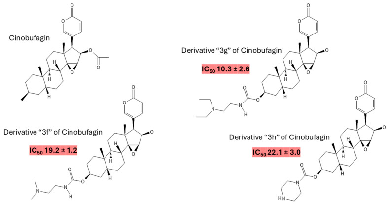
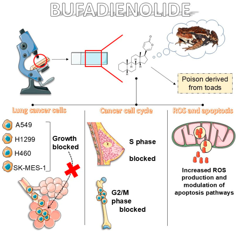
Cinobufagin (CB) (Figure 1), a bufadienolide found in traditional Chinese medicine [21] and derived from the parotid glands of toads [22], has recently garnered attention for its potential anticancer properties [23,24]. Bufadienolides are a class of steroidal compounds with documented biological activities [25], including anti-inflammatory [26], antiviral [27], and anticancer effects [28]. CB’s potential as an anticancer agent is supported by its ability to induce apoptosis [29], inhibit cell proliferation [30], and modulate vital molecular pathways involved in cancer progression [31]. CB derivatives also possess anticancer effectiveness against lung cancer cells, especially the carbamate derivatives [32] (Figure 1). Figure 2 illustrates the main anticancer effects of bufadienolides.
Figure 1.
Molecular structure of CB. CB is a bufadienolide with a molecular weight of 442.5 g/mol and an exact mass of 442.23553880 g/mol. The monoisotopic mass is the same, reflecting its precise atomic composition. Its lipophilicity is indicated by an XLogP3-AA value of 3.3, suggesting moderate solubility in lipids and fats. The compound has one hydrogen bond donor and six hydrogen bond acceptors. It features three rotatable bonds, which can contribute to its conformational flexibility. CB’s topological polar surface area (TPSA) is 85.4 Å2, which helps understand its interaction with polar environments. Additionally, CB has a heavy atom count of 32 and a complexity score of 923, reflecting its intricate structure. There are ten defined stereocenters, crucial for their three-dimensional orientation, while no undefined stereocenters or defined/undefined bond stereocenters exist. The compound is canonicalized, ensuring its chemical structure is represented in a standard format [33]. The figure also represents carbamate derivatives of CB with anticancer activity against A549 lung cancer cells alongside their respective IC50 values (nM) for 72 h [32].
Figure 2.
Main anticancer properties of bufadienolides.
CB’s ability to induce apoptosis in cancer cells is particularly noteworthy, as apoptosis is a critical mechanism for eliminating malignant cells and preventing tumor growth [34]. Additionally, CB influences cell cycle regulation, which can lead to cell cycle arrest [35] and inhibit the uncontrolled proliferation characteristic of cancer [36]. It also affects cancer cell migration [37] and invasion [24], indicating potential in reducing metastasis [38]. Furthermore, CB interacts with several crucial molecular pathways implicated in cancer. It modulates pathways such as LEF1 Lymphoid-enhancer-binding Factor 1 (LEF1) [39], Mitogen-activated Protein Kinase (MAPK) [40], Signal Transducer and Activator of Transcription 3 (STAT3) [41], Aurora Kinase A-Rapamycin Kinase–Eukaryotic Translation Initiation Factor 4E (AURKA-mTOR-eIF4E) [42], and Phosphoinositide 3-Kinase/Protein Kinase B (PI3K/Akt) [43]. These pathways play significant roles in regulating cell survival, growth, and metabolism, and CB’s impact on these pathways suggests a multifaceted approach to cancer treatment.
This manuscript provides a comprehensive review of CB’s anticancer effects, specifically in lung cancer, focusing on preclinical evidence due to the current absence of clinical studies. To our knowledge, it represents the first detailed analysis exclusively dedicated to CB’s impact across various types of lung cancer. Our review is critical because it offers an in-depth examination of CB’s effects using various preclinical models, enhancing the analysis’s breadth and depth. We assess CB’s influence on critical aspects such as lung cancer cell viability, apoptosis, migration, and molecular pathway modulation. This thorough examination aims to elucidate CB’s mechanisms of action and its potential as a therapeutic agent in lung cancer. In addition to presenting the current preclinical evidence, our manuscript will discuss the possible clinical implications of CB’s anticancer effects. We will also outline future research directions to advance understanding of CB’s role in cancer therapy. Moreover, we address the limitations of the included studies, providing a balanced perspective on the current evidence and identifying areas where further investigation is needed. Through this comprehensive approach, we seek to offer valuable insights that could guide future research and potentially inform clinical applications of CB in lung cancer therapy.
2. Literature Search Methodology
To investigate the efficacy and mechanisms of CB in preclinical models, we conducted a comprehensive literature search using several databases, including PubMed, Scopus, Web of Science, and Google Scholar. The search strategy incorporated keywords such as “CB”, “preclinical models”, “experimental studies”, “cancer”, “lung cancer”, and “metastasis”, alongside terms related to specific biological outcomes and processes like “apoptosis”, “cell proliferation”, “molecular signaling”, and “therapeutic efficacy”. The inclusion criteria were specifically tailored to focus on experimental studies involving CB and utilizing preclinical models of lung cancer. Studies were needed to present data on CB’s impact on lung cancer outcomes, relevant molecular mechanisms, and therapeutic potential. To ensure the relevance of the research, only studies that used experimental designs, reported transparent methodologies and results, and involved preclinical models of lung cancer were considered. Exclusion criteria included non-experimental papers, reviews, meta-analyses, and studies unrelated to CB or preclinical models. Studies published in languages other than English or those that did not meet the rigorous standards of experimental research were excluded. Two researchers (S.M.B. and L.F.L.) conducted data extraction using a standardized form to ensure consistency. Essential information collected included study design, details of CB administration (such as dosage and duration), outcomes measured, and any limitations reported. The quality of each study was assessed based on experimental design, sample size, and the clarity of result reporting, adhering to established guidelines for scientific quality. The data were synthesized qualitatively to summarize the effects of CB across various preclinical models, identify common findings, and discuss any limitations. This approach also sought to highlight gaps in the current knowledge and propose directions for further studies.
3. Literature Search Report
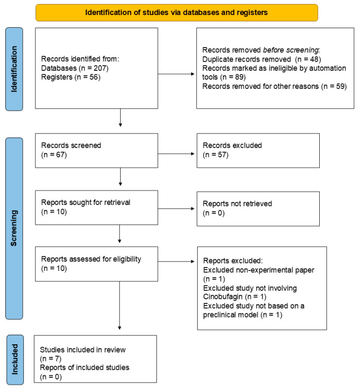
Records were sourced from multiple avenues to ensure comprehensive coverage of the topic. Specifically, 207 records were initially identified from various electronic databases, and an additional 56 records were obtained from research registers. Before screening, several records were removed to refine the dataset. A total of 48 duplicate records were eliminated to avoid redundancy. Furthermore, automation tools marked 89 records as ineligible based on predefined criteria, and 59 records were removed for other reasons, such as evident irrelevance to the research topic. After removing duplicates and ineligible records, 67 were screened based on their titles and abstracts. Of these, 57 records were excluded for not meeting the inclusion criteria. Ten reports were then sought for full-text retrieval to determine their suitability for inclusion in this review. All ten reports were successfully retrieved and assessed for eligibility. During this assessment, three reports were excluded: one was a non-experimental paper, one did not involve CB, and one was not based on a preclinical model. Ultimately, seven studies met all the inclusion criteria and were included in the review. This process ensured that the selected studies were relevant, experimental, focused on CB, utilized preclinical models, and were published in English, thus maintaining the rigor and quality of the review. Figure 3 provides a visual summary of the literature search process, illustrating the flow of records through the different stages of identification, screening, eligibility assessment, and inclusion.
Figure 3.
PRISMA flow diagram of the literature search process [44].
4. CB: Unveiling the Hidden Bufadienolide’s Promise in Combating Lung Cancer Development and Progression
Table 1 presents a comprehensive overview of various studies investigating the effects of CB on lung cancer models. The data include in vitro and in vivo experiments across multiple lung cancer cell lines and animal models, highlighting CB’s potential as a therapeutic agent. The findings showcase CB’s ability to reduce cell viability, induce apoptosis, and inhibit tumor growth and metastasis through various mechanisms, such as targeting specific apoptotic pathways and lipid raft components. Despite promising results, the limitations of each study—such as reliance on particular cell lines, limited in vivo validation, and challenges in translating results to clinical settings—underscore the need for further research. This comparative analysis aims to provide insights into CB’s therapeutic potential and guide future investigations toward effective and safe treatment strategies against lung cancer.
Table 1.
Exploring the efficacy and mechanisms of CB in lung cancer intervention and treatment: a comparative analysis of in vitro and in vivo studies.
| Model | Intervention | IC50 | Studies’ Outcomes | Studies’ Limitations | Possible Clinical Implications | Ref. |
|---|---|---|---|---|---|---|
| NSCLC cells (A549, H1299) and 16HBE cells | CB: 0.5, 1, and 2 µM | Not reported | CB significantly reduced viability in NSCLC cells; no significant effect on 16HBE cells. Increased apoptosis in NSCLC cells, dose-dependently; Bcl-2 levels decreased, Bax, cleaved caspase-3, and cleaved caspase-9 levels increased. Increased ROS levels in NSCLC cells. Redistributed cholesterol and sphingomyelin; reduced caveolin-1 and FLOT2 expression; inhibited p-Akt. | Results are based on in vitro models, which may not fully reflect in vivo tumor dynamics and immune interactions. Limited to two cell lines, which may not represent all NSCLC types. | CB could offer a novel treatment for NSCLC by targeting lipid rafts and caveolin-1, potentially overcoming drug resistance and enhancing therapy effectiveness. Clinical trials are needed for validation. | [45] |
| NSCLC cells (PC-9, H460) and mouse xenograft model | CB: 0.1, 0.2, and 0.5 µM for in vitro studies; 0.5–2 mg/kg/day via intraperitoneal injection for in vivo studies | 0.5624 μM and 0.04657 μM for PC-9 and H460 (NSCLC cells) | In vitro: CB significantly reduced cell viability, proliferation, and colony formation; induced apoptosis; inhibited migration and EMT. In vivo: CB significantly inhibited tumor growth in xenograft models; reduced phosphorylation of STAT3 and reversed IL-6-induced STAT3 nuclear translocation. No significant toxicity was observed. | Reliance on in vitro and a single xenograft model; the study does not address long-term safety or possible clinical applicability in humans. | CB effectively targets STAT3 signaling, showing promise as a therapeutic agent for NSCLC. Further clinical trials are necessary to confirm its efficacy and safety in human patients, and to explore potential in combination therapies. | [46] |
| H1299 NSCLC cell line | CB: 0.01, 0.04, and 0.16 µM | 0.035–0.008 μM for H1299 cells | CB inhibited H1299 cell proliferation. Significant antiproliferative activity was also observed in colony formation assays. CB significantly reduced DNA synthesis. CB significantly inhibited migration and invasion of H1299 cells. Concentration-dependent reduction in the expression of integrin α2, β-catenin, FAK, Src, c-Myc, and STAT3 was observed. | Limited to a single NSCLC cell line may not fully represent the heterogeneity of NSCLC. The study primarily focuses on in vitro assays, lacking in vivo validation. The long-term effects and safety profile of CB are not addressed. | CB shows potential as an effective agent against both the growth and metastasis of NSCLC by targeting critical molecular pathways involved in cell proliferation and migration. This suggests CB could be a valuable candidate for further development in NSCLC treatment strategies. | [47] |
| A549 NSCLC cell line, BEAS-2B normal cell line, mouse xenograft model | CB: 0–120 nM for in vitro studies; 10 mg/kg/day via intraperitoneal injection for in vivo studies | 25.15 ± 0.88 nM (24 h), 18.29 ± 1.28 μM (48 h), and 10.46 ± 1.79 nM (72 h) for A549 cells and 176.04 ± 31.50 nM (72 h) for BEAS-2B cells | In vitro: CB suppressed A549 cell proliferation in a dose- and time-dependent manner, with no cytotoxic effect on BEAS-2B cells. CB promoted apoptosis in A549 cells. CB significantly inhibited migration and invasion in A549 cells. Increased FOXO1 expression and decreased G9a expression in A549 cells. In vivo: CB inhibited A549 xenograft tumor growth, migration, and invasion while promoting apoptosis. | The study was limited to A549 cells and a single mouse model; BEAS-2B cells were used for cytotoxicity comparison but not for mechanistic insights. Mechanisms involving FOXO1 and G9a may not be universally applicable across all NSCLC subtypes. | CB could be a promising candidate for targeted cancer therapy in NSCLC by modulating FOXO1 and G9a. The findings highlight CB’s potential to inhibit NSCLC progression and suggest further investigation for its therapeutic application. | [48] |
| BEAS-2B, A549, and LLC cell lines, mouse xenograft model | CB-loaded PDA-based nanomedicine: 0–80 nM for in vitro studies; 1–2 mg/kg/day via intraperitoneal injection for in vivo studies | 61 nM for free CB and 32 nM for CB-loaded PDA-based nanomedicine in A549 cells; 74 nM for free CB and 39 nM for CB-loaded PDA-based nanomedicine in LLC cells | In vitro: The PDA nanomedicine demonstrated improved solubility and bioavailability of CB. Enhanced targeting of lung cancer cells via folic acid-receptor interaction was observed. In vivo: In a xenograft model, CB-loaded PDA nanomedicine inhibited tumor growth significantly compared to controls. | The study was conducted primarily in vitro and in animal models; the clinical translation of the nanomedicine’s effectiveness and safety needs further validation. Potential scalability issues and complex synthesis might affect practical application. | This targeted and responsive nanomedicine could enhance the effectiveness of CB in lung cancer therapy by improving its bioavailability and reducing side effects. The approach offers a promising strategy for delivering chemotherapeutic agents with precision and controlled release, potentially advancing treatment options for lung cancer. Further research and clinical trials must confirm its efficacy and safety in human subjects. | [49] |
| NSCLC cells (A549, NCI-H460, H1299, SK-MES-1, Calu-3) and BEAS-2B cells; nude mice xenograft model | CB: 0.5–4.0 μM for in vitro studies; 2.5–5.0 mg/kg/day via intraperitoneal injection for in vivo studies | Values ranging from 2.3 to 6.7 μM for A549, NCI-H460, H1299, SK-MES-1, and Calu-3; 22.3 μM for BEAS-2B | In vitro: CB significantly inhibited growth, migration, and invasion of A549 cells. CB-induced apoptosis through caspase activation and mitochondrial fragmentation. In vivo: CB demonstrated strong antitumor efficacy by inhibiting tumor growth and activating p53 phosphorylation. | The study results are based on various cell lines and animal models; further validation in clinical settings is needed. There are limited long-term effects and safety profile data. | CB shows potential as a selective and effective treatment for lung cancer, with promising in vivo antitumor activity. Further research is required to confirm clinical efficacy and safety. | [50] |
| NSCLC cell lines (A549, H1299, H460, and SK-MES-1) in vitro and NSG mice in vivo | CB: 0.6, 1.2, 2.5, 5, 10, and 20 μM for in vitro studies; 1.5, 5, and 10 mg/kg/day via intraperitoneal injection for in vivo studies | 2 μM for 40–50% inhibitive efficacy on the four cancer cells | In vitro: CB dose-dependently reduced viability in NSCLC cell lines effectively. CB induced significant cell cycle arrest at the G0/G1 phase and increased apoptosis in a dose- and time-dependent manner. Elevated ROS levels and decreased MMP were observed. In vivo: Significant inhibition of tumor growth in NSG mice was observed with CB, especially at 10 mg/kg/day. No significant cytotoxicity was observed in rat splenocytes. | In vitro models may not fully reflect in vivo tumor dynamics and immune interactions. The findings are based on only four NSCLC cell lines and one animal model, which may not accurately encompass all NSCLC types or predict human responses. The in vivo results are limited to a specific mouse model, and CB’s long-term effects and safety are not fully addressed. | CB demonstrates promising potential as a treatment for NSCLC by targeting specific apoptotic pathways, which could overcome drug resistance and improve therapeutic efficacy. Further clinical trials are necessary to validate these findings and assess the safety and effectiveness of CB in humans. | [51] |
Abbreviations: Bax, Bcl-2-associated X Protein; Bcl-2, B-cell lymphoma 2; CB, Cinobufagin; DNA, Deoxyribonucleic Acid; EMT, Epithelial–Mesenchymal Transition; FAK, Focal Adhesion Kinase; FLOT2, Flotillin-2; FOXO1, Forkhead box O1; G9a, Euchromatic Histone Lysine Methyltransferase 2; IL-6, Interleukin 6; MMP, Mitochondrial Transmembrane Potential; NSCLC, Non-Small-cell Lung Cancer; p53, Tumor Protein p53; p-Akt, Phosphorylated Protein Kinase B; PDA, polydopamine; ROS, Reactive Oxygen Species; Src, Proto-Oncogene Tyrosine-protein Kinase Src; STAT3, Signal Transducer and Activator of Transcription 3.
Xu et al. [45] explored the impact of CB on Non-Small-cell Lung Cancer (NSCLC) cell lines (A549, H1299) and the non-cancerous 16HBE cells. The study found that CB significantly reduced cell viability and induced apoptosis in NSCLC cells through mechanisms involving decreased B-cell lymphoma 2 (Bcl-2) levels and increased apoptotic markers such as Bcl-2-associated X Protein (Bax) and cleaved caspases. Additionally, CB affected lipid raft components by reducing caveolin-1 and Flotillin-2 (FLOT2) expression and inhibiting Protein Kinase B (Akt) phosphorylation. However, the study’s reliance on in vitro models and a limited number of cell lines might not entirely reflect in vivo tumor dynamics. Future research should address these limitations by incorporating more diverse NSCLC cell lines and utilizing advanced animal models. Furthermore, integrating immunological therapies, such as immune checkpoint inhibitors, with CB could enhance its therapeutic efficacy. Genetic studies could also explore the impact of CB on specific genetic mutations associated with NSCLC to tailor personalized treatment strategies.
Yan et al. [46] investigated CB’s effects in both in vitro NSCLC models (PC-9, H460) and an in vivo mouse xenograft model. The study demonstrated that CB effectively reduced cell viability, proliferation, and migration, inducing apoptosis and inhibiting tumor growth. However, the research is limited by its use of a single xenograft model and lack of long-term safety data. In order to build on these findings, future studies should explore the effects of CB in combination with existing chemotherapeutic agents and assess its potential in synergistic therapies. Additionally, incorporating radiotherapy alongside CB could provide a multi-modal approach to enhance overall treatment outcomes. Expanding research to include clinical trials will be essential to confirm CB’s efficacy and safety in diverse patient populations.
Sun et al. [47] focused on the H1299 NSCLC cell line, revealing that CB effectively inhibited cell proliferation, migration, and invasion with a low IC50 value. The study also identified CB’s impact on molecular pathways such as integrin α2, β-catenin, Focal Adhesion Kinase (FAK), and Proto-Oncogene Tyrosine-protein Kinase Src (Src), which are crucial for NSCLC metastasis. While the in vitro results are compelling, the study lacks in vivo validation and long-term safety data. Future research should address these gaps by incorporating diverse in vivo models and evaluating CB’s effects on tumor microenvironments. Additionally, exploring CB’s role in combination with targeted therapies and immunotherapies could provide a comprehensive approach to tackling NSCLC. Investigating genetic variations and their interaction with CB might offer insights into personalized treatment options.
Zhang et al. [48] evaluated CB’s effects on the A549 NSCLC cell line, the BEAS-2B standard cell line, and a mouse xenograft model. The study found that CB suppressed A549 cell proliferation in a dose- and time-dependent manner with no cytotoxic effect on BEAS-2B cells. CB promoted apoptosis in A549 cells, significantly inhibited migration and invasion, and modulated molecular pathways by increasing Forkhead box O1 (FOXO1) and decreasing Euchromatic Histone Lysine Methyltransferase 2 (G9a) expression. In vivo, CB inhibited A549 xenograft tumor growth, migration, and invasion while promoting apoptosis. However, the study was limited to A549 cells and a single mouse model, and BEAS-2B cells were used for cytotoxicity comparison but not for mechanistic insights. The mechanisms involving FOXO1 and G9a may not be universally applicable across all NSCLC subtypes. Future research should further explore CB’s potential as a targeted cancer therapy by examining its effects on other NSCLC subtypes and investigating the broader applicability of the FOXO1 and G9a pathways.
Li et al. [49] examined CB-loaded polydopamine (PDA) nanomedicine, which enhanced the solubility and targeting of CB in lung cancer cells. The study showed that this nanomedicine significantly inhibited tumor growth in xenograft models compared to controls. However, the clinical applicability remains uncertain due to the study’s focus on in vitro and animal models and potential scalability issues. Future research should prioritize scaling up the nanomedicine for clinical trials, focusing on its safety and efficacy in human subjects. Additionally, combining this nanomedicine approach with other therapeutic modalities, such as targeted gene therapies and immunological agents, could enhance its effectiveness. Investigating the potential for CB-loaded nanomedicines to improve the delivery and efficacy of existing chemotherapies and radiotherapies should also be a priority.
Peng et al. [50] conducted a comprehensive study on CB, evaluating its effects on various lung cancer cell lines and normal bronchial epithelial cells and its in vivo antitumor efficacy. Their findings revealed that CB exhibits substantial anticancer activity. CB effectively inhibited cell growth, migration, and invasion and induced apoptosis through caspase activation and mitochondrial fragmentation. The study also highlighted that CB significantly increased Reactive Oxygen Species (ROS) production in cancer cells, with ROS scavengers reducing its apoptotic effects, underscoring the role of oxidative stress in CB’s mechanism of action. In vivo, CB demonstrated strong antitumor efficacy by inhibiting tumor growth and activating Tumor Protein p53 (p53) phosphorylation in a nude mouse xenograft model. Despite these promising results, the study’s limitations include using a single animal model and focusing on specific cell lines, which may not fully represent the heterogeneity of NSCLC. Future research should aim to validate these findings across different NSCLC subtypes and incorporate long-term safety assessments. Additionally, exploring CB’s potential with other therapeutic modalities, such as targeted therapies and immunotherapies, could provide a more comprehensive treatment strategy for lung cancer.
Zhang et al. [51] evaluated CB’s effects on multiple NSCLC cell lines (A549, H1299, H460, SK-MES-1) and in NSG mice. The study found that CB effectively induced cell cycle arrest, increased apoptosis, and significantly inhibited tumor growth. However, the study’s limitations include using a single mouse model and a restricted range of cell lines, which may not fully represent NSCLC heterogeneity. Future research should include a broader array of preclinical models to capture the heterogeneity of NSCLC and evaluate CB in combination with existing treatments such as chemotherapy and targeted therapies. Investigating the potential of CB to enhance responses to immunotherapies and radiotherapies could provide a more comprehensive treatment strategy. Genetic studies could also explore the impact of CB on various genetic mutations and pathways involved in NSCLC, leading to more personalized treatment approaches.
In summary, the findings across these studies highlight CB’s potential as a novel treatment for lung cancer through its effects on apoptosis, signaling pathways, and tumor growth inhibition. To translate these results into effective clinical therapies, future research should address the limitations of current studies by incorporating diverse preclinical models, investigating synergistic therapies, and conducting comprehensive clinical trials. Emphasizing immunological therapies, genetic studies, and the integration of CB with existing and emerging treatment modalities will be crucial in advancing CB’s therapeutic potential and improving outcomes for lung cancer patients.
CB’s toxicity has garnered significant attention and concern regarding its safety as a potential drug candidate. However, little evidence is investigating this compound’s toxicological and pharmacokinetic characteristics. Wei et al. reported CB’s pharmacokinetics in rats by designing a method for rapidly and accurately determining CB’s blood concentration after intragastric administration of 20 mg/kg in rats. The maximum concentration was 45.83 ± 4.56 ng/mL, the time for maximum concentration was 0.083 ± 0 h, and the half-life was 2.79 ± 0.93 h. Although these authors did not evaluate the toxicological aspects of CB against normal, healthy cells, these results indicate that CB was rapidly absorbed and eliminated [52,53]. In a separate study, Baek et al. demonstrated that treatment with 0.5 and 1 μM of CB for 24 h promoted higher cytotoxicity against human multiple myeloma cells than in peripheral blood mononuclear cells, inhibiting cancer cell viability but not normal cells [40]. Similar results were found in Yin et al.’s study [54]. These researchers reported that CB exerted cytotoxicity against cultured osteosarcoma cells but less or no toxicity to human osteoblast cell lineage. These cells were treated with CB solutions for 12, 24, and 48 h.
Unlike other chemotherapeutic agents that demonstrate cytotoxicity against normal, healthy cells, CB does not induce oxidative Deoxyribonucleic Acid (DNA) damage and cytotoxicity against healthy cells at sublethal doses. Despite conventional DNA-damaging chemotherapies that target all cells, CB explores a cancer vulnerability, a common feature of most malignant transformed cells, which may be a metabolic vulnerability. It is well documented that cancer cells present additional sensitivity to oxidative insult due to high intrinsic oxidative pressure and diminished spare antioxidant capacity. However, cancerous lineages may survive toxic attacks by upregulating DNA repair to maintain genome functions, and CB-induced oxidative damage leads to replication stress and DNA damage response activation [55,56,57]. Combining or conjugating nontoxic doses of CB with chemotherapeutic agents that target DNA response to genotoxic attacks may be a potential avenue for future research endeavors for enhancing CB’s anticancer efficacy and reducing the potential side effects of synthetic chemotherapy drugs since lower doses of synthetic medications may be necessary due to the synergism with CB. Another point that must be raised is that CB can trigger anaphylactoid reactions by binding to the immunoglobulin E (IgE) receptor (IgE-R) and releasing β-hexosaminidase and histamine by this trigger [58]. Research has also shown that CB can interfere with the immune system, ultimately modulating the inflammatory phenotype of immune cells [59].
5. Conclusions and Future Research Directions
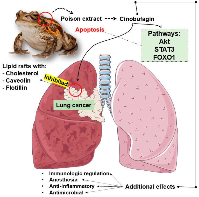
This manuscript has explored CB’s promising therapeutic potential in lung cancer treatment (Figure 4). Through various studies, CB has been shown to exert significant anticancer effects by inducing apoptosis, inhibiting tumor growth, and modulating vital molecular pathways in lung cancer progression. Despite these promising results, several critical areas warrant further investigation to fully realize CB’s potential as a treatment for lung cancer.
Figure 4.
CB’s promising therapeutic potential in lung cancer treatment. Akt, Protein Kinase B; FOXO1, Forkhead box O1; STAT3, Signal Transducer and Activator of Transcription 3.
Exploring CB’s impact on the tumor microenvironment (TME) is also a crucial direction for future research. Current studies have demonstrated CB’s direct cytotoxic effects on lung cancer cells, yet understanding how CB interacts with the TME could offer new insights into its overall therapeutic efficacy. The TME, which includes various cell types, extracellular matrix components, and soluble factors, significantly influences tumor progression and therapeutic resistance. Future research should investigate how CB affects immune cell infiltration, cytokine profiles, and interactions between tumor cells and stromal components. Such studies could pave the way for integrating CB with immunotherapies or other treatments to reshape the TME to enhance therapeutic outcomes.
Additionally, evaluating the long-term effects of CB and the potential development of resistance is crucial. Initial findings highlight CB’s effectiveness, but understanding how prolonged exposure might lead to resistance is essential. Research should focus on identifying molecular pathways involved in resistance and exploring genetic and epigenetic changes in lung cancer cells. This knowledge could guide the development of strategies to overcome resistance, potentially involving combination therapies or alternating regimens to maintain CB’s efficacy over time.
Another critical area for future research is assessing CB’s role in metastatic lung cancer. While current studies indicate CB’s effectiveness in inhibiting primary tumor growth, its effects on metastasis have not been thoroughly examined. Future investigations should explore how CB influences the tumor cell migration, invasion, and colonization of distant organs. Evaluating CB’s impact on metastatic pathways and circulating tumor cells could provide valuable insights into its potential as a treatment for metastatic disease. Combining CB with therapies targeting metastatic progression could further enhance its efficacy and improve patient outcomes.
Exploring the synergistic effects of CB with radiotherapy presents a promising research avenue. Radiotherapy is a standard treatment for lung cancer, and integrating CB could offer complementary benefits. Future studies should explore how CB interacts with radiation, including its impact on radiation-induced DNA damage repair and tumor cell apoptosis. Identifying optimal dosing schedules and treatment combinations could help maximize CB’s and radiotherapy’s therapeutic benefits, leading to more effective treatment regimens.
Developing advanced delivery systems for CB could also significantly enhance its therapeutic potential [60]. While initial studies have explored CB-loaded nanomedicines, future research should focus on refining these delivery methods to improve targeting accuracy and minimize off-target effects. Innovative delivery systems, such as ligand-targeted nanoparticles or encapsulation techniques, could enhance CB’s bioavailability and distribution within tumors. In targeted therapies, antibody–drug conjugate (ADC) strategies have been named the “biological missile” to target cancerous lineages effectively. ADC’s strategy is to combine a monoclonal antibody with a cytotoxic drug by a chemical linker. This results in advantages for precise targeting ability and potent killing effect, achieving an accurate and efficient elimination of cancerous cells [61]. Combining CB with monoclonal antibodies to target cancer lineages may be a potential avenue for future research endeavors since ADC is the leading targeted cancer therapy. Additionally, investigating patient-specific factors affecting drug delivery could lead to more personalized treatment approaches, optimizing CB’s benefits for individual patients.
Moreover, incorporating principles of green health into future research on CB can align therapeutic advancements with environmental sustainability. Green health emphasizes the development of eco-friendly and sustainable technologies, which is increasingly important in modern drug development. Research into environmentally responsible manufacturing processes for CB and its delivery systems could reduce the ecological footprint of cancer therapies. For instance, utilizing biodegradable materials in drug delivery systems or developing less toxic synthesis routes for CB could contribute to greener healthcare practices. This approach not only promotes the development of effective treatments but also supports broader goals of environmental stewardship and public health [62,63,64,65].
Finally, mitigating the potential toxicity of CB to leverage its therapeutic benefits for lung cancer treatment is entirely warranted. While CB demonstrates significant anticancer activity, its safety profile remains a critical concern. Investigating the mechanisms underlying CB’s toxicity and developing strategies to minimize adverse effects is essential. This could involve identifying biomarkers for early detection of toxicity, optimizing dosing regimens, and employing drug delivery systems that target tumors more precisely while reducing systemic exposure. Additionally, exploring combination therapies that enhance CB’s efficacy while mitigating its side effects could improve its safety and therapeutic index. Addressing these toxicity concerns through rigorous preclinical and clinical studies will be crucial for advancing CB from promising research findings to a viable and safe treatment option for lung cancer patients.
Exploring these research directions will be crucial in advancing CB as a therapeutic agent for lung cancer. By addressing these areas, we can potentially enhance treatment efficacy, overcome resistance, and improve outcomes for patients with lung cancer. Additionally, integrating green health principles into research and development efforts will ensure that advancements in cancer therapy are aligned with sustainable and environmentally friendly practices, promoting a holistic approach to cancer treatment and ecological responsibility. Concerning mechanisms of action, CB targeting lung cancer appears to involve the upstream or downstream of molecular pathways associated with cancer cell division, death, and metabolism, although these pathways are not seen to be modulated in all models concurrently. Table 2 demonstrates the genetic targets of CB to facilitate the presumptive assumptions of mechanisms of action’s interactions.
Table 2.
Genetic targets of CB against cancer cell lines.
| RELA | GSK3B | CCT3 | H2AX | CYP3A4 | ATAD5 |
| RPL6 | BHLHE40 | CDK2 | HSP90AB1 | DCTPP1 | FLOT2 |
| RRP9 | BIRC2 | CDK2 | JUN | EGFR | ODC1 |
| RUNX1 | BIRC5 | CDK9 | JUNB | FOS | PARP1 |
| SAT1 | CCNA2 | CDKN1A | KLF10 | GRWD1 | PCNA |
| TFB2M | TIMP2 | TGFB1 | MYC | TNF | TGFBR1 |
| TYMS | FOXO1 | STAT3 | FAK | G9a | SRC |
Acknowledgments
We would like to express our sincere gratitude to Servier Medical Art for providing the medical figures used to produce the images for this article. Their high-quality illustrations significantly enhanced the clarity and visual appeal of our work. Servier Medical Art is licensed under CC BY 4.0, allowing for their use with proper attribution. We appreciate their support and contribution to the advancement of scientific communication.
Author Contributions
S.M.B. and L.F.L. contributed to the conceptualization, methodology, validation, formal analysis, investigation, resources, data curation, writing—original draft preparation, writing—review and editing, visualization, supervision, project administration, and funding acquisition. K.T.P. contributed to the methodology, investigation, and writing—review and editing. E.P.d.L. contributed to the formal analysis, data curation, and writing—review and editing. J.d.S.C.O. contributed to the investigation and writing—review and editing. B.L.B. contributed to the methodology, validation, formal analysis, and writing—review and editing. A.C.A. contributed to the investigation and writing—review and editing. E.L.G. contributed to the resources, investigation, and writing—review and editing. R.E.G.R. contributed to the methodology, investigation, and writing—review and editing. D.A.M. contributed to the resources, investigation, and writing—review and editing. J.F.d.S.H. contributed to the methodology, validation, formal analysis, and writing—review and editing. V.M.C.S.C. contributed to the methodology, validation, formal analysis, and writing—review and editing. P.C.d.S.B. contributed to the methodology, validation, formal analysis, and writing—review and editing. E.d.S.B.M.P. contributed to the methodology, validation, formal analysis, and writing—review and editing. R.d.A.G. contributed to the methodology, validation, formal analysis, and writing—review and editing. All authors have read and agreed to the published version of the manuscript.
Data Availability Statement
No new data were created or analyzed in this study. Data sharing does not apply to this article.
Conflicts of Interest
The authors declare no conflicts of interest.
Funding Statement
This research received no external funding.
Footnotes
Disclaimer/Publisher’s Note: The statements, opinions and data contained in all publications are solely those of the individual author(s) and contributor(s) and not of MDPI and/or the editor(s). MDPI and/or the editor(s) disclaim responsibility for any injury to people or property resulting from any ideas, methods, instructions or products referred to in the content.
References
- 1.Sung H., Ferlay J., Siegel R.L., Laversanne M., Soerjomataram I., Jemal A., Bray F. Global Cancer Statistics 2020: GLOBOCAN Estimates of Incidence and Mortality Worldwide for 36 Cancers in 185 Countries. CA Cancer J. Clin. 2021;71:209–249. doi: 10.3322/caac.21660. [DOI] [PubMed] [Google Scholar]
- 2.Laurindo L.F., de Lima E.P., Laurindo L.F., Rodrigues V.D., Chagas E.F.B., de Alvares Goulart R., Araújo A.C., Guiguer E.L., Pomini K.T., Rici R.E.G., et al. The therapeutic potential of bee venom-derived Apamin and Melittin conjugates in cancer treatment: A systematic review. Pharmacol. Res. 2024;209:107430. doi: 10.1016/j.phrs.2024.107430. [DOI] [PubMed] [Google Scholar]
- 3.Brown J.S., Amend S.R., Austin R.H., Gatenby R.A., Hammarlund E.U., Pienta K.J. Updating the Definition of Cancer. Mol. Cancer Res. 2023;21:1142–1147. doi: 10.1158/1541-7786.MCR-23-0411. [DOI] [PMC free article] [PubMed] [Google Scholar]
- 4.El-Tanani M., Rabbani S.A., Babiker R., Rangraze I., Kapre S., Palakurthi S.S., Alnuqaydan A.M., Aljabali A.A., Rizzo M., El-Tanani Y., et al. Unraveling the tumor microenvironment: Insights into cancer metastasis and therapeutic strategies. Cancer Lett. 2024;591:216894. doi: 10.1016/j.canlet.2024.216894. [DOI] [PubMed] [Google Scholar]
- 5.Huang Y.F., Chiao M.T., Hsiao T.H., Zhan Y.X., Chen T.Y., Lee C.H., Liu S.Y., Liao C.H., Cheng W.Y., Yen C.M., et al. Genetic mutation patterns among glioblastoma patients in the Taiwanese population—Insights from a single institution retrospective study. Cancer Gene Ther. 2024;31:894–903. doi: 10.1038/s41417-024-00746-y. [DOI] [PMC free article] [PubMed] [Google Scholar]
- 6.Gotoh O., Sugiyama Y., Tonooka A., Kosugi M., Kitaura S., Minegishi R., Sano M., Amino S., Furuya R., Tanaka N., et al. Genetic and epigenetic alterations in precursor lesions of endometrial endometrioid carcinoma. J. Pathol. 2024;263:275–287. doi: 10.1002/path.6278. [DOI] [PubMed] [Google Scholar]
- 7.Iqbal M.J., Kabeer A., Abbas Z., Siddiqui H.A., Calina D., Sharifi-Rad J., Cho W.C. Interplay of oxidative stress, cellular communication and signaling pathways in cancer. Cell Commun. Signal. 2024;22:7. doi: 10.1186/s12964-023-01398-5. [DOI] [PMC free article] [PubMed] [Google Scholar]
- 8.Salvadores M., Supek F. Cell cycle gene alterations associate with a redistribution of mutation risk across chromosomal domains in human cancers. Nat. Cancer. 2024;5:330–346. doi: 10.1038/s43018-023-00707-8. [DOI] [PubMed] [Google Scholar]
- 9.Gholamzad A., Khakpour N., Khosroshahi E.M., Asadi S., Koohpar Z.K., Matinahmadi A., Jebali A., Rashidi M., Hashemi M., Sadi F.H., et al. Cancer stem cells: The important role of CD markers, Signaling pathways, and MicroRNAs. Pathol. Res. Pract. 2024;256:155227. doi: 10.1016/j.prp.2024.155227. [DOI] [PubMed] [Google Scholar]
- 10.O’Leary B., Skinner H., Schoenfeld J.D., Licitra L., Le Tourneau C., Esdar C., Schroeder A., Salmio S., Psyrri A. Evasion of apoptosis and treatment resistance in squamous cell carcinoma of the head and neck. Cancer Treat. Rev. 2024;129:102773. doi: 10.1016/j.ctrv.2024.102773. [DOI] [PubMed] [Google Scholar]
- 11.Swanton C., Bernard E., Abbosh C., André F., Auwerx J., Balmain A., Bar-Sagi D., Bernards R., Bullman S., DeGregori J., et al. Embracing cancer complexity: Hallmarks of systemic disease. Cell. 2024;187:1589–1616. doi: 10.1016/j.cell.2024.02.009. [DOI] [PubMed] [Google Scholar]
- 12.Liu J., Cundy T.P., Woon D.T.S., Lawrentschuk N. A Systematic Review on Artificial Intelligence Evaluating Metastatic Prostatic Cancer and Lymph Nodes on PSMA PET Scans. Cancers. 2024;16:486. doi: 10.3390/cancers16030486. [DOI] [PMC free article] [PubMed] [Google Scholar]
- 13.Laurindo L.F., Direito R., Bueno Otoboni A.M.M., Goulart R.A., Quesada K., Barbalho S.M. Grape Processing Waste: Effects on Inflammatory Bowel Disease and Colorectal Cancer. Food Rev. Int. 2024;40:336–369. doi: 10.1080/87559129.2023.2168281. [DOI] [Google Scholar]
- 14.Li C.I., Rogers S.C., Bult C.J., Guerra C.E., Talton A., Williams L.B., Law W. Executing plans to enhance diversity across cancer centers in the United States: Opportunities and challenges. J. Natl. Cancer Inst. 2024;116:1198–1205. doi: 10.1093/jnci/djae100. [DOI] [PMC free article] [PubMed] [Google Scholar]
- 15.França G.S., Baron M., King B.R., Bossowski J.P., Bjornberg A., Pour M., Rao A., Patel A.S., Misirlioglu S., Barkley D., et al. Cellular adaptation to cancer therapy along a resistance continuum. Nature. 2024;631:876–883. doi: 10.1038/s41586-024-07690-9. [DOI] [PubMed] [Google Scholar]
- 16.Laurindo L.F., Sosin A.F., Lamas C.B., de Alvares Goulart R., Dos Santos Haber J.F., Detregiachi C.R.P., Barbalho S.M. Exploring the logic and conducting a comprehensive evaluation of AdipoRon-based adiponectin replacement therapy against hormone-related cancers—A systematic review. Naunyn Schmiedebergs Arch. Pharmacol. 2024;397:2067–2082. doi: 10.1007/s00210-023-02792-z. [DOI] [PubMed] [Google Scholar]
- 17.Khan S.U., Fatima K., Aisha S., Malik F. Unveiling the mechanisms and challenges of cancer drug resistance. Cell Commun. Signal. 2024;22:109. doi: 10.1186/s12964-023-01302-1. [DOI] [PMC free article] [PubMed] [Google Scholar]
- 18.Katta B., Vijayakumar C., Dutta S., Dubashi B., Nelamangala Ramakrishnaiah V.P. The Incidence and Severity of Patient-Reported Side Effects of Chemotherapy in Routine Clinical Care: A Prospective Observational Study. Cureus. 2023;15:e38301. doi: 10.7759/cureus.38301. [DOI] [PMC free article] [PubMed] [Google Scholar]
- 19.Camerini A., Mazzoni F., Scotti V., Tibaldi C., Sbrana A., Calabrò L., Caliman E., Ciccone L.P., Bernardini L., Graziani J., et al. Efficacy and Safety of Chemotherapy after Immunotherapy in Patients with Advanced Non-Small-Cell Lung Cancer. J. Clin. Med. 2024;13:3642. doi: 10.3390/jcm13133642. [DOI] [PMC free article] [PubMed] [Google Scholar]
- 20.Laurindo L.F., de Maio M.C., Minniti G., de Góes Corrêa N., Barbalho S.M., Quesada K., Guiguer E.L., Sloan K.P., Detregiachi C.R.P., Araújo A.C., et al. Effects of Medicinal Plants and Phytochemicals in Nrf2 Pathways during Inflammatory Bowel Diseases and Related Colorectal Cancer: A Comprehensive Review. Metabolites. 2023;13:243. doi: 10.3390/metabo13020243. [DOI] [PMC free article] [PubMed] [Google Scholar]
- 21.Niu T., Zhao L., Lin X., Cai Y., Chen S., Wang M., Zhou L., Ding H., Yu X., Qin G. Cinobufagin, a bufadienolide from traditional Chinese medicine Bufo bufo gargarizans CANTOR, inhibits PC3 cell growth in vitro and in vivo. J. Tradit. Chin. Med. Sci. 2019;6:175–183. doi: 10.1016/j.jtcms.2019.03.005. [DOI] [Google Scholar]
- 22.Kowalski K., Marciniak P., Rychlik L. Proteins from toad’s parotoid macroglands: Do they play a role in gland functioning and chemical defence? Front. Zool. 2023;20:21. doi: 10.1186/s12983-023-00499-8. [DOI] [PMC free article] [PubMed] [Google Scholar]
- 23.AmeliMojarad M., AmeliMojarad M., Shariati P. Cinobufagin treatments suppress tumor growth by enhancing the expression of cuproptosis-related genes in liver cancer. Naunyn Schmiedebergs Arch. Pharmacol. 2024 doi: 10.1007/s00210-024-03349-4. [DOI] [PubMed] [Google Scholar]
- 24.Fang S., Peng L., Zhang M., Hou R., Deng X., Li X., Xin J., Peng L., Liu Z., Liu Y., et al. MiR-2110 induced by chemically synthesized cinobufagin functions as a tumor-metastatic suppressor via targeting FGFR1 to reduce PTEN ubiquitination degradation in nasopharyngeal carcinoma. Environ. Toxicol. 2024;39:3548–3562. doi: 10.1002/tox.24197. [DOI] [PubMed] [Google Scholar]
- 25.Silva E., Soares-da-Silva P. Chapter two—New Insights into the Regulation of Na+,K+-ATPase by Ouabain. In: Jeon K.W., editor. International Review of Cell and Molecular Biology. Volume 294. Academic Press; Cambridge, MA, USA: 2012. pp. 99–132. [DOI] [PubMed] [Google Scholar]
- 26.Zou D., Wang Q., Chen T., Sang D., Yang T., Wang Y., Gao M., He F., Li Y., He L., et al. Bufadienolides originated from toad source and their anti-inflammatory activity. Front. Pharmacol. 2022;13:1044027. doi: 10.3389/fphar.2022.1044027. [DOI] [PMC free article] [PubMed] [Google Scholar]
- 27.El-Seedi H.R., Yosri N., El-Aarag B., Mahmoud S.H., Zayed A., Du M., Saeed A., Musharraf S.G., El-Garawani I.M., Habib M.R., et al. Chemistry and the Potential Antiviral, Anticancer, and Anti-Inflammatory Activities of Cardiotonic Steroids Derived from Toads. Molecules. 2022;27:6586. doi: 10.3390/molecules27196586. [DOI] [PMC free article] [PubMed] [Google Scholar]
- 28.Deng L.J., Li Y., Qi M., Liu J.S., Wang S., Hu L.J., Lei Y.H., Jiang R.W., Chen W.M., Qi Q., et al. Molecular mechanisms of bufadienolides and their novel strategies for cancer treatment. Eur. J. Pharmacol. 2020;887:173379. doi: 10.1016/j.ejphar.2020.173379. [DOI] [PubMed] [Google Scholar]
- 29.Dai G., Zheng D., Guo W., Yang J., Cheng A.Y. Cinobufagin Induces Apoptosis in Osteosarcoma Cells Via the Mitochondria-Mediated Apoptotic Pathway. Cell Physiol. Biochem. 2018;46:1134–1147. doi: 10.1159/000488842. [DOI] [PubMed] [Google Scholar]
- 30.Hirasaki Y., Okabe A., Fukuyo M., Rahmutulla B., Mano Y., Seki M., Hoshii T., Namiki T., Kaneda A. Cinobufagin inhibits proliferation of acute myeloid leukaemia cells by repressing c-Myc pathway-associated genes. Chem. Biol. Interact. 2022;360:109936. doi: 10.1016/j.cbi.2022.109936. [DOI] [PubMed] [Google Scholar]
- 31.Qi Y., Wu H., Zhu T., Liu Z., Liu C., Yan C., Wu Z., Xu Y., Bai Y., Yang L., et al. Acetyl-cinobufagin suppresses triple-negative breast cancer progression by inhibiting the STAT3 pathway. Aging. 2023;15:8258–8274. doi: 10.18632/aging.204967. [DOI] [PMC free article] [PubMed] [Google Scholar]
- 32.Zhou Y., Hou J., Long H., Zhang Z., Lei M., Wu W. Design, synthesis and anti-tumor activities of carbamate derivatives of cinobufagin. Steroids. 2020;164:108749. doi: 10.1016/j.steroids.2020.108749. [DOI] [PubMed] [Google Scholar]
- 33.PubChem Bethesda (MD): National Library of Medicine (US), National Center for Biotechnology Information. 2004. PubChem Compound Summary for CID 11969542, Cinobufagin. [(accessed on 12 August 2024)]; Available online: https://pubchem.ncbi.nlm.nih.gov/compound/Cinobufagin.
- 34.Carneiro B.A., El-Deiry W.S. Targeting apoptosis in cancer therapy. Nat. Rev. Clin. Oncol. 2020;17:395–417. doi: 10.1038/s41571-020-0341-y. [DOI] [PMC free article] [PubMed] [Google Scholar]
- 35.Pan Z., Luo Y., Xia Y., Zhang X., Qin Y., Liu W., Li M., Liu X., Zheng Q., Li D. Cinobufagin induces cell cycle arrest at the S phase and promotes apoptosis in nasopharyngeal carcinoma cells. Biomed. Pharmacother. 2020;122:109763. doi: 10.1016/j.biopha.2019.109763. [DOI] [PubMed] [Google Scholar]
- 36.Afifi M.M., Crncec A., Cornwell J.A., Cataisson C., Paul D., Ghorab L.M., Hernandez M.O., Wong M., Kedei N., Cappell S.D. Irreversible cell cycle exit associated with senescence is mediated by constitutive MYC degradation. Cell Rep. 2023;42:113079. doi: 10.1016/j.celrep.2023.113079. [DOI] [PMC free article] [PubMed] [Google Scholar]
- 37.Jo S., Yang E., Lee Y., Jeon D., Namkung W. Cinobufagin Exerts Anticancer Activity in Oral Squamous Cell Carcinoma Cells through Downregulation of ANO1. Int. J. Mol. Sci. 2021;22:12037. doi: 10.3390/ijms222112037. [DOI] [PMC free article] [PubMed] [Google Scholar]
- 38.Zhu Z., Wang H., Qian X., Xue M., Sun A., Yin Y., Tang J., Zhang J. Inhibitory Impact Of Cinobufagin In Triple-Negative Breast Cancer Metastasis: Involvements Of Macrophage Reprogramming Through Upregulated MME and Inactivated FAK/STAT3 Signaling. Clin. Breast Cancer. 2024;24:e244–e257.e1. doi: 10.1016/j.clbc.2024.01.014. [DOI] [PubMed] [Google Scholar]
- 39.Kim G.H., Fang X.Q., Lim W.J., Park J., Kang T.B., Kim J.H., Lim J.H. Cinobufagin Suppresses Melanoma Cell Growth by Inhibiting LEF1. Int. J. Mol. Sci. 2020;21:6706. doi: 10.3390/ijms21186706. [DOI] [PMC free article] [PubMed] [Google Scholar]
- 40.Baek S.H., Kim C., Lee J.H., Nam D., Lee J., Lee S.G., Chung W.S., Jang H.J., Kim S.H., Ahn K.S. Cinobufagin exerts anti-proliferative and pro-apoptotic effects through the modulation ROS-mediated MAPKs signaling pathway. Immunopharmacol. Immunotoxicol. 2015;37:265–273. doi: 10.3109/08923973.2015.1027916. [DOI] [PubMed] [Google Scholar]
- 41.Bai Y., Wang X., Cai M., Ma C., Xiang Y., Hu W., Zhou B., Zhao C., Dai X., Li X., et al. Cinobufagin suppresses colorectal cancer growth via STAT3 pathway inhibition. Am. J. Cancer Res. 2021;11:200–214. [PMC free article] [PubMed] [Google Scholar]
- 42.Jin X., Wang J., Zou S., Xu R., Cao J., Zhang Y., Guo J., Wen X., Deng S., Zheng Y., et al. Cinobufagin Triggers Defects in Spindle Formation and Cap-Dependent Translation in Liver Cancer Cells by Inhibiting the AURKA-mTOR-eIF4E Axis. Am. J. Chin. Med. 2020;48:651–678. doi: 10.1142/S0192415X20500330. [DOI] [PubMed] [Google Scholar]
- 43.Xu Z., Bao J., Jin X., Li H., Fan K., Wu Z., Yao M., Zhang Y., Liu G., Wang D., et al. The Effects of Cinobufagin on Hepatocellular Carcinoma Cells Enhanced by MRT68921, an Autophagy Inhibitor. Am. J. Chin. Med. 2023;51:1595–1611. doi: 10.1142/S0192415X23500726. [DOI] [PubMed] [Google Scholar]
- 44.Page M.J., McKenzie J.E., Bossuyt P.M., Boutron I., Hoffmann T.C., Mulrow C.D., Shamseer L., Tetzlaff J.M., Akl E.A., Brennan S.E., et al. The PRISMA 2020 statement: An updated guideline for reporting systematic reviews. BMJ. 2021;372:n71. doi: 10.1136/bmj.n71. [DOI] [PMC free article] [PubMed] [Google Scholar]
- 45.Xu Z., Li J., Fang S., Lian M., Zhang C., Lu J., Sheng K. Cinobufagin disrupts the stability of lipid rafts by inhibiting the expression of caveolin-1 to promote non-small cell lung cancer cell apoptosis. Arch. Med. Sci. 2024;20:887–908. doi: 10.5114/aoms/174578. [DOI] [PMC free article] [PubMed] [Google Scholar]
- 46.Yan S., Ma C., Zhou F., Zheng H., Yang L., Xiao Z., Zhu J., Zhao H., Zhao C., Xu X. Cinobufagin exerts an antitumor effect in non-small-cell lung cancer by blocking STAT3 signaling. J. Cancer. 2023;14:3309–3320. doi: 10.7150/jca.86544. [DOI] [PMC free article] [PubMed] [Google Scholar]
- 47.Sun M., Huang D., Liu Y., Chen H., Yu H., Zhang G., Chen Q., Chen H., Zhang J. Effects of Cinobufagin on the Proliferation, Migration, and Invasion of H1299 Lung Cancer Cells. Chem. Biodivers. 2023;20:e202200961. doi: 10.1002/cbdv.202200961. [DOI] [PubMed] [Google Scholar]
- 48.Zhang L., Liang B., Xu H., Gong Y., Hu W., Jin Z., Wu X., Chen X., Li M., Shi L., et al. Cinobufagin induces FOXO1-regulated apoptosis, proliferation, migration, and invasion by inhibiting G9a in non-small-cell lung cancer A549 cells. J. Ethnopharmacol. 2022;291:115095. doi: 10.1016/j.jep.2022.115095. [DOI] [PubMed] [Google Scholar]
- 49.Li J., Zhang Z., Deng H., Zheng Z. Cinobufagin-Loaded and Folic Acid-Modified Polydopamine Nanomedicine Combined With Photothermal Therapy for the Treatment of Lung Cancer. Front. Chem. 2021;9:637754. doi: 10.3389/fchem.2021.637754. [DOI] [PMC free article] [PubMed] [Google Scholar]
- 50.Peng P., Lv J., Cai C., Lin S., Zhuo E., Wang S. Cinobufagin, a bufadienolide, activates ROS-mediated pathways to trigger human lung cancer cell apoptosis in vivo. RSC Adv. 2017;7:25175–25181. doi: 10.1039/C7RA01085K. [DOI] [Google Scholar]
- 51.Zhang G., Wang C., Sun M., Li J., Wang B., Jin C., Hua P., Song G., Zhang Y., Nguyen L.L., et al. Cinobufagin inhibits tumor growth by inducing intrinsic apoptosis through AKT signaling pathway in human nonsmall cell lung cancer cells. Oncotarget. 2016;7:28935–28946. doi: 10.18632/oncotarget.7898. [DOI] [PMC free article] [PubMed] [Google Scholar]
- 52.Wei W., Li H., Li Z., Qu H., Da J., Jin X., Wang Y., Wu W., Guo D.-A. Simultaneous determination of cinobufagin and its five metabolites in rat plasma by LC-MS/MS for characterization of metabolic profiles and pharmacokinetic study. Anal. Methods. 2019;11:5464–5471. doi: 10.1039/C9AY00807A. [DOI] [Google Scholar]
- 53.Zhang H., Jian B., Kuang H. Pharmacological Effects of Cinobufagin. Med. Sci. Monit. 2023;29:e940889. doi: 10.12659/MSM.940889. [DOI] [PMC free article] [PubMed] [Google Scholar]
- 54.Yin J.Q., Wen L., Wu L.C., Gao Z.H., Huang G., Wang J., Zou C.Y., Tan P.X., Yong B.C., Jia Q., et al. The glycogen synthase kinase-3β/nuclear factor-kappa B pathway is involved in cinobufagin-induced apoptosis in cultured osteosarcoma cells. Toxicol. Lett. 2013;218:129–136. doi: 10.1016/j.toxlet.2012.11.006. [DOI] [PubMed] [Google Scholar]
- 55.Christmann M., Kaina B. Transcriptional regulation of human DNA repair genes following genotoxic stress: Trigger mechanisms, inducible responses and genotoxic adaptation. Nucleic Acids Res. 2013;41:8403–8420. doi: 10.1093/nar/gkt635. [DOI] [PMC free article] [PubMed] [Google Scholar]
- 56.Niu J., Wang J., Zhang Q., Zou Z., Ding Y. Cinobufagin-induced DNA damage response activates G(2)/M checkpoint and apoptosis to cause selective cytotoxicity in cancer cells. Cancer Cell Int. 2021;21:446. doi: 10.1186/s12935-021-02150-0. [DOI] [PMC free article] [PubMed] [Google Scholar]
- 57.Lee S., Lee Y., Choi Y.J., Han K.S., Chung H.W. Cyto-/genotoxic effects of the ethanol extract of Chan Su, a traditional Chinese medicine, in human cancer cell lines. J. Ethnopharmacol. 2014;152:372–376. doi: 10.1016/j.jep.2014.01.023. [DOI] [PubMed] [Google Scholar]
- 58.Han S., Lv Y., Kong L., Sun Y., Fu J., Li L., He L. Simultaneous identification of the anaphylactoid components from traditional Chinese medicine injections using rat basophilic leukemia 2H3 and laboratory of allergic disease 2 dual-mixed/cell membrane chromatography model. Electrophoresis. 2018;39:1181–1189. doi: 10.1002/elps.201700457. [DOI] [PubMed] [Google Scholar]
- 59.Xie S., Spelmink L., Codemo M., Subramanian K., Pütsep K., Henriques-Normark B., Olliver M. Cinobufagin Modulates Human Innate Immune Responses and Triggers Antibacterial Activity. PLoS ONE. 2016;11:e0160734. doi: 10.1371/journal.pone.0160734. [DOI] [PMC free article] [PubMed] [Google Scholar]
- 60.Laurindo L.F., de Carvalho G.M., de Oliveira Zanuso B., Figueira M.E., Direito R., de Alvares Goulart R., Buglio D.S., Barbalho S.M. Curcumin-Based Nanomedicines in the Treatment of Inflammatory and Immunomodulated Diseases: An Evidence-Based Comprehensive Review. Pharmaceutics. 2023;15:229. doi: 10.3390/pharmaceutics15010229. [DOI] [PMC free article] [PubMed] [Google Scholar]
- 61.Fu Z., Li S., Han S., Shi C., Zhang Y. Antibody drug conjugate: The “biological missile” for targeted cancer therapy. Signal Transduct. Target. Ther. 2022;7:93. doi: 10.1038/s41392-022-00947-7. [DOI] [PMC free article] [PubMed] [Google Scholar]
- 62.Imaizumi V.M., Laurindo L.F., Manzan B., Guiguer E.L., Oshiiwa M., Otoboni A., Araujo A.C., Tofano R.J., Barbalho S.M. Garlic: A systematic review of the effects on cardiovascular diseases. Crit. Rev. Food Sci. Nutr. 2023;63:6797–6819. doi: 10.1080/10408398.2022.2043821. [DOI] [PubMed] [Google Scholar]
- 63.Laurindo L.F., Barbalho S.M., Araújo A.C., Guiguer E.L., Mondal A., Bachtel G., Bishayee A. Açaí (Euterpe oleracea Mart.) in Health and Disease: A Critical Review. Nutrients. 2023;15:989. doi: 10.3390/nu15040989. [DOI] [PMC free article] [PubMed] [Google Scholar]
- 64.Minniti G., Laurindo L.F., Machado N.M., Duarte L.G., Guiguer E.L., Araujo A.C., Dias J.A., Lamas C.B., Nunes Y.C., Bechara M.D., et al. Mangifera indica L., By-Products, and Mangiferin on Cardio-Metabolic and Other Health Conditions: A Systematic Review. Life. 2023;13:2270. doi: 10.3390/life13122270. [DOI] [PMC free article] [PubMed] [Google Scholar]
- 65.Nishikito D.F., Borges A.C.A., Laurindo L.F., Otoboni A., Direito R., Goulart R.A., Nicolau C.C.T., Fiorini A.M.R., Sinatora R.V., Barbalho S.M. Anti-Inflammatory, Antioxidant, and Other Health Effects of Dragon Fruit and Potential Delivery Systems for Its Bioactive Compounds. Pharmaceutics. 2023;15:159. doi: 10.3390/pharmaceutics15010159. [DOI] [PMC free article] [PubMed] [Google Scholar]
Associated Data
This section collects any data citations, data availability statements, or supplementary materials included in this article.
Data Availability Statement
No new data were created or analyzed in this study. Data sharing does not apply to this article.


