Abstract
The commercial use of Si3N4 ceramics in the biomedical field dates back to the early 1980s and, initially, did not show promising results, which is why their biocompatibility was not then investigated further until about 10 years later. Over the years, a change in trend has been observed; more and more studies have shown that this material could possess high biocompatibility and antibacterial properties. However, the relevant literature struggles to find mechanisms that can incontrovertibly explain the reasons behind the biological activity of Si3N4. The proposed mechanisms are often pure hypotheses or are not substantiated by comprehensive analyses. This review begins by studying the early references to the biological activity of Si3N4 and then reviews the literature regarding the bioactivity of this ceramic over time. An examination of the early insights into surface chemistry and biocompatibility lays the foundation for a detailed examination of the chemical reactions that Si3N4 undergoes in biological environments. Next, the analysis focuses on the mechanisms of bioactivity and antipathogenicity that the material exhibits both alone and in combination with modern bioglass. However, it is highlighted that despite the general consensus on the biocompatibility and bioactivity of Si3N4 ceramics, sometimes the proposed biological mechanisms behind its behavior are discordant or unsupported by the direct evaluation of specific biochemical activities. This review highlights both the reliable information in the literature and the gaps in research that need to be filled in order to fully understand the reasons behind the biological properties of this material.
Keywords: Si3N4, biocompatibility, silicon nitride, biological activity, history
1. Introduction
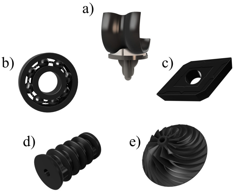
Silicon nitride ceramics (Si3N4) are recognized for their characteristics like strong durability and resistance to wear which make them suitable for use in different areas such as in the manufacturing of cutting tools, in industrial components, and in electronics and engine components where their high hardness, thermal stability, and electrical properties provide significant advantages over traditional materials (Figure 1). Beyond these applications, however, they have received a lot of interest also in the biomedical field lately as they transitioned from being used in high tech industries to becoming promising materials, for healthcare applications as well. Their ability to withstand harsh conditions while remaining compatible with the body makes them a favorable choice for various medical settings requiring both toughness and biocompatibility.
Figure 1.
Examples of common industrial applications of Si3N4 making use of its properties: (a) femoral component of a knee implant (biocompatibility), (b) ball bearing (wear resistance), (c) lathe insert (hardness), (d) insulator (electrical resistance), (e) turbine pump impeller (thermal resistance).
Silicon nitride stands out among ceramics not for its mechanical strength but also for its fascinating biological properties that researchers have discovered to be beneficial, for supporting the vital cellular functions necessary for seamless integration into biological environments. Studies indicate that silicon nitride exhibits interactions with cell types such as osteoblasts by encouraging their attachment and proliferation. This dynamic relationship plays a role in enhancing the material’s capacity to fuse with neighboring tissues and foster the processes of healing and regeneration.
Nevertheless, it is worth noting that while there have been discoveries in the field of biomaterials related to silicon nitride material properties and their effects, there is still a lack of understanding regarding the underlying biological mechanisms at work. The outcomes of the various studies conducted on this matter are different. These inconsistencies pose inquiries into the impact of manufacturing techniques and material characteristics on functionality.
This review aims, by synthesizing the findings from a broad spectrum of studies, to provide a comprehensive evaluation of the current state of knowledge regarding the biological activity of silicon nitride ceramics; with a focus on critically analyzing the gaps in the current research, we seek to clarify the role of silicon nitride in biomedical contexts and highlight the areas for further understanding its place in medicine.
Chronological Development of Biological Perspectives on Silicon Nitride Ceramics
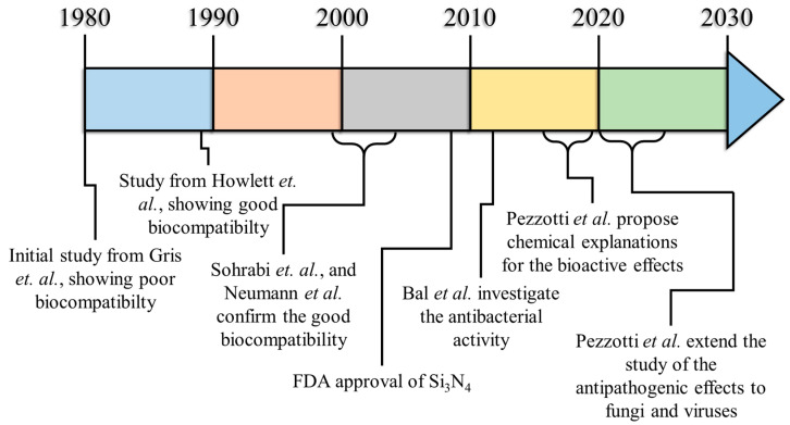
The first published research involving the biocompatibility testing of Si3N4 ceramics dates back to 1980 (Figure 2, Table 1), when Griss et al. published “Alumina, ceramic, bioglass and silicon nitride: a comparative biocompatibility study” showing that, overall, it was the least compatible material when compared to stainless steel, Al2O3, and bioglass [1]. After Griss, only a few other research articles from the 1980s mention the use of Si3N4 as a structural biomaterial. In the two volumes of Metal and Ceramic Biomaterials, Vol. I, Structure and Vol. II, Strength and Surface, published in 1984 by CRC Press (Taylor and Francis Group) [2,3], silicon nitride is already mentioned as a biomedical material with outstanding mechanical performances, but its actual biocompatibility was not further questioned. The idea behind the applicability of Si3N4, at the time, was probably based on the misconception that it could be completely bio inert because of its intrinsic chemical resistance at high temperatures and in aggressive media. This idea is based on the scarce biomedical literature of the time: silicon nitride has been used as an insulating layer to protect various biomedical devices, in particular electrodes [4,5,6,7], and no mention of its bioactivity can be found in the scientific literature before 1989.
Figure 2.
Research timeline: the evolution of Si3N4 as a biomaterial.
Table 1.
List of major works examining Si3N4 biocompatibility in different forms and chemical compositions.
| Authors | Composition/Producer | Ref | Bio Compatibility |
Mechanism |
|---|---|---|---|---|
| Griss et al. | 2.94 micron, meno 0.01 MgO, 0.073 CaO, 0.006 Fe2O3, 0.03 ZrO2 | [1] | Bad | Unknown |
| Howlett et al. | Unknown | [8] | Good | Unknown |
| Griss et al. | 2.94 micron, meno 0.01 MgO, 0.073 CaO, 0.006 Fe2O3, 0.03 ZrO2 | [9,10] | Bad | Unknown |
| Sohrabi et al. | Unknown | [11] | Good | Unknown |
| Sohrabi et al. | Conducting Materials, Columbia, MD | [12] | Good | Roughness, chemistry |
| Neumann et al. | Mg 0.1, Al 1.7, O 4.3, N 55.6, Si 37.3, Y 1.0 | [13] | Good | Unknown |
| Neumann et al. | Mg 0.1, Al 1.7, O 4.3, N 55.6, Si 37.3, Y 1.0 | [14] | Good | Unknown |
| Neumann et al. | N 55.6, Mg 0.1, Al 1.7, Si 37.3, Y 1.0, O 4.3 N 50.8, Al 2.4, Si 40.7, Y 1.0, O 5.1 N 53.8, Mg 0.1, Al 1.1, Si 39.8, Y 1.5, O 3.8 N 48.9, Mg 0.5, Al 2.4, Si 41.5, Y 1.1, O 5.6 N 55.1, Mg 0.1, Al 1.7, Si 38.3, Y 0.9, O 4.0 |
[15] | Good | Unknown |
| Neumann et al. | Unknown | [16] | Good | Surface structures, chemistry |
| Bal et al. | N 53.30, Si 39.87, Y 1.1, O 4.1 | [17] | Unknown | Unknown |
| Bal et al. | N 53.30, Si 39.87, Y 1.1, O 4.1 | [18] | Unknown | Unknown |
| Bal et al. | N 53.30, Si 39.87, Y 1.1, O 4.1 | [19] | Unknown | Unknown |
| Cappi et al. | 10% Y3Al5O12 | [20] | Good | Substrate chemistry and grain size but not roughness |
| Wang et al. | Si-O-N with high N/O ratio | [21] | Good | Substrate chemistry |
| Gustavsson et al. | Unknown | [22] | Good | Unknown |
| Yamamoto et al. | particles (700 µm) | [23] | Bad | Particle size and concentration |
| Bogner et al. | 6 wt.% Y2O3 and 4 wt.% Al2O3 | [24] | Bad | Unknown |
| Anderson et al. | Unknown | [25] | Good | Unknown |
| Webster et al. | Si3N4, Y2O3, Al2O3 | [26] | Good | Unknown |
| Pezzotti et al. | Si3N4, Y2O3, Al2O3 As-fabricated Si 35.1 N 35.5 O 17.5 Al 2.1 Y 0.1 C 9.7 HF-etched Si 31.6 N 35.2 O 8.4 Al 0.9 Y 0.1 C 21.8 Oxidized Si 32.7 N 0.1 O 57.7 Al 2.9 Y 1.3 C 5.4 Thermal treatment in N2 Si 32.7 N 33.3 O 16.6 Al 5.1 Y 2.1 C 10.3 |
[27] | Good | Substrate chemistry |
| Sun et al. | Unknown | [28] | Bad | Unknown |
| Dion et al. | Unknown | [29] | Good | Unknown |
| Aydin et al. | Unknown | [30] | Bad | Particle size and concentration |
| Pezzotti et al. | Si3N4, Y2O3, Al2O3 | [31] | Good | Substrate chemistry |
| Marin et al. | Si-rich Si3N4 N-rich |
[32] | Good | Substrate chemistry |
| Marin et al. | 6 wt.% Y2O3, 4 wt.% Al2O3, 90 wt.% Si3N4 | [33] | Good | Surface roughness and chemistry |
| Zanocco et al. | 6 wt.% Y2O3, 4 wt.% Al2O3, 90 wt.% Si3N4 | [34] | Good | Surface roughness and chemistry |
| Marin et al. | Bioglass doped with 5 wt.% and 10 wt.% Si3N4 | [35] | Good | Substrate chemistry |
| Ahuja et al. | 6 wt.% Y2O3, 4 wt.% Al2O3, 90 wt.% Si3N4 | [36] | Good | Substrate chemistry |
| Awad et al. | 6 wt.% Y2O3, 4 wt.% Al2O3, 90 wt.% Si3N4 | [37] | Good | Surface chemistry and wettability |
| Santos et al. | Unknown | [38] | Bad | Unknown |
| Frajkorová et al. | Si3N4-bioglass composite (100 wt.%, 90–10 wt.% and 70–30 wt.%) | [39] | Good | Bioglass addition |
| Amaral et al. | Si3N4-bioglass composite (70–30 wt.%) | [40] | Good | Unknown |
In the pioneering article, “The effect of silicon nitride ceramic on Rabbit Skeletal Cells and Tissue—an in vitro and in vivo investigation” [8], Howlett et al. presented a morphological assessment of the effect of silicon nitride ceramic (Si3N4) on rabbit marrow stromal cells and their differentiation when grown in vitro and in vivo. According to the authors, Fresh marrow and marrow stromal cells formed bone, cartilage and fibrous tissue in contact with Si3N4. Moreover, Si3N4 devices implanted in adult rabbits were enclosed by a stable cuff of bone within four months of implantation and remained unchanged during the rest of the animal’s life. Despite the authors concluding that “Si3N4 has the potential of an important ceramic for use in osseous reconstruction”, the research in this field would proceed slowly and without continuity for more than two decades.
In a letter to the editor of Clinical Orthopaedics and Related Research [9], Prof. Peter Griss (author of the paper from 1980), raised concerns about Si3N4’s actual biocompatibility, claiming that Howlett et al. tested the ceramic without a comparative material of established good or bad biocompatibility, which considerably reduces the validity of the conclusions drawn from the experiments. Moreover, in the letter, Griss reports that in a new femur implantation model, Si3N4 shows a similar lack of biocompatibility. Griss concludes the letter stating that Si3N4 should not be recommended as a candidate endoprosthesis material, but that more experimental quantitative data were needed for a final statement.
The results of the femur implantation model of prof. Griss would be ultimately published in 1992 [10]: again, Si3N4 (as well as SiC) resulted in being less biocompatible than the reference material (alumina).
A few years later, Sohrabi et al. studied the pro-inflammatory cytokine expression by human osteoblast-like cells upon their exposure to silicon nitride. The results showed that Si3N4 is biocompatible, and its exposure to a wide range of silicon nitride particle concentrations (1, 10, 100mg/mL) does not hinder the proliferation or induce pro-inflammatory cytokine expression of human osteoblast-like cells [11]. In the same year, the same research group also found that silicon nitride stimulates the proliferation and the osteocalcin production of human osteoblast-like cells [12], further supporting the hypothesis that Si3N4 is biocompatible and bioactive, but again without providing a direct comparison with an adequate reference material, such as alumina.
While the in vivo results from Griss showed an overall inadequate biocompatibility of Si3N4 in vivo, similar experiments conducted by Neumann et al. [13] showed good osseointegration and a lack of immuno-inflammatory reactions, suggesting that Si3N4 has similar biocompatibility features as the Al2O3 control. Neumann et al. further noted that “During the period of 4 to 8 weeks after implantation, there was a significant decrease of bone-implant attachment for Al2O3 compared to Si3N4” [14].
In a later attempt to extinguish the existing controversy between Griss and Howlett, Neumann et al. performed a series of comparisons of silicon nitride and alumina samples with different chemical compositions [15]. All the silicon nitride and alumina samples resulted in being biocompatible in vitro, with no sign of cytotoxicity. Neumann further noted that “The comparison of cell counts with the distribution of elements in the different silicon nitride samples shows no correlation”, but it is important to note that all the Si3N4 specimens used by Neumann had overall similar chemical compositions, with Mg between 0.1% and 0.5%, Al between 1.1 and 2.4%, and Y between 0.9 and 1.5%. Considering the innate variability of the in vitro assessments, the effects of these elements could have been well below the detection limit for the method used. Neumann et al. also tested a prototype of a silicon nitride mini-plate osteofixation system for the midface [16], observing satisfactory results in terms of intraoperative handling, mechanical stability, and biocompatibility, in particular when compared to Al2O3. Similar results were also obtained by Ruan et al., working on endosteal implants in dogs [41].
A more recent, in vitro comparison of oxide and non-oxide materials was published by Cappi et al. in 2009 [20]. In their experiments, cold isostatic pressed and hot isostatic pressed Si3N4 were compared to Al2O3, ZrO2, and SiC. The study came to the conclusion that the non-oxide ceramic materials, Si3N4 and SiC, either produced via pressureless sintering or hot-pressing, are cytocompatible for human mesenchymal stem cells, and allow for osteogenic differentiation.
In the early 2000s, further insights into the potential bioactivity of Si3N4 emerged also from the field of microelectromechanical systems: in an in vitro test using platelet rich-plasma, the authors observed that thin films of Si3N4, Si1.0N1.1, and Si cause more platelet adhesion when compared to SiO2, increasing the risk of thrombogenesis [42]. Reducing the risk required a drastic modification of the surface chemistry of Si3N4, for example going towards silicon oxynitride films [21]. The further functionalization of the Si3N4 thin film surfaces, for example with primary amine functional groups (NH2), stimulated the adhesion and spreading of an osteoblast-like cell line [22] but, remarkably, the highest spontaneous alkaline phosphatase activity was observed for cells grown on un-functionalized Si3N4 substrates.
Another common method to estimate the cytotoxicity of a material is to use particulates. Unlike bulk materials, particulates offer a higher surface-to-volume ratio, strongly exacerbating chemical and biological reactions. Furthermore, particulate cytotoxicity is also strongly affected by the average particle diameter (nanometric particles can penetrate cellular membranes) and shape (particles with sharp edges are usually more cytotoxic than round particles. The earliest assessment of Si3N4 particulate cytotoxicity can be found in a 2003 paper from Yamamoto et al. [23], for L929 and J744A.1 cell lines. As observed before by Neumann et al. for bulk ceramic discs, the behavior of Si3N4 resulted in being similar to that of Al2O3 and, overall, more cytotoxic at lower concentrations with respect to TiO2 and SiC.
In 2006, new research from Bogner et al. on biocompatible microelectronic materials tested on Caco-2 cells partially contradicted previous results, showing that in p-Si wafers coated with Si3N4 (as well as Au, Al, and ITO), the alkaline phosphatase activities only reached between 15% and 35% [24].
By 2008, Bal et al. were testing silicon nitride ceramic bearings for total hip arthroplasty [17,18,19]. The components, obtained via sintering and HIPping, showed superior fracture toughness and flexural strength than the reference Al2O3, and did not change their properties after autoclaving. In another paper from 2012, Bal et al. [43] noted that Si3N4 could be a superior competitor to the more common Al2O3-based orthopedic ceramics on the market (Zirconia-toughened alumina), a prediction that, at least from the tribological point of view, has been contested by other researchers [44].
Titanium and PEEK, both possessing hydrophobic characteristics with net negative surface charges, were compared to Si3N4, which exhibited a hydrophilic surface with a net positive charge. Notably, two surface finishes of Si3N4 were explored: as-fired and polished. The study revealed a decreased biofilm formation and fewer live bacteria on both the as-fired and polished Si3N4 surfaces compared to Ti and PEEK. These observations suggested that the differential surface chemistry and nanostructured topography of Si3N4 played a pivotal role in impeding bacterial biofilm formation, colonization, and growth.
The investigation extended beyond the bacterial response to examine protein adsorption on material surfaces, specifically focusing on fibronectin, vitronectin, and laminin. Remarkably, Si3N4 demonstrated significantly greater amounts of these proteins adhering to its surface compared to Ti or PEEK. This finding highlighted the influence of the surface properties on the adsorption of physiologic proteins, suggesting a potential correlation with the observed in-vitro differences in the bacterial affinity for the respective biomaterials.
The findings of this study propose a novel strategy in designing future orthopedic implants based on the intrinsic biomaterial properties. The hydrophilic nature, positive surface charge, and enhanced protein adsorption on Si3N4 present an encouraging prospect for developing implants with inherent resistance to bacterial colonization. This paradigm shifts the open avenues for advancing orthopedic implant technology by exploiting the surface properties of biomaterials to mitigate the risks associated with bacterial infections. The differential responses observed in bacterial affinity and protein adsorption suggest that the unique surface properties of Si3N4 hold promise for the development of orthopedic implants with enhanced resistance to bacterial colonization.
Following the groundbreaking results of the 2012 in vitro study on the biological activity of silicon nitride (Si3N4) in orthopedic implants, the subsequent research endeavors sought to validate these findings through in vivo testing [26]. In the same year, a pivotal study investigated the performance of Si3N4 implants in comparison to poly(ether ether ketone) (PEEK) and titanium (Ti) implants [45], emphasizing the critical aspects of bone formation and bacterial resistance in orthopedic applications.
Dense implants made of Si3N4, PEEK, or Ti were surgically implanted into rat calvarial defects to simulate real-world orthopedic scenarios. Bacterial infection was induced by injecting Staphylococcus epidermidis, and control animals received saline only. The rats were sacrificed at intervals of 3, 7, and 14 days post-surgery, as well as at 3 months, to examine new bone formation and the presence or absence of bacteria.
Three months after surgery in the absence of bacterial injection, Si3N4 demonstrated an impressive ∼69% new bone formation around the implant, outperforming both PEEK (24%) and Ti (36%). In the presence of bacteria, Si3N4 maintained superior performance with new bone formation at 41%, compared to 26% for Ti and 21% for PEEK. Notably, live bacteria were identified around the PEEK (88%) and Ti (21%) implants, while none were present adjacent to Si3N4.
Despite not being the first published articles to demonstrate the bioactivity of Si3N4, the in vitro and in vivo studies of 2012 provided compelling evidence supporting the favorable attributes of Si3N4 orthopedic implants and can be considered as the starting point for the industrial interest for bioactive silicon nitride ceramics. The superior new bone formation and resistance to bacterial infection observed for Si3N4, as compared to Ti and PEEK, underscore the potential clinical significance of Si3N4 as a biomaterial for orthopedic implants.
However, as can be seen from Table 1 and Table 2, which list the most significant works regarding the biocompatibility and antipathogenic activity of Si3N4, respectively, there is no unified consensus on the mechanisms related to bioactivity.
Table 2.
List of major works examining Si3N4 antipathogen activity in different forms and chemical compositions.
| Authors | Composition/Producer | Ref | Antipathogen Activity | Mechanism |
|---|---|---|---|---|
| Bal et al. | Si3N4, Y2O3, Al2O3 As Fired and Polished surfaces |
[45] | Staphylococcus Epidermidis (Good), Staphylococcus. Aureus (Good), Pseudomonas aeruginosa (Good), Escherichia coli (Good), Enterococcus (Good) |
Hydrophilicity and surface chemistry |
| Webster et al. | Si3N4, Y2O3, Al2O3 | [26] | Staphylococcus Epidermidis (Good), | Hydrophilicity and surface net charge |
| Pezzotti et al. | Si3N4, Y2O3, Al2O3 | [46] | Porphyromonas gingivalis (Good) | Peroxynitrite formation |
| Bock et al. | 6 wt.% Y2O3, 4 wt.% Al2O3, 90 wt.% Si3N4 As Fired N2-Annealed (SiYAlON excess on the surface) SiYAlON glazed Oxidized (Si-OH excess on the surface) |
[47] | Staphylococcus epidermidis (Good) | Peroxynitrite formation |
| Pezzotti et al. | PMMA-βSi3N4 composite (6, 8, 10, 15, and 30 vol.%, no info on Si3N4 composition) |
[48] | Candida albicans (Good) | Chemical and osmotic stress |
| Pezzotti et al. | 6 wt.% Y2O3, 4 wt.% Al2O3, 90 wt.% Si3N4 | [49] | Human herpesvirus 1 (Good) | Peroxynitrite formation |
| Ishikawa et al. | 6 wt.% Y2O3, 4 wt.% Al2O3, 90 wt.% Si3N4 | [50] | Staphylococcus. Aureus (Good), | Peroxynitrite formation and surface morphology |
| Fang et al. | Unknown | [51] | Sulfate-reducing bacteria (Bad) | Weak adhesion |
| Yao et al. | Unknown | [52] | Pseudomonas aeruginosa (Bad) Enterococcus hirae (Bad) |
Weak adhesion |
| Pinar Gordesli et al. | Unknown | [53] | Listeria monocytogenes (Bad) | Weak adhesion |
| Boonaert et al. | Unknown | [54] | Phanerochaete chrysosporium (Bad) Lactococcus lactis (Bad) |
Weak adhesion |
| Park et al. | Unknown | [55] | Listeria monocytogenes (Bad) | Unknown |
| Pezzotti et al. | 6 wt.% Y2O3, 4 wt.% Al2O3, 90 wt.% Si3N4 | [56] | Plasmopara viticola (Good) | Peroxynitrite formation |
2. Early Notes on Surface Chemistry and Biocompatibility
It is important to note that, up to 2012, all the investigations into the biocompatibility of Si3N4 did not provide meaningful insights about its chemical structure, to the point that most research articles do not even specify the chemical composition of the ceramic utilized.
A paper from Bock et al. [57], dating back to 2015, is arguably the first piece of scientific literature to properly investigate the chemical structure of the surface of as-fabricated and polished silicon nitride subjected to different post treatments. The authors found that both the surface chemistry and morphology of Si3N4 can be varied through conventional thermal, chemical, and mechanical treatments, with as-fabricated Si3N4 exhibiting anisotropic grains covered with a thin Si2N2O passivation layer, strongly negative charging at biological pH, and moderate hydrophilicity. Etching in HF produced a surface composition with a higher N/Si and lower O/Si ratios than as-fabricated materials, strong negative surface charging at homeostatic pH, and moderate hydrophilicity. Thermal treatment in N2 produced a surface coated in crystalline β-Si(Y)AlON precipitates and exhibited extreme hydrophilicity. Thermal treatment in an oxidizing atmosphere resulted in a surface composition effectively comparable to amorphous SiO2. It also exhibited extremely low wetting angles and charging behavior that mimicked pure silica [58]. The isoelectric points of these variously treated samples increased with decreasing O/Si and with increasing N/Si atomic ratio, as the surfaces transitioned from resembling pure SiO2 to pure Si3N4.
When tested with human osteosarcoma cells (SaOS-2), the same surface treatments resulted in marked differences in the amounts of bone tissue produced, with the N2-annealed specimen reaching about 40% higher bone tissue volumes per unit of area. The author concluded that “alterations of its lattice defects promote proliferation of osteoblasts and the subsequent generation of natural hydroxyapatite. Surface modulation of Si3N4 illustrates the concept that engineering of atomically defective biomaterials can lead to remarkable osteoconductive characteristics”. A more detailed explanation was published in a follow-up paper [27], where the authors claimed that the peculiar SiYAlON phase on the surface of these specimens contained an abundance of positively charged N-vacancies and N–N bonds which modulated the negative surface charge developed by the deprotonation of amphoteric silanols. The various hypotheses for the improved bioactivity of Si3N4 will be discussed in detail in the following section, but this paper represents the first attempt to rationalize the biological behavior of silicon nitride from a chemical point of view.
The same research group tested surface-modulated Si3N4 samples against Porphyromonas gingivalis [46], showing that the oxidized (hydroxyl-rich) Si3N4 was less effective in inducing PG lysis when compared to as-sintered and HF-etched (amine-rich) Si3N4. The N/Si (O/Si) ratios were 1.22 (0.17), 1.05 (0.15), and 0.09 (1.98) for the as-sintered (polished), HF-etched, and oxidized samples, respectively. Unfortunately, the authors did not provide any quantitative analysis, but the fluorescence imaging shows a weak green CFDA signal for the oxidized surface, which the authors interpreted as a sign of weaker bacterial lysis.
3. Crystal Structure of Biomedical Si3N4
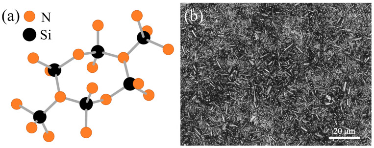
Silicon nitride (Si3N4) exists in two primary crystalline forms, alpha (α) and beta (β), which differ in their atomic arrangements and, consequently, their properties. The α-phase has a hexagonal structure that incorporates stacking faults, resulting in lower thermodynamic stability, while the β-phase adopts a more stable hexagonal lattice (Figure 3a) with elongated grains that interlock, conferring superior mechanical properties such as toughness and fracture resistance. In biomedical applications, β-Si3N4 is almost exclusively used due to these characteristics, which are essential for load-bearing implants and other medical devices where structural integrity and durability are critical. Additionally, the β-phase has been reported to exhibit favorable biocompatibility and bioactivity, promoting bone growth and integration, making it the preferred choice in regenerative and orthopedic applications.
Figure 3.
(a) Structure of β-Si3N4 and (b) typical microstructure of an as-sintered β-Si3N4 specimen, as observed at 150×.
The microstructure of β-Si3N4 is characterized by the presence of elongated, acicular (needle-like) grains that interlock to form a tough, resilient network (Figure 3b). These grains play a crucial role in enhancing the material’s toughness: they act as crack-bridging elements, forcing cracks to deflect or deviate around them rather than following a straight path. This crack deflection mechanism significantly increases the energy required for crack propagation, imparting exceptional fracture toughness to β-Si3N4. To achieve this structure during sintering, specific additives—typically oxides like yttria (Y2O3) and alumina (Al2O3)—are essential [59]. These additives promote liquid-phase sintering by lowering the temperature required for densification, enabling the growth and bonding of β-phase grains into a strong, interlocking microstructure.
Interestingly, early investigations on α- and β-Si3N4-reinforced PEEK showed that, despite the lower stability of α-Si3N4, its antibacterial effect is negligible, even though its biocompatibility is comparable to, if not slightly superior to, that of β-Si3N4 [60].
4. Chemical Reactions of Si3N4 In Vivo
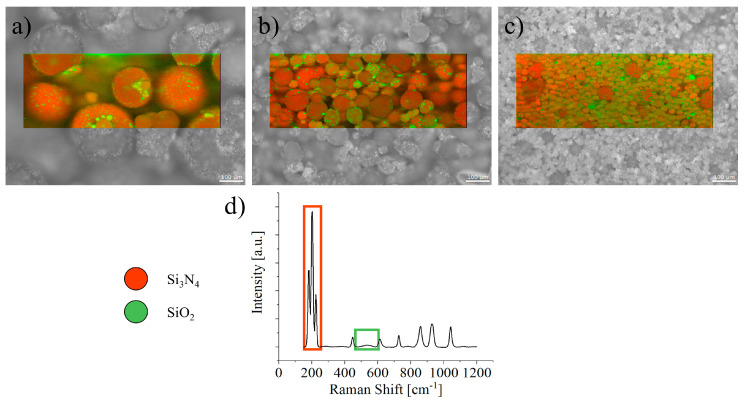
Silicon nitride (Si3N4) naturally undergoes surface oxidation when exposed to humid environments, forming a thin silicon oxide layer that plays a crucial role in its stability and activity. This oxidation effect is particularly pronounced for Si3N4 particles, with an increasingly significant impact as the particle size decreases. In smaller particles, the larger surface-area-to-volume ratio enhances the exposure to atmospheric moisture, leading to accelerated oxidation (Figure 4). This intensified oxidation in fine particles can influence their reactivity, making them more susceptible to environmental changes and potentially affecting their performance in applications such as biomedical implants or wear-resistant coatings.
Figure 4.
SiO2 and Si3N4 surface coverage on Si3N4 particles of different nominal sizes, (a) 300, (b) 50 and (c) 20 μm, as measured by Raman spectroscopy using the bands in (d) [61].
Low-temperature dissolution of Si3N4 particulates has been described in detail multiple times. The hydrolysis of silicon nitride is often described by the simplified reaction:
assuming that silica is the main hydrolysis product. Considering that Si3N is the highest species formed on the surface of Si3N4, we can instead expect the following hydrolysis reactions [62]:
And we know from previous studies that silanol (Si-OH) is the major surface group on silica, while on silicon nitride, the major surface groups are amine (Si2-NH) and silanol (Si-OH), which are produced by a spontaneous oxidation of silicon nitride [63]. These surface groups can acquire a charge in aqueous solution according to the following reactions. The zeta potential of silicon nitride is higher due to the positively charged amine groups on the silicon nitride surface:
Which are responsible for the amphoteric behavior of the Si3N4 surface, as previously reported [64]. FTIR analysis performed on Si3N4 powder evidenced the presence of –NH2 surface groups, further supporting this hypothesis [65,66]. Additional studies on silicon nitride powder have also shown that the oxidized surface layer dissolves in the same fashion as silica [67,68]:
While, depending on the pH of the environment, ammonia can be converted to ammonium ions and vice versa [69]:
The total ammonia () is the sum of + and the pK of this ammonia/ammonium ion reaction is around 9.5. The amounts of each of the two species can be calculated from the Henderson–Hasselbalch equation if the pH and appropriate pK are known [69]:
Molecular dynamics simulations also suggest that hydrolysis may proceed through the nucleophilic attack of water with the formation of an intermediate molecular complex involving a penta-coordinated silicon [11,12].
One last chemical compound that has been associated with the dissolution of Si3N4 in biological environments is peroxynitrite (), a reactive nitrogen species usually formed when nitric oxide () reacts with the superoxide anion (). It was first identified as a mediator of cell death in animals but was later shown to act as a positive regulator of cell signaling, mainly through the posttranslational modification of proteins by tyrosine nitration [70]. The first (and only) experimental observation of the formation of peroxynitrite on Si3N4 was performed by Pezzotti et al. using Raman spectroscopy [46], but without providing any further validation using a consolidated detection method. The band was reported to be located at 1044 cm−1, and can be easily confused with one of the bands of β-Si3N4 (1040 cm−1). Moreover, careful observations of the spectra provided confirmed that the actual band is located at 1032 cm−1, casting further doubts on the original observation and weakening the hypothesis of peroxynitrite formation on Si3N4. Nevertheless, the claim has been subsequently referenced various times in the literature, without providing any additional supporting evidence [47,48,49,50,71,72].
Despite the apparently simple surface chemistry, the dissolution of Si3N4 can proceed at different paces and follow different paths depending on parameters such as pH, oxidation status (in particular the ratio between Si-OH and Si2-NH), specific surface area and chemical composition, as evidenced by the variations in the isoelectric point reported in the literature [57,73,74,75]. The complexity of these interactions, and the lack of a comprehensive prediction model might be responsible for the variability in the biological responses observed, in particular in the early literature.
5. Bioactive Mechanisms
Multiple literature references support the hypothesis that Si3N4 (as well as Si and SiO2) is not cytotoxic [28,29] unless in the form of nano-particulates [30], in which case it might induce changes in the genes related to apoptosis, DNA damage or repair, and oxidative stress. It should be observed that, in many cases, when tested in vitro, Si3N4 performed as well as the bio-inert controls, but did not exhibit an enhanced cellular proliferation [11,76].
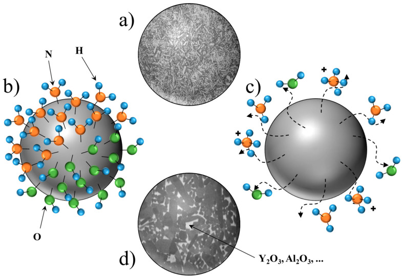
Despite the numerous proposals from researchers, the exact origin of the bioactivity of silicon nitride (Si3N4) remains unconfirmed, leading to an ongoing debate in the scientific community. The hypotheses surrounding this phenomenon can be categorized into four main areas: the first focuses on the effect of surface roughness (Figure 5a), suggesting that the variations in texture may enhance cellular adhesion and proliferation. The second category examines the influence of the surface functional groups (Figure 5b), positing that specific chemical functionalities on the Si3N4 surface may play a crucial role in mediating the interactions with biological tissues. The third hypothesis revolves around the release of bioactive species (Figure 5c), proposing that the ions or compounds leaching from Si3N4 could contribute to its bioactive properties. Finally, the fourth category considers the role of bioactive additives (Figure 5d), where the incorporation of specific materials may augment the bioactivity of Si3N4 in biomedical applications.
Figure 5.
The four chemical explanations for the bioactive behavior of Si3N4, according to the literature: (a) surface roughness, (b) functional groups on the surface, (c) release of bioactive species, and (d) presence of bioactive additives. Dashed arrows indicate release of molecules from the particle, solid lines indicate the specific ion/molecule. A “+” sign indicates that the molecule is in ionic form.
According to Pezzotti et al., both Si and N available at the interface with Si3N4 are associated with the upregulation of the metabolic activity of osteoblasts [31], which in turn results in rapid and efficient bone growth. In preliminary research from the same authors, the amount of hydroxyapatite (mineralized bone) formed is higher for samples containing extra nitrogen [27], a result that, apparently, contradicts the observations made regarding Si- and N-rich PVD coatings [32], where N-rich Si3N4 showed higher cellular proliferation and better antimicrobial effects, while Si-rich Si3N4 showed higher amounts of bone tissue formed. Further investigations of Si-rich laser-cladded coatings of Si3N4, showed results comparable to the titanium control, but superior to both zirconia and polyethylene [33]. In yet another manuscript from the same research group, the authors noted that both cellular proliferation and bone growth diminish with decreasing nitrogen content in Si3N4 [34].
The nitrogen released from the surface of Si3N4 takes the form of ammonia or ammonium ions, depending on the environmental pH. In normal cellular metabolism, free ammonium ions are produced and consumed regularly. Glutamine synthetase utilizes free ammonium ions to produce glutamine in the cytosol, whereas glutaminase and glutamate dehydrogenase generate free ammonium ions in the mitochondria from glutamine and glutamate, respectively [77]. Ammonia and bicarbonate are condensed in the liver mitochondria to yield carbamoylphosphate, initiating the urea cycle, the major mechanism of ammonium removal in humans [77]. Martinelle and Häggström [78] observed that there is a clear difference between the ammonia/ammonium that is added to the cells and which is formed by the cells during the metabolism of amino acids, especially glutamine and glutamate, with the former leading to predictable intracellular and extracellular pH (pHe) changes. The authors postulated that one important toxic effect of ammonia/ammonium is an increased demand for maintenance energy, caused by the need to maintain the ion gradients over the cytoplasmic membrane. Other researchers found that ammonia induces the overproduction of ROS, decreases MMP, interrupts Ca2+ homeostasis, and subsequently causes cell apoptosis, via the P53-BAX-BCL2 and mitochrondial apoptotic pathways [79,80].
The previous literature on the bone tissue formed on Si3N4 biomaterials hypothesized that nitrogen can be substituted to oxygen in both the (PO4)3− of and (SiO4)4− tetrahedra and the OH group as well, but the hypothesis is not supported by the XPS analysis provided by the authors, as no additional bond, such as Ca–N, are shown to have formed, either from the Ca or the N binding energy intervals [31].
Based on the previous literature references, there seems not to be enough strong evidence to support the hypothesis that the nitrogen released from the surface of Si3N4 can have a beneficial effect on the cellular proliferation and/or metabolism, but this does not mean that nitrogen makes no positive contribution to the biocompatibility of Si3N4. As previously observed, in aqueous environments, the surface of Si3N4 is mainly composed of two species, hydroxides and secondary amines. Of these two structures, the secondary amines are more likely to exhibit bioactive properties, in particular towards collagen [81], resulting in an increased cellular adhesion [82,83]. Moreover, NH2 functionalized bioglasses were also reported to increase cellular proliferation, when tested with human bone marrow-derived mesenchymal stem cells [84]
Nitrogen elution from Si3N4 is regulated by the environmental pH, specific surface area, and surface chemistry. Compared to the “friendly nitrogen kinetics” hypothesis [71], which would require a case-by-case material design optimization to avoid potential cytotoxic effects, the “bioactive surface amines” hypothesis finds more support in the literature, as bioactivity was observed in a wide range of Si3N4 compositions, surface morphologies, and experimental settings. Despite the claims, no experimental proof has been provided so far for the positive effects of nitrogen elution from Si3N4.
Apart from surface amines, the chemical composition of the ceramic is also likely to play a crucial role in the biocompatibility, in particular considering the results from the early testing on Si3N4 ceramics with low amounts of additives, such as the ones reported by Griss [9,10]. Most of the Si3N4 materials that are used nowadays for biomedical applications contain a not negligible amount of Y2O3, used as a sintering aid. The recent literature results showed that Y2O3 increases the cellular proliferation and bone tissue formation in vitro, even if in very low concentrations [85,86]. Honma et al. performed a direct comparison between Si3N4 and Y2O3 powders, showing that when the two materials are mixed together, osteoblasts preferentially grow on Y2O3, where they also produce higher amounts of mineralized bone tissue [87]. The authors hypothesized that this could be due to the antioxidant action of Y2O3, while Xiang et al. observed that Y2O3 particles contribute to the inhibition of the ROS/NF-κB pathway, which plays a role in the immune response, and cell proliferation and differentiation [88]. The absence of Y2O3 might also explain why Griss et al. reported such poor biological performances for Si3N4 [9,10] when compared to subsequent research [10,89]. A correlation between the presence of Y and the bioactivity of ceramics would also explain the in vitro results obtained for the SiYAlON phase by various independent research groups [27,90,91].
Other additives in Si3N4 ceramics, such as CaO and Ca3(PO4)2, have been shown to influence both structural and biological properties, supporting the hypothesis of a significant contribution by these additives to bioactivity. Studies have demonstrated that such additives can enhance the bioactivity by creating glassy intergranular phases and promoting surface porosity. For example, flame-treated Si3N4 ceramics incorporating Ca3(PO4)2 exhibit increased cell viability and bone integration by forming bioactive layers rich in hydroxyapatite and calcium silicate phases, which favor cellular adhesion and proliferation [92]. Moreover, silicon-rich compositions enhance the bone mineralization, suggesting a synergistic effect of composition and structure [32]. Apart from calcium-based compounds, the addition of other oxides such as SrO, MgO, and SiO2 improved the cellular proliferation and differentiation [93]. Positive biological responses were also observed when adding ZnO [94] or SiAlON [95]. These findings indicate that the composition of Si3N4 could be further optimized for biological applications by exploring alternative additives to replace the conventional ones.
According to the literature, surface amines are effective in stimulating collagen, and in vitro testing performed using SAOS-2 osteosarcoma cells showed that the addition of Si3N4 to bioglass stimulated the production of more biological matrix compared to mineralized bone [35]. This effect has subsequently been confirmed also for bulk Si3N4 materials [36,37]. The results also indicate the upregulation of osteogenic transcription factors such as RUNX2, SP7, collagen type I, and osteocalcin.
Nitrogen and silicon elution, as observed by Pezzotti et al., are likely to occur on most Si3N4-based materials; however, a review of the literature suggests that these are not the key factors in regulating the biocompatibility. Spontaneously formed surface amines and osteo-inductive yttrium-rich phases are more likely to play the main role.
6. Anti-Pathogenic Mechanisms
Like previously discussed for bioactivity, the anti-pathogenic effects of Si3N4 have also been associated with the surface roughness, functional groups, release of antibacterial drugs, and presence of antibacterial secondary phases due to the introduction of additives. The bioactivity of Si3N4 can be explained via its surface chemistry, considering both the presence of surface amines and yttrium, while nitrogen elution is still the most likely mechanism responsible for the anti-pathogenic effects observed on most Si3N4 ceramics and Si3N4-based composites.
Unlike cells, bacteria are prokaryotic, meaning that they lack a complex internal structure like the nucleus and membrane-bound organelles found in eukaryotic human cells. This simplified structure makes them generally less equipped to handle the toxic effects of ammonia compared to human cells. On the other hand, many bacteria possess enzymes like urease that can detoxify ammonia by converting it into less harmful compounds. Certain human cells also have these enzymes, albeit to a lesser extent. A higher tolerance to ammonia/ammonium would also explain why, under normal testing circumstances, Si3N4 does not show the same cytotoxic effects as human cells, but the magnitude of this effect on bacteria should be evaluated case by case, as the tolerance varies greatly between species [96] and strains.
Gorth et al. compared the activity of several bacteria exposed to the surface of Si3N4 and other biomaterials [45]. Despite being the slowest to grow, Enterococcus faecium, a highly ammonia-tolerant bacteria often studied for its ammonia reduction potential [97], was the only strain to show some degree of resistance to the antibacterial activity of Si3N4. On the other hand, despite S. epidermidis, S. aureus, E. coli, and P. aeruginosa being all able to produce the enzyme, urease, its activity is generally limited compared to other urease-positive bacteria. Unfortunately, only a limited number of bacteria have been used for the in vitro testing of Si3N4 so far, and the list does not include other bacteria with strong urease activity, such as Helicobacter pylori [98], Proteus mirabilis, or Staphylococcus saprophyticus, meaning that a hypothetical correlation between urease activity and the antibacterial efficiency of Si3N4 cannot be demonstrated yet.
Some additional insights into the anti-pathogenic potential of ammonia released from Si3N4 can be deduced from the literature in vitro testing with Candida strains. Despite lacking the urease enzyme, Candida albicans uses urea amidolyase to hydrolyze urea and produce ammonia, which is its only source of nitrogen for the production of aminoacids [99]. Excesses of ammonium are then expelled from the cell and used for environmental pH modulation [100], but if the pH reaches a certain threshold, it might cause the inactivation of cell membrane enzymes resulting in a loss of biological activity [101]. Still, Si3N4 showed remarkable antifungal properties when tested against C. albicans, suggesting that ammonia release is not its main and/or only anti-pathogenic mechanism [48]. Tests have been conducted using amine-functionalized polymers [102,103], silica nanoparticles [104], and carbon nanotubes [105], showing exceptional fungicidal capabilities against Candida albicans.
As for the bioactive mechanisms discussed in the previous chapters, two possible pathways have emerged for the interpretation of the antibacterial capabilities of Si3N4, either due to ammonia/ammonium release or surface amines, with the latter being limited to the direct contact of the pathogen with the ceramic. One easy way to discriminate between the two mechanisms would be to perform a disk-diffusion antimicrobial susceptibility test on Agar plates and measure the inhibition area surrounding Si3N4: for the contact-based mechanism, the area would be negligible. Despite its simplicity and common use, such tests were never reported in the scientific literature. The authors are aware of such an investigation having been performed at least once between late 2014 and early 2015 with Porphyromonas gingivalis, Treponema denticola, and Tannerella forsythia, but the results were never published, as no antibacterial activity could be measured using this method on Si3N4 [106].
Another important remark from Gorth et al. is that the antibacterial activity of Si3N4 seems to be independent of the Gram classification [45]. This assertion has two potential consequences: Si3N4 might have a broader range of effectiveness compared to treatments that target specific cell wall structures and the Si3N4 antibacterial mechanism might not directly target the cell wall. Vancomycin, for example, is a glycopeptide antibiotic that works by interfering with the synthesis of the bacterial cell wall of Gram-positive bacteria, in particular targeting the peptidoglycan [107], but is ineffective against Gram-negative bacteria due to the presence of an outer membrane containing lipopolysaccharides.
Boschetto et al. studied the response of E. coli to Si3N4 surfaces by observing the metabolic alterations, in particular concerning the RNA and DNA structures within the bacterium. The authors reported “extensive disruption of the cell membrane”, suggesting that Si3N4 does attack cell walls directly in both Gram-positive and Gram-negative groups, but the claim is based on indirect spectroscopic alterations only and should be further validated through direct microscopic observation and biochemical analysis. The study concludes that “a high ammonium concentration outside the bacterial cell, usually referred to as “ammonium shock”, leads to lysis”, but it should be noted that this mechanism is not mentioned in the reference mentioned by the authors [108] or by the other recent scientific literature. Ammonium shock does cause the disruption of the metabolic processes and cellular functions, but this does not result in bacterial lysis [109,110,111]. Osmotic pressure, which was also observed by the authors [71], would be a more likely mechanism, that would also equally affect Gram-positive bacteria [112,113].
We can gain further insights into the antibacterial properties of Si3N4 by examining the extensive body of literature on AFM testing, which includes the studies on parameters like cell elasticity and adhesion performed with a Si3N4-coated tip [51,52,54,114]. The results suggest that the adhesion forces between Si3N4 and bacteria cell walls are present, but weak, while antibacterial effects or cell wall rupturing were never observed. Aucapina et al. observed that these forces increase over time, and eventually grow stronger than bacteria–bacteria interactions [115]. Bacteria adhesion to Si3N4 was shown to greatly vary even within the same bacteria species [55], and was ultimately postulated to be stronger for virulent strains, as a survival mechanism. Si3N4 was also shown to adhere to other micro-organisms’ membranes, such as the sporangia of the pathogenic oomycete Plasmopara viticola, leading to its death [56].
The role of additives in the anti-pathogenic mechanisms of Si3N4 has not been extensively explored in the literature. Du et al. noticed that the addition of rare earth oxides increases the antimicrobial properties [116], but the biocompatibility of such materials has yet to be investigated. Zeng et al. and Liu et al. observed an increased antibacterial efficiency when ZnO nanowires or whiskers are added to Si3N4 [94,117].
7. Si3N4 in Bioglasses
Due to its mechanical properties, Si3N4 has also been used as a strengthening agent for other bioactive ceramics creating composites. One of the first studies conducted by Santos et al. showed how adding a 30% proportion of Si3N4 in Bioglass®45S5 drastically improved the hardness and the fracture toughness with values complying with those usually reported for cortical bone. Consequently, in vitro bioactivity tests using SBF demonstrated how a thin layer of apatite was formed, suggesting how Si3N4 presence did not alter the bioactive effect of the bioglass [38]. An improvement of the mechanical properties was observed also by Amaral et al., who established the suitable conditions to fabricate almost fully dense silicon nitride biocomposites by hot pressing at 1350 °C–40 min–30 MPa [118]. A further confirmation of the improvement in the mechanical properties combined with a good bioactivity has been proved also by Frajkorova et al. Despite the composite being fabricated via sintering at 980 °C for 1 h in a nitrogen atmosphere, the specimen containing different concentrations of bioglass (10% and 30%, respectively) showed a better bioactivity via the immersion of composites in simulated body fluid for different time periods whereby the hydroxyapatite layer was developed on their surface [39]. The Si3N4 bulk used as a reference material exhibited no bioactivity, which clearly confirmed the positive effect of the bioglass addition. The first study analyzing the bioactivity of the composite Si3N4-bioglass through in vitro cells treatment was performed by Amaral et al. in 2002 [40]. Human bone marrow cells (MG63) were seeded on the composite specimen, and the performances were evaluated via proliferation (MTT assay), mineralization (SEM, ALP, and Alizarin Red assays), and protein level tests. The results showed a significant series of events associated with the rapid protein absorption of the surface, changes in the concentration of ionized Ca and P release in the medium, and high viability and proliferation of MG63. Also, the high ALP level and mineralized extracellular matrix produced by the cells confirmed the high bioactivity of the composites in bone formation. However, a Si3N4 bulk as reference was missing, and this did not provide information and data about the improvement associated with the use of Bioglass®.
Another study focused on improving the surface chemistry and topography of Zirconia-toughened alumina (ZTA) by using a powder mixture of silicon nitride and Bioglass® was conducted by Marin et al. [35]. Initially the surface of the ZTA was treated via laser patterning, and the wells formed were filled with Bioglass® or composites containing different concentrations of silicon nitride in Bioglass® (5% and 10%, respectively). The results, after in vitro tests with the SaOS-2 cell line, showed how the presence of the bioactive powder and composites improved the cells’ adherence and bone formation on the surface. No differences in terms of extracellular matrix area coverage and volume were observed between the samples presenting Bioglass® or composites, indicating how different amounts of silicon nitride did not affect the mineralization. Between the samples presenting fillers, only a variation in the mineral to matrix ratio by using FTIR was observed; by increasing the silicon nitride concentration, the ratio decreased, indicating a higher signal associated with Amide I, the organic component of the extracellular matrix. For the further confirmation of this effect provided by Si3N4, it would have been necessary to test the samples of ZTA presenting Si3N4 powder without Bioglass® and also provide other tests straightly direct to analyze the collagen component (for example, the immunofluorescence staining of Col I) without basing the conclusion only on the FTIR spectra of a limited area of the specimens.
Summarizing the studies present in the literature, Si3N4 did not affect the bioactivity provided by Bioglass®, and concurrently improved the mechanical properties. However, despite the well-known antibacterial properties of Si3N4, no studies or data in the literature about the bacteriostatic behavior of composites such as Si3N4-Bioglass® have been reported.
8. What Is Missing?
While other biomedical materials have advanced significantly through the use of additive manufacturing to better meet patient-specific needs, silicon nitride (Si3N4) is still lagging behind. Currently, Si3N4 implants are primarily produced using traditional ceramic processes, which involve powder compaction and high-temperature sintering. Though effective, these methods are costly, require extensive post-fabrication machining, and offer limited design flexibility [119]. Some progress has been made with additive manufacturing techniques like robocasting, which could enable custom Si3N4 implants with complex architectures and tailored porosity [120,121]. However, despite these advancements, Si3N4 has yet to fully embrace the additive techniques to the extent seen with other materials, leaving a gap in its application for patient-specific orthopedic solutions.
Si3N4 has shown potential in both spinal fusion and joint replacement applications, but its adoption in orthopedics remains limited due to several concerns. In spinal implants, while studies suggest that Si3N4 may offer advantages over other biomaterials, the variability in the clinical factors such as implant size, patient population, and biomaterial designs makes it difficult to generalize the findings. Moreover, the lack of randomized control trials and inconsistent definitions of subsidence complicate the assessment of Si3N4’s true efficacy [122]. In joint replacement, regulatory approval has not been granted, and concerns about its wear performance persist. Specifically, the surface oxide layer of Si3N4 may flake off, potentially leading to third-body wear, which could compromise the longevity of implants [72]. This uncertainty, coupled with the reliance on the integrity of the SiO2 film to maintain low friction, underscores the need for further investigation before Si3N4 can be widely adopted in orthopedic applications.
The biological applications of Si3N4 have been discussed numerous times in the literature, but despite the volume of scientific data and the general scientific consensus supporting the idea that Si3N4 bioceramics are biocompatible and bioactive, crucial information about the biological mechanisms involved is still missing.
The gap has been partially bridged by the integration of complementary techniques, in particular Raman spectroscopy. However, a potential over-reliance on Raman data has emerged in some fields. While spectroscopic techniques offer a powerful window into the chemical composition of biological samples, they inherently provide an indirect measure of the complex biological processes. Spectral data reflect the collective presence of various molecules, often lacking the specificity to distinguish between closely related compounds or functional groups.
In the case of Si3N4, Raman spectroscopy has been often used as the main investigative tool, with little support from more conventional techniques. Unfortunately, so far, researchers have shown little interest in supporting or disproving these findings that are referenced based primarily on Raman data. This lack of investigation with complementary methods raises concerns about the generalizability and definitiveness of these conclusions.
Another critical aspect that has not been sufficiently discussed is the influence of the chemical composition. Commercial Si3N4 formulations often contain significant amounts of additives, some of which possess inherent bioactivity. However, this crucial factor is frequently disregarded, with some studies in the literature neglecting to even report the chemical composition of their Si3N4 samples. This lack of transparency hinders a comprehensive understanding of the observed effects and makes it difficult to distinguish between the contributions of Si3N4 itself and the potential bioactivity of its additives.
In addition, most of the chemical and biological mechanisms reported in the literature appear to be very simplistic, based on circumstantial tests, and can be generated by the simple misinterpretation of spectra. Moreover, they often do not take into account the conditions that occur in vivo. In the absence of a scientific consensus on the surface chemistry, stoichiometry, and kinetics of the reactions of Si3N4 in biological or humid environments, it is impossible to achieve a comprehensive understanding of the biological mechanisms involved.
Another factor to consider is the fact that many studies tend to contradict each other, at least in what parameters determine the biocompatibility of silicon nitride. In recent years, there has been a growing body of literature focusing on the biological activity of silicon nitride (Si3N4) ceramics, highlighting their potential applications in biomedical fields. Several studies have provided evidence of silicon nitride’s favorable interactions with osteoblasts, promoting cell adhesion, proliferation, and mineralization. For instance, Neumann et al. found good osseointegration and minimal inflammatory reactions in vivo, supporting silicon nitride as a viable alternative to the established materials like alumina. However, contrasting findings from Griss et al. raised questions about its actual biocompatibility, with some studies reporting that Si3N4 exhibited inferior compatibility compared to other biomaterials.
Moreover, the differences in study outcomes can be attributed to various factors, including the specific processing methods employed, the composition of silicon nitride, and the presence of additives such as yttria (Y2O3). For instance, it has been suggested that the addition of yttria enhances the mechanical properties and bioactivity of silicon nitride ceramics, as noted by Bal et al., who found that yttria-containing Si3N4 exhibited superior cellular proliferation and mineralization compared to Si3N4 without additives. Conversely, other studies have indicated that the lack of standardization in sample preparation and testing conditions may contribute to the inconsistencies in the reported biocompatibility and bioactivity.
The antibacterial properties of silicon nitride also reflect a complex landscape of findings. Some researchers, such as Gorth et al., emphasized the material’s hydrophilicity and surface chemistry as critical factors for its antibacterial activity, while others have pointed to the role of nitrogen species released from the material, which may disrupt bacterial metabolism. Yet, the evidence remains inconclusive; the specific mechanisms underlying the antibacterial effects of silicon nitride are not fully understood, and there is a need for comparative studies that evaluate the efficacy of Si3N4 against a broader range of bacterial strains.
Additionally, many studies have focused on in vitro assays that, while informative, may not accurately reflect in vivo behavior. This variability underscores the necessity for more extensive investigations to validate the observed biological activities of silicon nitride ceramics. Collectively, these studies illuminate the complexities surrounding the biological responses to silicon nitride, emphasizing the importance of considering the interplay between material composition, surface characteristics, and the physiological context in which these ceramics are utilized.
One last concern that must be raised is the low number of animal studies that have been performed on Si3N4 and published in the scientific literature. While Raman spectroscopy offers non-destructive insights into the molecular vibrations and material composition, its limitations, such as potential misinterpretations of spectra and difficulties in analyzing complex biomaterials, necessitate complementary techniques. In vitro evaluations play a critical role in the preliminary assessment of biomaterials, involving cultured cells to investigate the cytotoxicity, cell adhesion, and proliferation. For instance, cytotoxicity assays, such as MTT or Alamar Blue, measure the cell viability and metabolic activity, providing crucial insights into how materials affect the cellular health.
However, in vitro evaluations are limited by their inability to fully replicate the complexity of living systems. Consequently, in vivo studies become indispensable. Such studies provide essential information on the tissue integration, systemic effects, and long-term biocompatibility, revealing how materials perform under physiological conditions. For example, in vivo assessments can highlight the formation of fibrous capsules around implants and the inflammatory responses that may occur, thereby offering insights into the biomaterial’s safety and efficacy. To gain a comprehensive understanding of the biological function, spectroscopic methods should be considered as complementary to the direct assessments of specific biochemical activities. Biological assays, for example, can quantify the enzymatic activity, gene expression, or protein–protein interactions, providing crucial information.
The few studies at our disposal are not sufficient to generate a solid statistical analysis. Furthermore, the existing studies present conflicting results, with some demonstrating very good bioactivity and others showing an almost complete lack thereof. This inconsistency in the findings further complicates the analysis and necessitates a more comprehensive research effort.
9. Conclusions
This review delved deeply into the intricate biological dynamics of Si3N4, scrutinizing its biocompatibility and antipathogenic potential across a spectrum of the literature spanning from works in the 1980s to contemporary studies. Despite a prevailing consensus on its biocompatibility and bioactivity, this analysis uncovered nuanced complexities. The shift in perspective from early studies, which dismissed any semblance of biocompatibility, to more recent findings demonstrating the cell growth on Si3N4 surfaces prompts the suspicion of a fundamental change in the ceramic’s chemistry over time. The production of advanced ceramics has evolved, and although the basic chemistry of the material has remained essentially unchanged, the effect of any additives has never been investigated. It is fair to assume that the chemical composition often takes precedence and that the role of additives like yttria remains underexplored yet is potentially influential in determining the Si3N4 biocompatibility.
Moreover, the hypotheses surrounding Si3N4’s mechanisms, especially in the case of anti-pathogen behavior, are sometimes discordant. The rationale behind certain behaviors is occasionally attributed to the sample morphology, while at other times, it is linked to the chemistry of the material. Yet a definitive consensus and standardization of the sample preparation methods remain elusive and supported only by complementary techniques such as Raman spectroscopy, lacking cohesive validation through conventional methods. While in vitro findings hold promise, the scarcity and discordance of in vivo experiments underscore the imperative for collaborative, more comprehensive investigations.
Author Contributions
Conceptualization, E.M., F.B. and A.R.; methodology, E.M., F.B. and A.R.; validation, E.M., F.B. and A.R.; formal analysis, E.M., F.B. and A.R.; investigation, A.R. and F.B.; resources, E.M.; data curation, E.M. and A.R.; writing—original draft preparation, E.M.; writing—review and editing, F.B. and A.R.; supervision, E.M.; project administration, E.M. All authors have read and agreed to the published version of the manuscript.
Institutional Review Board Statement
Not applicable.
Informed Consent Statement
Not applicable.
Data Availability Statement
Data are contained within the article.
Conflicts of Interest
The authors declare no conflicts of interest.
Funding Statement
This research received no external funding.
Footnotes
Disclaimer/Publisher’s Note: The statements, opinions and data contained in all publications are solely those of the individual author(s) and contributor(s) and not of MDPI and/or the editor(s). MDPI and/or the editor(s) disclaim responsibility for any injury to people or property resulting from any ideas, methods, instructions or products referred to in the content.
References
- 1.Griss P., Werner E., Heimke J. Alumina, Ceramic, Bioglass and Silicon Nitride: A Comparative Biocompatibility Study. In: Hastings G., Williams D.F., editors. Mechanical Properties of Biomaterials. John Wiley & Sons; Milton, QLD, Australia: 1980. [Google Scholar]
- 2.Ducheyne P., Hastings G.W. Metal and Ceramic Biomaterials: Volume I Structure. Volume 2. CRC Press; Boca Raton, FL, USA: 1984. [Google Scholar]
- 3.Kousbroek R. Metal and Ceramic Biomaterials: Volume II Strength and Surface. Volume 2. CRC Press; Boca Raton, FL, USA: 1984. [Google Scholar]
- 4.Normann R.A., Campbell P.K., Li W.P. Silicon Based Microstructures Suitable for Intracortical Electrical Stimulation (Visual Prosthesis Application); Proceedings of the Annual International Conference of the IEEE Engineering in Medicine and Biology Society; New Orleans, LA, USA. 4–7 November 1988; Piscataway, NJ, USA: IEEE; 1988. pp. 714–715. [Google Scholar]
- 5.Prohaska O.J., Olcaytug F., Pfundner P., Dragaun H. Thin-Film Multiple Electrode Probes: Possibilities and Limitations. IEEE Trans. Biomed. Eng. 1986;BME-33:223–229. doi: 10.1109/TBME.1986.325894. [DOI] [PubMed] [Google Scholar]
- 6.Bergveld P., Wiersma J., Meertens H. Extracellular Potential Recordings by Means of a Field Effect Transistor without Gate Metal, Called OSFET. IEEE Trans. Biomed. Eng. 1976;BME-23:136–144. doi: 10.1109/TBME.1976.324574. [DOI] [PubMed] [Google Scholar]
- 7.Edell D.J., Churchill J.N., Gourley I.M. Biocompatibility of a Silicon Based Peripheral Nerve Electrode. Biomater. Med. Devices Artif. Organs. 1982;10:103–122. doi: 10.3109/10731198209118775. [DOI] [PubMed] [Google Scholar]
- 8.Howlett C.R., McCartney E., Ching W. The Effect of Silicon Nitride Ceramic on Rabbit Skeletal Cells and Tissue. An in Vitro and in Vivo Investigation. Clin. Orthop. Relat. Res. 1989;244:293–304. doi: 10.1097/00003086-198907000-00032. [DOI] [PubMed] [Google Scholar]
- 9.Griss P. Silicon Nitride Ceramic (Si3N4) on Rabbit Skeletal Cells and Tissue. Clin. Orthop. Relat. Res. 1990;256:306–308. doi: 10.1097/00003086-199007000-00042. [DOI] [PubMed] [Google Scholar]
- 10.Orth J., Ludwig M., Piening W., Wilke A., Griss P. Bioceramics and the Human Body. Springer; Dordrecht, The Netherlands: 1992. Biocompatibility of Siliconcarbide and Siliconnitride Ceramics. Results of an Animal Experiment; pp. 372–377. [Google Scholar]
- 11.Sohrabi A., Holland C., Kue R., Nagle D., Hungerford D.S., Frondoza C.G. Proinflammatory Cytokine Expression of IL-1beta and TNF-Alpha by Human Osteoblast-like MG-63 Cells upon Exposure to Silicon Nitride in Vitro. J. Biomed. Mater. Res. 2000;50:43–49. doi: 10.1002/(SICI)1097-4636(200004)50:1<43::AID-JBM7>3.0.CO;2-A. [DOI] [PubMed] [Google Scholar]
- 12.Kue R., Sohrabi A., Nagle D., Frondoza C., Hungerford D. Enhanced Proliferation and Osteocalcin Production by Human Osteoblast-like MG63 Cells on Silicon Nitride Ceramic Discs. Biomaterials. 1999;20:1195–1201. doi: 10.1016/S0142-9612(99)00007-1. [DOI] [PubMed] [Google Scholar]
- 13.Neumann A., Kramps M., Ragoß C., Maier H.R., Jahnke K. Histological and Microradiographic Appearances of Silicon Nitride and Aluminum Oxide in a Rabbit Femur Implantation Model. Materwiss. Werksttech. 2004;35:569–573. doi: 10.1002/mawe.200400778. [DOI] [Google Scholar]
- 14.Neumann A., Kramps M., Ragoss C., Jahnke K. R052: Silicon Nitride as Biomaterial: A Comparative In Vivo Investigation. Otolaryngol. Head Neck Surg. 2003;129:P171–P172. doi: 10.1016/S0194-5998(03)01050-7. [DOI] [Google Scholar]
- 15.Neumann A., Reske T., Held M., Jahnke K., Ragoß C., Maier H.R. Comparative investigation of the biocompatibility of various sili-con nitride ceramic qualities in vitro. J. Mater. Sci. Mater. Med. 2004;15:1135–1140. doi: 10.1023/B:JMSM.0000046396.14073.92. [DOI] [PubMed] [Google Scholar]
- 16.Neumann A., Unkel C., Werry C., Herborn C.U., Maier H.R., Ragoß C., Jahnke K. Prototype of a silicon nitride ceramic-based miniplate osteofixation system for the midface. Otolaryngol. Head Neck Surg. 2006;134:923–930. doi: 10.1016/j.otohns.2006.01.022. [DOI] [PubMed] [Google Scholar]
- 17.Bal B.S., Khandkar A., Lakshminarayanan R., Clarke I., Hoffman A.A., Rahaman M.N. Testing of Silicon Nitride Ceramic Bearings for Total Hip Arthroplasty. J. Biomed. Mater. Res. B Appl. Biomater. 2008;87B:447–454. doi: 10.1002/jbm.b.31123. [DOI] [PubMed] [Google Scholar]
- 18.Bal B.S., Khandkar A., Lakshminarayanan R., Clarke I., Hoffman A.A., Rahaman M.N. Fabrication and testing of silicon nitride bearings in total hip arthroplasty: Winner of the 2007 “HAP” PAUL award. J. Arthroplast. 2009;24:110–116. doi: 10.1016/j.arth.2008.01.300. [DOI] [PubMed] [Google Scholar]
- 19.Bal B.S., Rahaman M. Advances in Ceramics-Electric and Magnetic Ceramics, Bioceramics, Ceramics and Environment. IntechOpen; London, UK: 2011. The rationale for silicon nitride bearings in orthopaedic applications; pp. 421–432. [Google Scholar]
- 20.Cappi B., Neuss S., Salber J., Telle R., Knüchel R., Fischer H. Cytocompatibility of High Strength Non-Oxide Ceramics. J. Biomed. Mater. Res. A. 2010;93:67–76. doi: 10.1002/jbm.a.32527. [DOI] [PubMed] [Google Scholar]
- 21.Wang Q.Y., Yang P., Huang J., Liang J., Sun H., Huang N. Optimization of Hemocompatibility of Silicon Oxynitride Films. Adv. Mater. Res. 2009;79:727–730. doi: 10.4028/www.scientific.net/AMR.79-82.727. [DOI] [Google Scholar]
- 22.Gustavsson J., Altankov G., Errachid A., Samitier J., Planell J.A., Engel E. Modulation of Biological Properties of Silicon Nitride for Biosensor Applications by Self-Assembled Monolayers. Adv. Sci. Technol. 2006;53:122–127. [Google Scholar]
- 23.Yamamoto A., Honma R., Sumita M., Hanawa T. Cytotoxicity Evaluation of Ceramic Particles of Different Sizes and Shapes. J. Biomed. Mater. Res. A. 2004;68:244–256. doi: 10.1002/jbm.a.20020. [DOI] [PubMed] [Google Scholar]
- 24.Bogner E., Dominizi K., Hagl P., Bertagnolli E., Wirth M., Gabor F., Brezna W., Wanzenboeck H.D. Bridging the Gap—Biocompatibility of Microelectronic Materials. Acta Biomater. 2006;2:229–237. doi: 10.1016/j.actbio.2005.10.006. [DOI] [PubMed] [Google Scholar]
- 25.Anderson M.C., Olsen R. Bone Ingrowth into Porous Silicon Nitride. J. Biomed. Mater. Res. A. 2009;92:1598–1605. doi: 10.1002/jbm.a.32498. [DOI] [PubMed] [Google Scholar]
- 26.Webster T.J., Patel A.A., Rahaman M.N., Sonny Bal B. Anti-Infective and Osteointegration Properties of Silicon Nitride, Poly(Ether Ether Ketone), and Titanium Implants. Acta Biomater. 2012;8:4447–4454. doi: 10.1016/j.actbio.2012.07.038. [DOI] [PubMed] [Google Scholar]
- 27.Pezzotti G., McEntire B.J., Bock R., Zhu W., Boschetto F., Rondinella A., Marin E., Marunaka Y., Adachi T., Yamamoto T., et al. In Situ Spectroscopic Screening of Osteosarcoma Living Cells on Stoichiometry-Modulated Silicon Nitride Bioceramic Surfaces. ACS Biomater. Sci. Eng. 2016;2:1121–1134. doi: 10.1021/acsbiomaterials.6b00126. [DOI] [PubMed] [Google Scholar]
- 28.Sun T., Park W.-T., Tsang J.W.M., Yee T.B., Je M. Cytocompatibility Assessment of Si, Plasma Enhanced Chemical Vapor Deposition-Formed SiO2 and Si3N4 Used for Neural Prosthesis: A Comparative Study. Nanosci. Nanotechnol. Lett. 2013;5:916–920. doi: 10.1166/nnl.2013.1629. [DOI] [Google Scholar]
- 29.Dion I., Bordenave L., Lefebvre F., Bareille R., Baquey C., Monties J.-R., Havlik P. Physico-Chemistry and Cytotoxicity of Ceramics. J. Mater. Sci. Mater. Med. 1994;5:18–24. doi: 10.1007/BF00121148. [DOI] [PubMed] [Google Scholar]
- 30.Aydin E., Türkez H., Hacimüftüoğlu F. In Vitro Cytotoxicity and Molecular Effects Related to Silicon Nanoparticles Exposures. Afyon Kocatepe Üniversitesi Fen Ve Mühendislik Bilim. Derg. 2017;17:10–17. doi: 10.5578/fmbd.53781. [DOI] [Google Scholar]
- 31.Pezzotti G., Oba N., Zhu W., Marin E., Rondinella A., Boschetto F., McEntire B., Yamamoto K., Bal B.S. Human Osteoblasts Grow Transitional Si/N Apatite in Quickly Osteointegrated Si3N4 Cervical Insert. Acta Biomater. 2017;64:411–420. doi: 10.1016/j.actbio.2017.09.038. [DOI] [PubMed] [Google Scholar]
- 32.Marin E., Boschetto F., Zanocco M., Zhu W., Adachi T., Kanamura N., Yamamoto T., McEntire B.J., Jones E.N., Powell C., et al. Biological Responses to Silicon and Nitrogen-Rich PVD Silicon Nitride Coatings. Mater. Today Chem. 2021;19:100404. doi: 10.1016/j.mtchem.2020.100404. [DOI] [Google Scholar]
- 33.Marin E., Zanocco M., Boschetto F., Yamamoto T., Kanamura N., Zhu W., McEntire B.J., Bal B.S., Ashida R., Mazda O., et al. In Vitro Comparison of Bioactive Silicon Nitride Laser Claddings on Different Substrates. Appl. Sci. 2020;10:9039. doi: 10.3390/app10249039. [DOI] [Google Scholar]
- 34.Zanocco M., Marin E., Rondinella A., Boschetto F., Horiguchi S., Zhu W., McEntire B.J., Bock R.M., Bal B.S., Pezzotti G. The Role of Nitrogen Off-Stoichiometry in the Osteogenic Behavior of Silicon Nitride Bioceramics. Mater. Sci. Eng. C. 2019;105:110053. doi: 10.1016/j.msec.2019.110053. [DOI] [PubMed] [Google Scholar]
- 35.Marin E., Adachi T., Boschetto F., Zanocco M., Rondinella A., Zhu W., Bock R., McEntire B., Bal S.B., Pezzotti G. Biological Response of Human Osteosarcoma Cells to Si3N4-Doped Bioglasses. Mater. Des. 2018;159:79–89. doi: 10.1016/j.matdes.2018.08.020. [DOI] [Google Scholar]
- 36.Ahuja N., Awad K.R., Brotto M., Aswath P.B., Varanasi V. A Comparative Study on Silicon Nitride, Titanium and Polyether Ether Ketone on Mouse Pre-Osteoblast Cells. Med. Devices Sens. 2021;4:e10139. doi: 10.1002/mds3.10139. [DOI] [PMC free article] [PubMed] [Google Scholar]
- 37.Awad K.R., Ahuja N., Shah A., Tran H., Aswath P.B., Brotto M., Varanasi V. Silicon Nitride Enhances Osteoprogenitor Cell Growth and Differentiation via Increased Surface Energy and Formation of Amide and Nanocrystalline HA for Craniofacial Reconstruction. Med. Devices Sens. 2019;2:e10032. doi: 10.1002/mds3.10032. [DOI] [PMC free article] [PubMed] [Google Scholar]
- 38.José D., Santos M., Amaral S.M., Oliveira M.A., Lopez R.F. Silva Silicon Nitride—Bioglass® Composite for Biomedical Applications. Key Eng. 2000;192–195:589–592. [Google Scholar]
- 39.Frajkorová F., Bodišová K., Boháč M., Bartoníčková E., Sedláček J. Preparation and Characterisation of Porous Composite Biomaterials Based on Silicon Nitride and Bioglass. Ceram. Int. 2015;41:9770–9778. doi: 10.1016/j.ceramint.2015.04.049. [DOI] [Google Scholar]
- 40.Amaral M., Costa M.A., Lopes M.A., Silva R.F., Santos J.D., Fernandes M.H. Si3N4-Bioglass Composites Stimulate the Proliferation of MG63 Osteoblast-like Cells and Support the Osteogenic Differentiation of Human Bone Marrow Cells. Biomaterials. 2002;23:4897–4906. doi: 10.1016/S0142-9612(02)00249-1. [DOI] [PubMed] [Google Scholar]
- 41.Ruan S., Li S., Shi S. Histomorphometry of silicon nitride composite endosteal implant in dogs. Zhonghua Kou Qiang Yi Xue Za Zhi. 1999;34:223–225. [PubMed] [Google Scholar]
- 42.Weisenberg B.A., Mooradian D.L. Hemocompatibility of Materials Used in Microelectromechanical Systems: Platelet Adhesion and Morphology in Vitro. J. Biomed. Mater. Res. 2002;60:283–291. doi: 10.1002/jbm.10076. [DOI] [PubMed] [Google Scholar]
- 43.Bal B.S., Rahaman M.N. Orthopedic Applications of Silicon Nitride Ceramics. Acta Biomater. 2012;8:2889–2898. doi: 10.1016/j.actbio.2012.04.031. [DOI] [PubMed] [Google Scholar]
- 44.Yorifuji M., Affatato S., Tateiwa T., Takahashi Y., Shishido T., Marin E., Zanocco M., Zhu W., Pezzotti G., Yamamoto K. Wear Simulation of Ceramic-on-Crosslinked Polyethylene Hip Prostheses: A New Non-Oxide Silicon Nitride versus the Gold Standard Composite Oxide Ceramic Femoral Heads. Materials. 2020;13:2917. doi: 10.3390/ma13132917. [DOI] [PMC free article] [PubMed] [Google Scholar]
- 45.Gorth D.J., Puckett S., Ercan B., Webster T.J., Rahaman M., Bal B.S. Decreased Bacteria Activity on Si3N4 Surfaces Compared with PEEK or Titanium. Int. J. Nanomed. 2012;7:4829–4840. doi: 10.2147/IJN.S35190. [DOI] [PMC free article] [PubMed] [Google Scholar]
- 46.Pezzotti G., Bock R.M., McEntire B.J., Jones E., Boffelli M., Zhu W., Baggio G., Boschetto F., Puppulin L., Adachi T., et al. Silicon Nitride Bioceramics Induce Chemically Driven Lysis in Porphyromonas gingivalis. Langmuir. 2016;32:3024–3035. doi: 10.1021/acs.langmuir.6b00393. [DOI] [PubMed] [Google Scholar]
- 47.Bock R.M., Jones E.N., Ray D.A., Sonny Bal B., Pezzotti G., McEntire B.J. Bacteriostatic Behavior of Surface Modulated Silicon Nitride in Comparison to Polyetheretherketone and Titanium. J. Biomed. Mater. Res. Part A. 2017;105:1521–1534. doi: 10.1002/jbm.a.35987. [DOI] [PubMed] [Google Scholar]
- 48.Pezzotti G., Asai T., Adachi T., Ohgitani E., Yamamoto T., Kanamura N., Boschetto F., Zhu W., Zanocco M., Marin E., et al. Antifungal Activity of Polymethyl Methacrylate/Si3N4 Composites against Candida albicans. Acta Biomater. 2021;126:259–276. doi: 10.1016/j.actbio.2021.03.023. [DOI] [PubMed] [Google Scholar]
- 49.Pezzotti G., Ohgitani E., Ikegami S., Shin-Ya M., Adachi T., Yamamoto T., Kanamura N., Marin E., Zhu W., Okuma K., et al. Instantaneous Inactivation of Herpes Simplex Virus by Silicon Nitride Bioceramics. Int. J. Mol. Sci. 2023;24:12657. doi: 10.3390/ijms241612657. [DOI] [PMC free article] [PubMed] [Google Scholar]
- 50.Ishikawa M., de Mesy Bentley K.L., McEntire B.J., Bal B.S., Schwarz E.M., Xie C. Surface Topography of Silicon Nitride Affects Antimicrobial and Osseointegrative Properties of Tibial Implants in a Murine Model. J. Biomed. Mater. Res. Part A. 2017;105:3413–3421. doi: 10.1002/jbm.a.36189. [DOI] [PMC free article] [PubMed] [Google Scholar]
- 51.Fang H.H.P., Chan K.-Y., Xu L.-C. Quantification of Bacterial Adhesion Forces Using Atomic Force Microscopy (AFM) J. Microbiol. Methods. 2000;40:89–97. doi: 10.1016/S0167-7012(99)00137-2. [DOI] [PubMed] [Google Scholar]
- 52.Yao X., Walter J., Burke S., Stewart S., Jericho M.H., Pink D., Hunter R., Beveridge T.J. Atomic Force Microscopy and Theoretical Considerations of Surface Properties and Turgor Pressures of Bacteria. Colloids Surf. B Biointerfaces. 2002;23:213–230. doi: 10.1016/S0927-7765(01)00249-1. [DOI] [Google Scholar]
- 53.Gordesli F.P., Abu-Lail N.I. Combined Poisson and Soft-Particle DLVO Analysis of the Specific and Nonspecific Adhesion Forces Measured between L. Monocytogenes Grown at Various Temperatures and Silicon Nitride. Environ. Sci. Technol. 2012;46:10089–10098. doi: 10.1021/es300653w. [DOI] [PubMed] [Google Scholar]
- 54.Boonaert C.J.P., Rouxhet P.G., Dufrêne Y.F. Surface Properties of Microbial Cells Probed at the Nanometre Scale with Atomic Force Microscopy. Surf. Interface Anal. 2000;30:32–35. doi: 10.1002/1096-9918(200008)30:1<32::AID-SIA774>3.0.CO;2-Y. [DOI] [Google Scholar]
- 55.Park B.-J., Haines T., Abu-Lail N.I. A Correlation between the Virulence and the Adhesion of Listeria monocytogenes to Silicon Nitride: An Atomic Force Microscopy Study. Colloids Surf. B Biointerfaces. 2009;73:237–243. doi: 10.1016/j.colsurfb.2009.05.027. [DOI] [PubMed] [Google Scholar]
- 56.Pezzotti G., Fujita Y., Boschetto F., Zhu W., Marin E., Vandelle E., McEntire B.J., Bal S.B., Giarola M., Polverari A. Activity and Mechanism of Action of the Bioceramic Silicon Nitride as an Environmentally Friendly Alternative for the Control of the Grapevine Downy Mildew Pathogen Plasmopara viticola. Front. Microbiol. 2020;11:610211. doi: 10.3389/fmicb.2020.610211. [DOI] [PMC free article] [PubMed] [Google Scholar]
- 57.Bock R.M., McEntire B.J., Bal B.S., Rahaman M.N., Boffelli M., Pezzotti G. Surface Modulation of Silicon Nitride Ceramics for Orthopaedic Applications. Acta Biomater. 2015;26:318–330. doi: 10.1016/j.actbio.2015.08.014. [DOI] [PubMed] [Google Scholar]
- 58.Jin J., Wang X., Wick C.D., Dang L.X., Miller J.D. Silica surface states and their wetting characteristics. Surf. Innov. 2019;8:145–157. doi: 10.1680/jsuin.19.00053. [DOI] [Google Scholar]
- 59.Loehman R.E., Rowcliffe D.J. Sintering of Si3N4-Y2O3-Al2O3. J. Am. Ceram. Soc. 1980;63:144–148. doi: 10.1111/j.1151-2916.1980.tb10679.x. [DOI] [Google Scholar]
- 60.Pezzotti G., Marin E., Adachi T., Lerussi F., Rondinella A., Boschetto F., Zhu W., Kitajima T., Inada K., McEntire B.J., et al. Incorporating Si3 N4 into PEEK to Produce Antibacterial, Osteocondutive, and Radiolucent Spinal Implants. Macromol. Biosci. 2018;18:e1800033. doi: 10.1002/mabi.201800033. [DOI] [PubMed] [Google Scholar]
- 61.Marin E., Boschetto F., Honma T., Yang Q., Ajunwa O.M., Marsili E., Zhu W., Pezzotti G. Particulate Reinforced Bone Cements: The Potential Antibacteric Role of Nitrides. Materialia. 2023;28:101748. doi: 10.1016/j.mtla.2023.101748. [DOI] [Google Scholar]
- 62.Tamai K., Yasui A., Morinaga H., Doi T.K., Kurokawa S. Effect of Particle-Substrate Interaction on the Polishing Rate; Proceedings of the International Conference on Planarizaiton/CMP Technology; Fukuoka, Japan. 19–21 November 2009. [Google Scholar]
- 63.Bu K.-H., Moudgil B.M. Colloidal Silica Based High Selectivity Shallow Trench Isolation (STI) Chemical Mechanical Polishing (CMP) Slurry. Mater. Res. Soc. Symp. Proc. 2005;867:W8-5. doi: 10.1557/PROC-867-W8.5. [DOI] [Google Scholar]
- 64.Pezzotti G. Silicon Nitride as a Biomaterial. J. Ceram. Soc. Jpn. 2023;131:398–428. doi: 10.2109/jcersj2.23057. [DOI] [Google Scholar]
- 65.Hien T.T.T., Ishizaki C., Ishizaki K. NHx/OH Surface Groups in Commercial Si3N4 Powders Analyzed by Diffuse Reflectance Infrared Fourier Transform (DRIFT) Spectroscopy. J. Ceram. Soc. Jpn. 2003;111:28–32. doi: 10.2109/jcersj.111.28. [DOI] [Google Scholar]
- 66.Dufour L.-C., Perdereau M. Surfaces and Interfaces of Ceramic Materials. Springer; Dordrecht, The Netherlands: 1989. Small Particles and Thin Films of Metals on Ceramic Oxides; pp. 419–448. [Google Scholar]
- 67.Bergström L., Bostedt E. Surface Chemistry of Silicon Nitride Powders: Electrokinetic Behaviour and ESCA Studies. Colloids Surf. 1990;49:183–197. doi: 10.1016/0166-6622(90)80101-9. [DOI] [Google Scholar]
- 68.Zhmud B.V., Bergström L. Dissolution Kinetics of Silicon Nitride in Aqueous Suspension. J. Colloid Interface Sci. 1999;218:582–584. doi: 10.1006/jcis.1999.6425. [DOI] [PubMed] [Google Scholar]
- 69.Ip Y.K., Chew S.F., Randall D.J. Fish Physiology. Volume 20. Academic Press; Cambridge, MA, USA: 2001. Ammonia Toxicity, Tolerance, and Excretion; pp. 109–148. Nitrogen Excretion. [Google Scholar]
- 70.Vandelle E., Delledonne M. Peroxynitrite Formation and Function in Plants. Plant Sci. 2011;181:534–539. doi: 10.1016/j.plantsci.2011.05.002. [DOI] [PubMed] [Google Scholar]
- 71.Pezzotti G. Silicon Nitride: A Bioceramic with a Gift. ACS Appl. Mater. Interfaces. 2019;11:26619–26636. doi: 10.1021/acsami.9b07997. [DOI] [PubMed] [Google Scholar]
- 72.Rahaman M., Xiao W. Silicon Nitride Bioceramics in Healthcare. Int. J. Appl. Ceram. Technol. 2018;15:861–872. doi: 10.1111/ijac.12836. [DOI] [Google Scholar]
- 73.Zhang J., Ye F., Jiang D., Iwasa M. Dispersion of Si3N4 Powders in Aqueous Media. Colloids Surf. A Physicochem. Eng. Asp. 2005;259:117–123. doi: 10.1016/j.colsurfa.2005.02.006. [DOI] [Google Scholar]
- 74.Galassi C., Rastelli E., Roncari E., Ardizzone S., Cattania M.G. Characterization and Stabilization of Si3N4 Suspensions. J. Mater. Res. 1995;10:339–344. doi: 10.1557/JMR.1995.0339. [DOI] [Google Scholar]
- 75.Greil P., Nitzsche R., Friedrich H., Hermel W. Evaluation of Oxygen Content on Silicon Nitride Powder Surface from the Measurement of the Isoelectric Point. J. Eur. Ceram. Soc. 1991;7:353–359. doi: 10.1016/0955-2219(91)90058-8. [DOI] [Google Scholar]
- 76.Lee S.S., Huber S., Ferguson S.J. Comprehensive in vitro comparison of cellular and osteogenic response to alternative biomaterials for spinal implants. Mater. Sci. Eng. C. 2021;127:112251. doi: 10.1016/j.msec.2021.112251. [DOI] [PubMed] [Google Scholar]
- 77.Adeva M.M., Souto G., Blanco N., Donapetry C. Ammonium Metabolism in Humans. Metabolism. 2012;61:1495–1511. doi: 10.1016/j.metabol.2012.07.007. [DOI] [PubMed] [Google Scholar]
- 78.Martinelle K., Häggström L. Mechanisms of Ammonia and Ammonium Ion Toxicity in Animal Cells: Transport across Cell Membranes. J. Biotechnol. 1993;30:339–350. doi: 10.1016/0168-1656(93)90148-G. [DOI] [PubMed] [Google Scholar]
- 79.Wang F., Chen S., Jiang Y., Zhao Y., Sun L., Zheng B., Chen L., Liu Z., Zheng X., Yi K., et al. Effects of Ammonia on Apoptosis and Oxidative Stress in Bovine Mammary Epithelial Cells. Mutagenesis. 2018;33:291–299. doi: 10.1093/mutage/gey023. [DOI] [PubMed] [Google Scholar]
- 80.Han Q., Zhang J., Sun Q., Xu Y., Teng X. Oxidative Stress and Mitochondrial Dysfunction Involved in Ammonia-Induced Nephrocyte Necroptosis in Chickens. Ecotoxicol. Environ. Saf. 2020;203:110974. doi: 10.1016/j.ecoenv.2020.110974. [DOI] [PubMed] [Google Scholar]
- 81.Coelho N.M., González-García C., Salmerón-Sánchez M., Altankov G. Arrangement of Type IV Collagen on NH2 and COOH Functionalized Surfaces. Biotechnol. Bioeng. 2011;108:3009–3018. doi: 10.1002/bit.23265. [DOI] [PubMed] [Google Scholar]
- 82.Zhang J., Liu C., Li Y., Sun J., Wang P., Di K., Chen H., Zhao Y. Effect of yttrium ion on the proliferation, differentiation and mineralization function of primary mouse osteoblasts in vitro. J. Rare Earths. 2010;28:466–470. doi: 10.1016/S1002-0721(09)60135-6. [DOI] [Google Scholar]
- 83.Arima Y., Iwata H. Effects of Surface Functional Groups on Protein Adsorption and Subsequent Cell Adhesion Using Self-Assembled Monolayers. J. Mater. Chem. 2007;17:4079–4087. doi: 10.1039/b708099a. [DOI] [PubMed] [Google Scholar]
- 84.Zhao S., Zhang J., Zhu M., Zhang Y., Liu Z., Ma Y., Zhu Y., Zhang C. Effects of Functional Groups on the Structure, Physicochemical and Biological Properties of Mesoporous Bioactive Glass Scaffolds. J. Mater. Chem. B Mater. Biol. Med. 2015;3:1612–1623. doi: 10.1039/C4TB01287A. [DOI] [PubMed] [Google Scholar]
- 85.Griss P. Alumina Ceramic, Bioglass and Silicon Nitride: A Comparative Biocompatibility Study. Mecahnical Prop. Biomater. 1980 [Google Scholar]
- 86.Marin E., Bassi G., Yoshikawa O., Boschetto F., Zhu W., Rossi A., Lanzutti A., Xu H., Montesi M., Panseri S., et al. The Role of Y2O3 in the Bioactivity of YSZ/PLLA Composites. J. Mater. Sci. 2023;58:11218–11234. doi: 10.1007/s10853-023-08608-y. [DOI] [Google Scholar]
- 87.Honma T., Marin E., Boschetto F., bin Idrus M.D., Mizuno K., Miyamoto N., Adachi T., Yamamoto T., Kanamura N., Zhu W., et al. In Vitro Osteoconductivity of PMMA-Y2O3 Composite Resins. Adv. Ind. Eng. Polym. Res. 2023;6:24–38. doi: 10.1016/j.aiepr.2022.08.003. [DOI] [Google Scholar]
- 88.Song X., Shang P., Sun Z., Lu M., You G., Yan S., Chen G., Zhou H. Therapeutic Effect of Yttrium Oxide Nanoparticles for the Treatment of Fulminant Hepatic Failure. Nanomedicine. 2019;14:2519–2533. doi: 10.2217/nnm-2019-0154. [DOI] [PubMed] [Google Scholar]
- 89.Howlett C.R. Reply to Griss. Clin. Orthop. Relat. Res. 1990;256:306. doi: 10.1097/00003086-199007000-00043. [DOI] [PubMed] [Google Scholar]
- 90.Li M., Zhang L., Zhang C., Xu E., Liu X., Zhao F., Sun X., Chen H., Gao J. Effect of Y2O3 on the Physical Properties and Biocompatibility of β–SiAlON Ceramics. Ceram. Int. 2020;46:23427–23432. doi: 10.1016/j.ceramint.2020.06.112. [DOI] [Google Scholar]
- 91.Huang Y., Zhai X., Ma T., Zhang M., Pan H., Weijia Lu W., Zhao X., Sun T., Li Y., Shen J., et al. Rare Earth-Based Materials for Bone Regeneration: Breakthroughs and Advantages. Coord. Chem. Rev. 2022;450:214236. doi: 10.1016/j.ccr.2021.214236. [DOI] [Google Scholar]
- 92.Hnatko M., Hičák M., Labudová M., Galusková D., Sedláček J., Lenčéš Z., Šajgalík P. Bioactive Silicon Nitride by Surface Thermal Treatment. J. Eur. Ceram. Soc. 2020;40:1848–1858. doi: 10.1016/j.jeurceramsoc.2019.12.053. [DOI] [Google Scholar]
- 93.Fu L., Xiong Y., Carlsson G., Palmer M., Örn S., Zhu W., Weng X., Engqvist H., Xia W. Biodegradable Si3N4 Bioceramic Sintered with Sr, Mg and Si for Spinal Fusion: Surface Characterization and Biological Evaluation. Appl. Mater. Today. 2018;12:260–275. doi: 10.1016/j.apmt.2018.06.002. [DOI] [Google Scholar]
- 94.Zeng X., Sipaut C.S., Ismail N.M., Liu Y., Farm Y.Y., Peng B., He J. Fabrication of 3D Printed Si3N4 Bioceramics with Superior Comprehensive Performance through ZnO Nanowires Doping. Ceram. Int. 2024;50:34457–34466. doi: 10.1016/j.ceramint.2024.06.156. [DOI] [Google Scholar]
- 95.Mu J., Zhang L., Zhang C., Xu E., Wang L., Liu X., Chang G., Sun X., Ma C., Yuan H., et al. Improved Sintering Performance of β-SiAlON-Si3N4 and Its Osteogenic Differentiation Ability by Adding β-SiAlON. J. Biomater. Appl. 2022;36:1652–1663. doi: 10.1177/08853282211054323. [DOI] [PubMed] [Google Scholar]
- 96.Konieczna I., Zarnowiec P., Kwinkowski M., Kolesinska B., Fraczyk J., Kaminski Z., Kaca W. Bacterial Urease and Its Role in Long-Lasting Human Diseases. Curr. Protein Pept. Sci. 2012;13:789–806. doi: 10.2174/138920312804871094. [DOI] [PMC free article] [PubMed] [Google Scholar]
- 97.Xiao S., Zhang H., Zhu R., Liao X., Wu Y., Mi J., Wang Y. Ammonia Reduction by the GdhA and GlnA Genes from Bacteria in Laying Hens. Ecotoxicol. Environ. Saf. 2021;222:112486. doi: 10.1016/j.ecoenv.2021.112486. [DOI] [PubMed] [Google Scholar]
- 98.Mobley H.L., Hu L.T., Foxal P.A. Helicobacter Pylori Urease: Properties and Role in Pathogenesis. Scand. J. Gastroenterol. Suppl. 1991;187:39–46. doi: 10.3109/00365529109098223. [DOI] [PubMed] [Google Scholar]
- 99.Ghosh S., Navarathna D.H.M.L.P., Roberts D.D., Cooper J.T., Atkin A.L., Petro T.M., Nickerson K.W. Arginine-Induced Germ Tube Formation in Candida Albicans Is Essential for Escape from Murine Macrophage Line RAW 264.7. Infect. Immun. 2009;77:1596–1605. doi: 10.1128/IAI.01452-08. [DOI] [PMC free article] [PubMed] [Google Scholar]
- 100.Vylkova S., Carman A.J., Danhof H.A., Collette J.R., Zhou H., Lorenz M.C. The Fungal Pathogen Candida Albicans Autoinduces Hyphal Morphogenesis by Raising Extracellular PH. MBio. 2011;2:e00055-11. doi: 10.1128/mBio.00055-11. [DOI] [PMC free article] [PubMed] [Google Scholar]
- 101.Kangarlou A., Sofiabadi S., Yadegari Z., Asgary S. Antifungal Effect of Calcium Enriched Mixture Cement against Candida Albicans. Iran. Endod. J. 2009;4:101–105. [PMC free article] [PubMed] [Google Scholar]
- 102.El-Zahed M.M., Kiwaan H.A., Farhat A.A.M., Moawed E.A., El-Sonbati M.A. Anticandidal Action of Polyurethane Foam: A New Modifier with Functionalized Isothiouronium Group. Iran. Polym. J. 2023;32:71–79. doi: 10.1007/s13726-022-01112-1. [DOI] [Google Scholar]
- 103.Kreutzwiesner E., Noormofidi N., Wiesbrock F., Kern W., Rametsteiner K., Stelzer F., Slugovc C. Contact Bactericides and Fungicides on the Basis of Amino-Functionalized Poly(Norbornene)s. J. Polym. Sci. A Polym. Chem. 2010;48:4504–4514. doi: 10.1002/pola.24241. [DOI] [Google Scholar]
- 104.Montazeri M., Razzaghi-Abyaneh M., Nasrollahi S.A., Maibach H., Nafisi S. Enhanced Topical Econazole Antifungal Efficacy by Amine-Functionalized Silica Nanoparticles. Bull. Mater. Sci. 2020;43:13. doi: 10.1007/s12034-019-1974-2. [DOI] [Google Scholar]
- 105.Aggarwal M., Husain S., Kumar B. Functionalized Carbon Nanotubes for Biomedical Applications. Wiley; Hoboken, NJ, USA: 2023. Role of Functionalized Carbon Nanotubes in Antimicrobial Activity; pp. 377–411. [Google Scholar]
- 106.Baggio G. Master’s Theis. University of Venice; Venice, Italy: 2016. Spectroscopic Investigations on the Possible Antibacterial Activity of Differently Surface-Treated Silicon Nitrides for Dental Applications. [Google Scholar]
- 107.Marsot A., Boulamery A., Bruguerolle B., Simon N. Vancomycin: A Review of Population Pharmacokinetic Analyses. Clin. Pharmacokinet. 2012;51:1–13. doi: 10.2165/11596390-000000000-00000. [DOI] [PubMed] [Google Scholar]
- 108.Nitrogen Assimilation in Escherichia coli: Putting Molecular Data into a Systems Perspective—PubMed. [(accessed on 5 March 2024)]; doi: 10.1128/MMBR.00025-13. Available online: https://pubmed.ncbi.nlm.nih.gov/24296575/ [DOI] [PMC free article] [PubMed]
- 109.Yan Y., Ping S., Peng J., Han Y., Li L., Yang J., Dou Y., Li Y., Fan H., Fan Y., et al. Global Transcriptional Analysis of Nitrogen Fixation and Ammonium Repression in Root-Associated Pseudomonas Stutzeri A1501. BMC Genom. 2010;11:11. doi: 10.1186/1471-2164-11-11. [DOI] [PMC free article] [PubMed] [Google Scholar]
- 110.Frank C.S., Langhammer P., Fuchs B.M., Harder J. Ammonium and Attachment of Rhodopirellula Baltica. Arch. Microbiol. 2011;193:365–372. doi: 10.1007/s00203-011-0681-1. [DOI] [PubMed] [Google Scholar]
- 111.Hesketh A., Fink D., Gust B., Rexer H.-U., Scheel B., Chater K., Wohlleben W., Engels A. The GlnD and GlnK Homologues of Streptomyces Coelicolor A3(2) Are Functionally Dissimilar to Their Nitrogen Regulatory System Counterparts from Enteric Bacteria. Mol. Microbiol. 2002;46:319–330. doi: 10.1046/j.1365-2958.2002.03149.x. [DOI] [PubMed] [Google Scholar]
- 112.Bremer E., Krämer R. Responses of Microorganisms to Osmotic Stress. Annu. Rev. Microbiol. 2019;73:313–334. doi: 10.1146/annurev-micro-020518-115504. [DOI] [PubMed] [Google Scholar]
- 113.Du X., Lee S.S., Blugan G., Ferguson S.J. Silicon Nitride as a Biomedical Material: An Overview. Int. J. Mol. Sci. 2022;23:6551. doi: 10.3390/ijms23126551. [DOI] [PMC free article] [PubMed] [Google Scholar]
- 114.Abu-Lail N.I., Camesano T.A. Specific and nonspecific interaction forces between Escherichia coli and silicon nitride, determined by Poisson statistical analysis. Langmuir. 2006;22:7296–7301. doi: 10.1021/la0533415. [DOI] [PubMed] [Google Scholar]
- 115.Aucapina R., Ouedraogo N., Ferguson M.A. Examining Bacterial Cell Interactions Using Atomic Force Microscopy. Biophys. J. 2015;108:633a. doi: 10.1016/j.bpj.2014.11.3441. [DOI] [Google Scholar]
- 116.Du S., Li F., Zhang J., Chen Z., Zhang S., Zhao S., Zhao D., Fan B., Chen K., Liu G. Effects of Sintering Additives and Sintering Methods on the Mechanical, Antimicrobial and Optical Properties of Si3N4 Bioceramics. J. Mech. Behav. Biomed. Mater. 2024;154:106529. doi: 10.1016/j.jmbbm.2024.106529. [DOI] [PubMed] [Google Scholar]
- 117.Liu T., Zhou X., Qi F., Li L., Li Q., Shi G., Zhang L., Wang Z., Dong S., Zhu Y. The Effect of T-ZnOw Addition on the Microstructure, Mechanical and Antibacterial Properties of Si3N4 Ceramics for Biomedical Applications. Ceram. Int. 2019;45:2393–2399. doi: 10.1016/j.ceramint.2018.10.158. [DOI] [Google Scholar]
- 118.Amaral M., Lopes M.A., Silva R.F., Santos J.D. Densification Route and Mechanical Properties of Si3N4–Bioglass Biocomposites. Biomaterials. 2002;23:857–862. doi: 10.1016/S0142-9612(01)00194-6. [DOI] [PubMed] [Google Scholar]
- 119.McEntire B.J., Lakshminarayanan R., Thirugnanasambandam P., Sampson J.S., Bock R., Brien D.O. Processing and Characterization of Silicon Nitride Bioceramics. Bioceram. Dev. Appl. 2016;6:1–9. doi: 10.4172/2090-5025.1000093. [DOI] [Google Scholar]
- 120.Zhao S., Xiao W., Rahaman M.N., O’Brien D., Seitz-Sampson J.W., Sonny Bal B. Robocasting of Silicon Nitride with Controllable Shape and Architecture for Biomedical Applications. Int. J. Appl. Ceram. Technol. 2017;14:117–127. doi: 10.1111/ijac.12633. [DOI] [Google Scholar]
- 121.Çalışkan F., Demir A., Tatlı Z. Fabrication of Si3N4 Preforms from Si3N4 Produced via CRN Technique. J. Porous Mater. 2013;20:1501–1507. doi: 10.1007/s10934-013-9736-9. [DOI] [Google Scholar]
- 122.Ament J.D., Vokshoor A., Yee R., Johnson J.P. A Systematic Review and Meta-Analysis of Silicon Nitride and Biomaterial Modulus as It Relates to Subsidence Risk in Spinal Fusion Surgery. N. Am. Spine Soc. J. 2022;12:100168. doi: 10.1016/j.xnsj.2022.100168. [DOI] [PMC free article] [PubMed] [Google Scholar]
Associated Data
This section collects any data citations, data availability statements, or supplementary materials included in this article.
Data Availability Statement
Data are contained within the article.


