Abstract
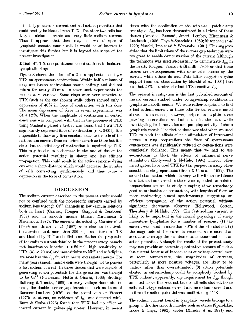
1. Fast inward currents were elicited in freshly isolated sheep lymphatic smooth muscle cells by depolarization from a holding potential of -80 mV using the whole-cell patch-clamp technique. The currents activated at voltages positive to -40 mV and peaked at 0 mV. 2. When sodium chloride in the bathing solution was replaced isosmotically with choline chloride inward currents were abolished at all potentials. 3. These currents were very sensitive to tetrodotoxin (TTX). Peak current was almost abolished at 1 microM with half-maximal inhibition at 17 nM. 4. Examination of the voltage dependence of steady state inactivation showed that more than 90% of the current was available at the normal resting potential of these cells (-60 mV). 5. The time course of recovery from inactivation was studied using a double-pulse protocol and showed that recovery was complete within 100 ms with a time constant of recovery of 20 ms. 6. Under current clamp, action potentials were elicited by depolarizing current pulses. These had a rapid upstroke and a short duration and could be blocked with 1 microM TTX. 7. Spontaneous contractions of isolated rings of sheep mesenteric lymphatic vessels were abolished or significantly depressed by 1 microM TTX.
Full text
PDF


Selected References
These references are in PubMed. This may not be the complete list of references from this article.
- Amédée T., Renaud J. F., Jmari K., Lombet A., Mironneau J., Lazdunski M. The presence of Na+ channels in myometrial smooth muscle cells is revealed by specific neurotoxins. Biochem Biophys Res Commun. 1986 Jun 13;137(2):675–681. doi: 10.1016/0006-291x(86)91131-9. [DOI] [PubMed] [Google Scholar]
- Bezanilla F., Armstrong C. M. Inactivation of the sodium channel. I. Sodium current experiments. J Gen Physiol. 1977 Nov;70(5):549–566. doi: 10.1085/jgp.70.5.549. [DOI] [PMC free article] [PubMed] [Google Scholar]
- Brading A., Bülbring E., Tomita T. The effect of sodium and calcium on the action potential of the smooth muscle of the guinea-pig taenia coli. J Physiol. 1969 Feb;200(3):637–654. doi: 10.1113/jphysiol.1969.sp008713. [DOI] [PMC free article] [PubMed] [Google Scholar]
- Cotton K. D., Hollywood M. A., McHale N. G., Thornbury K. D. Outward currents in smooth muscle cells isolated from sheep mesenteric lymphatics. J Physiol. 1997 Aug 15;503(Pt 1):1–11. doi: 10.1111/j.1469-7793.1997.001bi.x. [DOI] [PMC free article] [PubMed] [Google Scholar]
- Garnier D., Rougier O., Gargouïl Y. M., Coraboeuf E. Analyse électrophysiologique du plateau des réponses myocardiques, mise en évidence d'un courant lent entrant en absence d'ions bivalents. Pflugers Arch. 1969;313(4):321–342. doi: 10.1007/BF00593957. [DOI] [PubMed] [Google Scholar]
- Hollywood M. A., McHale N. G. Mediation of excitatory neurotransmission by the release of ATP and noradrenaline in sheep mesenteric lymphatic vessels. J Physiol. 1994 Dec 1;481(Pt 2):415–423. doi: 10.1113/jphysiol.1994.sp020450. [DOI] [PMC free article] [PubMed] [Google Scholar]
- Jmari K., Mironneau C., Mironneau J. Selectivity of calcium channels in rat uterine smooth muscle: interactions between sodium, calcium and barium ions. J Physiol. 1987 Mar;384:247–261. doi: 10.1113/jphysiol.1987.sp016453. [DOI] [PMC free article] [PubMed] [Google Scholar]
- Mironneau J., Martin C., Arnaudeau S., Jmari K., Rakotoarisoa L., Sayet I., Mironneau C. High-affinity binding sites for [3H]saxitoxin are associated with voltage-dependent sodium channels in portal vein smooth muscle. Eur J Pharmacol. 1990 Aug 10;184(2-3):315–319. doi: 10.1016/0014-2999(90)90624-f. [DOI] [PubMed] [Google Scholar]
- Muraki K., Imaizumi Y., Watanabe M. Sodium currents in smooth muscle cells freshly isolated from stomach fundus of the rat and ureter of the guinea-pig. J Physiol. 1991 Oct;442:351–375. doi: 10.1113/jphysiol.1991.sp018797. [DOI] [PMC free article] [PubMed] [Google Scholar]
- Nonomura Y., Hotta Y., Ohashi H. Tetrodotoxin and manganese ions: effects on electrical activity and tension in taenia coli of guinea pig. Science. 1966 Apr 1;152(3718):97–98. doi: 10.1126/science.152.3718.97. [DOI] [PubMed] [Google Scholar]
- Ohya Y., Sperelakis N. Fast Na+ and slow Ca2+ channels in single uterine muscle cells from pregnant rats. Am J Physiol. 1989 Aug;257(2 Pt 1):C408–C412. doi: 10.1152/ajpcell.1989.257.2.C408. [DOI] [PubMed] [Google Scholar]
- Okabe K., Kitamura K., Kuriyama H. The existence of a highly tetrodotoxin sensitive Na channel in freshly dispersed smooth muscle cells of the rabbit main pulmonary artery. Pflugers Arch. 1988 Apr;411(4):423–428. doi: 10.1007/BF00587722. [DOI] [PubMed] [Google Scholar]
- Rougier O., Vassort G., Stämpfli R. Voltage clamp experiments on frog atrial heart muscle fibres with the sucrose gap technique. Pflugers Arch Gesamte Physiol Menschen Tiere. 1968;301(2):91–108. doi: 10.1007/BF00362729. [DOI] [PubMed] [Google Scholar]
- Shuba M. F. The effect of sodium-free and potassium-free solutions, ionic current inhibitors and ouabain on electrophysiological properties of smooth muscle of guinea-pig ureter. J Physiol. 1977 Jan;264(3):837–851. doi: 10.1113/jphysiol.1977.sp011697. [DOI] [PMC free article] [PubMed] [Google Scholar]
- Sperelakis N., Inoue Y., Ohya Y. Fast Na+ channels and slow Ca2+ current in smooth muscle from pregnant rat uterus. Mol Cell Biochem. 1992 Sep 8;114(1-2):79–89. [PubMed] [Google Scholar]
- Vassort G. Voltage-clamp analysis of transmembrane ionic currents in guinea-pig myometrium: evidence for an initial potassium activation triggered by calcium influx. J Physiol. 1975 Nov;252(3):713–734. doi: 10.1113/jphysiol.1975.sp011167. [DOI] [PMC free article] [PubMed] [Google Scholar]


