Abstract
Avian influenza (AI) is an extremely contagious viral disease of domestic and wild birds that can spread rapidly among bird populations, inducing serious economic losses in the poultry industry. During the winter season 2021–2022, we isolated seventeen highly pathogenic avian influenza (HPAI) H5N8 viruses from outbreaks involving ducks in Egypt, occurring in both backyard and farm settings. The aim of this study was to pinpoint genetic key substitutions (KSs) that could heighten the risk of a human pandemic by influencing the virus’s virulence, replication ability, host specificity, susceptibility to drugs, or transmissibility. To understand their evolution, origin, and potential risks for a human pandemic, whole-genome sequencing and phylogenetic analysis were conducted. Our analysis identified numerous distinctive mutations in the Egyptian H5N8 viruses, suggesting potential enhancements in virulence, resistance to antiviral drugs, and facilitation of transmission in mammals. In this study, at least five genotypes within one genome constellation of H5N8 viruses were identified, raising concerns about the potential emergence of novel viruses with altered characteristics through reassortment between different genotypes and distinct groups. These findings underscore the role of ducks in the virus’s evolutionary process and emphasize the urgent need for enhanced biosecurity measures in domestic duck farms to mitigate pandemic risk.
Keywords: avian influenza virus, ducks, H5N8, influenza, whole-genome sequencing, reassortment
1. Introduction
Avian influenza (AI) is an extremely contagious viral disease of domestic and wild birds that can spread rapidly among bird populations, inducing serious economic losses in the poultry industry [1]. The disease is caused by avian influenza virus (IAV), which is a member of the influenza A viruses (IAVs) within the family Orthomyxoviridae. IAV is an enveloped, negative-sense, single-stranded, segmented RNA virus [2,3]. IAVs are categorized based on the surface proteins hemagglutinin (HA) and neuraminidase (NA) into 16 HA subtypes, denoted as H1 to H16, and 9 NA subtypes, denoted as N1 to N9 [4]. Based on the virus pathogenicity in chicken, IAVs are classified into highly and low-pathogenic IAVs (HPIAVs and LPIAVs), respectively. LPIAVs include all subtypes, whereas HPIAVs include some of the H5 and the H7 subtypes [5].
Wild birds, particularly of the Anseriformes and Charadriiformes orders, serve as the natural reservoirs of IAVs, where these viruses typically circulate asymptomatically [6,7]. HPAI viruses, specifically of the H5 subtype, pose a significant threat to both poultry and human health. In 1996, in a domestic goose in Guangdong China (Gs/GD), the lineage of (HPAI) A(H5N1) viruses was initially detected. Since then, these viruses have continued to circulate and spread, with their hemagglutinin (HA) genes diversifying into multiple genetic clades. The H5 Gs/GD lineage clade 2.3.4.4, particularly the H5N8 subtype, was first identified in domestic poultry in China in 2010. By 2014, it had caused numerous outbreaks in domestic and wild birds in South Korea, Japan, China, Europe, and North America [8]. Two distinct clusters of HPAI A(H5N8) viruses emerged during these outbreaks: group A viruses were detected in China, South Korea, Japan, Taiwan, Canada, the United States, and Europe (Buan-like), while group B viruses were identified only in China and South Korea in 2013–2014 and have since spread globally (Gochang-like virus) [9]. In 2016/2017, a novel reassortant virus of subtype H5N8 within clade 2.3.4.4 was discovered in wild birds at the Russian–Mongolian border during the summer of 2016. This virus subsequently spread to numerous countries across Europe, Asia, and Africa, including Egypt in the autumn of 2016 [10,11]. Some strains of H5N8 displayed decreased virulence, lower spreading ability, and a longer mean death time compared to the original HPAI strain of H5N1. The virus caused high death rates in both wild and domestic birds and crossed species boundaries, infecting mammals like humans, foxes, and seals in multiple countries [12,13,14]. In recent times in Europe, the EuroII-2020 subclade, belonging to clade 2.3.4.4b, has become the most prevalent H5N8 variant. This subclade is has been closely connected to counterparts identified in Iraq, Russia, and Kazakhstan since May 2020. Additionally, diverse genotypes have emerged through multiple reassortment events with LPAI viruses found in wild birds across Eurasia [15].
Egypt is considered a hotspot for IAVs owing to its geographical positioning in the convergence of two migratory bird flyways via the Mediterranean-Black Sea and East Africa–West Asia flyways which overlap with the more regional Rift Valley–Red Sea flyway and the endemicity of different IAV subtypes in bird populations [16]. In late 2016, HPAI H5N8 clade 2.3.4.4b virus was initially detected in migratory avian species, particularly common coot and green-winged teal, in Egypt [11]. A comprehensive surveillance initiative launched from late 2016 to 2021 revealed six distinct genotypes of the HPAI H5N8 virus in both migratory and domestic avian populations. The rapid transmission of the virus within domestic poultry across different regions in Egypt posed a significant threat to the poultry industry [17]. By 2021, a subclade called EuroII-2020 H5N8 was discovered in ostriches in Egypt [18]. Various H5 subtypes, including H5N1 and H5N5, all belonging to clade 2.3.4.4b, were identified in wild birds in Egypt [19,20]. A recent study indicated that H5N8 viruses from Egypt also belong to clade 2.3.4.4b and exhibit close genetic relationships with strains from China, Iran, and Iraq, sharing over 98% nucleotide-sequence similarity. The pathogenicity of these H5N8 strains has been confirmed through multiple assays, demonstrating a high risk to domestic birds, even those that have been vaccinated [21].
Another recent finding suggested that H5N8 can adapt to mammalian hosts through specific genetic markers, which enhance its growth and virulence in these species. A comprehensive model demonstrated that as mutations accumulate, the risk score for mammalian adaptation increases significantly. Notably, a strain isolated from migratory birds exhibited high pathogenicity in mice, with a lethal dose indicating severe effects on multiple organs. Although H5N8 primarily circulates among birds, its adaptability and emerging evidence of mammalian infections underscore the need for heightened monitoring and research to address potential zoonotic threats [22].
The current study focused on the genome sequence analysis of 17 H5N8 viruses isolated from duck farms and backyards. The primary objective was to identify key genetic signatures (KSs) associated with an increased risk of a human pandemic. These potential signatures include modifications affecting host specificity, virulence, replication capability, transmissibility, or drug susceptibility. The present study also aimed to enhance our understanding regarding the genetic attributes and origins of the recently emerged highly pathogenic avian influenza (HPAI) H5N8 viruses in Egypt, along with exploring their genetic and phylogenetic relationships.
2. Materials and Methods
2.1. Samples and Study Area
The present study was performed during the winter season 2021/2022 on 100 duck farms located in four governorates within the delta region of Egypt. These farms consisted of 60 breeder farms and 40 small and backyard farms. To determine the presence of avian influenza, samples were collected under aseptic conditions from ducks suspected of being infected. These samples included oropharyngeal and cloacal swabs, as well as tissue specimens from the liver, pancreas, spleen, brain, tracheas, and lungs. The collection of these samples was performed carefully as per the guidelines established by the World Health Organization. To form a single working sample per farm, individual bird samples from 10 ducks were combined from each farm (10 ducks/flock/1 pool/1 sample). The birds, ranging in age from two weeks to one year, were promptly transported to the laboratory following their demise. Virus isolation, real-time RT-qPCR, and biopsy were conducted at the Animal Health Research Institute’s Reference Laboratory for Veterinary Quality Control on Poultry Production. In Table S1, epidemiological information regarding samples that were taken and the samples positive for H5N8 avian influenza is detailed. Table S2 summarizes the genotyping of Egyptian IAV H5N8 genes analyzed in the current study with their accession numbers in GenBank.
2.2. Virus Detection and Isolation
The process of detecting and identifying viruses was performed using real-time RT-qPCR. The sample was prepared for analysis by extracting the viral RNA in accordance with the QIAamp viral RNA mini kit’s (Qiagen, GmbH, Hilden, Germany) instructions. A 25 µL final volume of the QuantiTect RT Mix was employed to perform real-time RT-PCR amplification in accordance with the instructions provided with the reagent. Metabion (Germany) supplied the primers and probes utilized in the subtyping of H5N8 by means of real-time RT-qPCR. These components were derived as previously described by Domingo, E. [23]. Samples were suspended in phosphate-buffered saline (PBS; pH 7.2) treated with antibiotics. These suspensions were then inoculated into 9–11-day-old specific-pathogen-free (SPF) embryonated chicken eggs, which were incubated at 37 °C for 4–5 days. The presence of avian influenza virus (IAV) in the allantoic fluid was detected using a hemagglutination (HA) assay with chicken erythrocytes [24].
2.3. cDNA Synthesis
In this analysis, viral RNA was extracted from allantoic fluid using the QIAamp Viral RNA Mini kit’s (Qiagen, GmbH, Hilden, Germany), following the manufacturer’s protocol. Once extracted, the viral gene-positive RNA was reverse-transcribed to generate complementary DNA (cDNA). For this step, the RevertAid First Strand cDNA Synthesis Kit (Thermo Fisher Scientific, Waltham, MA, USA) was used, ensuring efficient and accurate cDNA synthesis from the RNA template.
2.4. Sequence Analysis of IAV Genes
The nucleotide sequences of all IAV gene segments were determined by nanopore sequencing using a MinION Mk1B (Oxford Nanopore Technologies, Oxford, UK), as previously described [25,26]. Briefly, viral genes were amplified from synthesized cDNA by KOD One PCR Master Mix Blue (TaKaRa Bio Inc., Otsu, Japan) with gene segment-specific and subtype-specific primer sets [27]. PCR amplicons were purified using a Wizard SV gel and PCR clean-up system kit (Promega, Madison, WI, USA). Adapter ligation was performed using a direct cDNA Sequencing Kit (Oxford Nanopore Technologies) with a Native Barcoding Expansion Kit (Oxford Nanopore Technologies). Sequencing was processed using a Flongle flow cell (Oxford Nanopore Technologies). The consensus sequences for each gene segment were generated using Geneious Prime v.2021.1.1 (Biomatters Ltd., Auckland, New Zealand).
2.5. Mutational Analysis of Genes
The gene mutations in Egyptian H5N8 viruses were analyzed utilizing computational tools and approaches. Nucleotide homology and protein sequences were analyzed using the program MegAlign (DNAStar, Madison, WI, USA). Glycosylation sites of NA and HA were determined using the NetNGlyc 1.0 Server (http://www.cbs.dtu.dk/services/NetNGlyc/) (accessed on 30 April 2023), and mutations at antigenic and receptor-binding sites were identified through alignment of genes from Egyptian isolates. FluSurver (https://flusurver.bii.a-star.edu.sg/) (accessed on 30 April 2023). was employed to detect mutations in influenza virus proteins that have the potential to impact a multitude of biological attributes, such as drug resistance, virulence, host specificity, and others. Additionally, we used the CDC inventory to annotate amino acid positions by comparing them with the 390 A/Vietnam/1203/2004 reference and the original A/H5N1 goose/Guangdong reference from 1996 [28].
2.6. Phylogenetic Analysis
The consensus sequences were aligned using the MAFFT v7 online server (https://mafft.cbrc.jp/alignment/server/) (accessed on 30 April 2023).manually assembled using the BioEdit software package version 5.0.9 [29]. The open-source BLAST program (National Center for Biotechnology Information, Bethesda, MD, USA, http://blast.ncbi.nlm.nih.gov/Blast.cgi) (accessed on 30 April 2023), was used to identify the closest related sequences and analyze genetic similarities. The consensus sequences were then compared with sequences from the most closely related virus strains available in the GISAID and NCBI databases. Maximum-likelihood phylogenetic trees for each gene segment were generated using IQTREE v1.6.6 (https://github.com/iqtree/iqtree1) (accessed on 30 April 2023), with the best-fitted model selected dependent on the Akaike criterion: (TIM + F + G4) for HA, (K3Pu + F + R2) for NA, (K3P + G4) for M, (K3Pu + F + G4) PA, (TIM + F + G4) NP, (TN + F + G4) for PB1, (GTR + F + G4) for PB2, and (TPM2u + F + G4) for NS. Their robustness was assessed through ultrafast bootstrap resampling analysis with 1000 replications [30,31]. The resulting phylogenetic trees were visualized using FigTreev1.4.4 software (http://tree.bio.ed.ac.uk/software/figtree/) (accessed on 30 April 2023).
2.7. Genotype Analyses
To understand the genetic diversity of influenza A viruses (IAVs) in Egypt, the analysis involved defining clusters for each gene segment based on specific criteria. These criteria included sharing a phylogenetic cluster with a minimum bootstrap value of 70 and sequences having more than 97% nucleotide-sequence identities. Each genotype was determined by the combination of the cluster assignment of eight gene segments.
3. Results
3.1. Clinical Signs and Pathological Examination
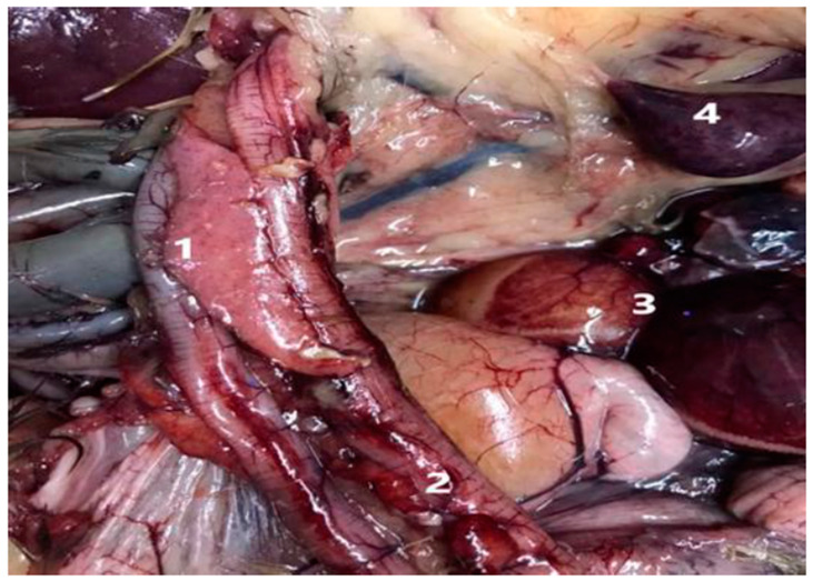
The bird populations exhibited symptoms and physical characteristics that suggested the presence of avian influenza, including a lack of appetite, reduced egg production in vaccinated farms, discharge from the eyes and nose, depression, greenish-white diarrhea, and varying degrees of high death rates. Some birds that were not vaccinated displayed nervous signs such as full reluctance to move, prostration, wing paralysis, opisthotonus, torticollis, and abnormal gait. The main postmortem lesions were generalized congestion, encephalitis, tracheitis, pneumonia, degeneration and multifocal necrosis in the liver, multifocal necrotic pancreatitis, and petechial hemorrhage in the pancreas, duodenitis, congested ovaries, and oviduct and necrotic foci in spleen with congestion (Figure 1).
Figure 1.
Pathological outcomes in the suspected case 1. Pancreatitis with petechial hemorrhage in pancreas. 2. Duodenitis. 3. Congested ovary and oviduct 4. Necrotic foci in spleen with congestion.
3.2. Virus Detection and Isolation
From a total of 100 samples collected from duck farms and backyards, only 26 tested positive for HA activity and M gene real-time PCR (RT-PCR). Of these 26 H5N8-positive samples, 17 were successfully isolated on embryonated chicken eggs (ECEs). Whole-genome sequencing was then performed on these 17 H5N8-positive specimens (Tables S1 and S2).
3.3. Genetic Analysis of the Viral Genome of Egyptian H5N8 Viruses
In the HA protein, twenty-two viruses in this study share a common and characteristic polybasic cleavage site motif “PLREKRRKR/GLF”, which is typical of Egyptian H5N8 viruses isolated from 2016 to 2022. The presence of a polybasic cleavage site is a significant virulence determinant and is associated with the highly pathogenic avian influenza (HPAI) phenotype. This motif is crucial in the virus’s ability to cause severe disease and is a key factor in its pathogenicity [32].
Comparing the HA protein of our Egyptian H5N8 viruses with Flusurver reference strain A/Sichuan/26221/2014 (H5N6) revealed significant evolutionary mutations. These mutations have altered the receptor-binding and antigenic binding sites of the HA protein. Mutations such as T110S, T139H, N205T, T139P, A172V, N199T, and R512K are found in critical regions that interact with terminal sialic acids on the surface glycans of host cells. These regions are essential for receptor binding, and alterations in these sites can significantly impact how the virus recognizes and binds to host cell receptors. Understanding these changes is important for predicting shifts in host range and could aid in assessing the risk of cross-species transmission (Table 1 and Table 2) [33].
Table 1.
Amino acid markers associated with specific phenotypic effects found in the genome of HPAI H5N8 viruses from Egypt sequenced in the current study by comparing them with the 390 A/Vietnam/1203/2004 reference and the original A/H5N1 goose/Guangdong reference from 1996.
| Protein | Amino Acid Change(s) H5 Numbering | Detections in Egypt | Phenotypic Consequences | Ref |
|---|---|---|---|---|
| HA | D94N | Yes | Increased virus binding to α2–6 | [33] |
| S133A | Yes (17/17) 100% | [34] | ||
| S154N | Yes (17/17) 100% | [35] | ||
| T156A | Yes (17/17) | [36] | ||
| V182N | Yes (17/17) 100% | [37] | ||
| K189R/T | Yes (1/17) K189T | [38] | ||
| S107R, T108I | Yes (17/17) | [39] | ||
| 323 to 330 (R-X-R, K-R) |
Yes PLREKRRKR/GLF |
HPAI cleavage site | [40] | |
| NA | I314V | Yes (17/17) | Reduced susceptibility to oseltamivir | [41] |
| M | N30D | Yes (17/17) | Increased virulence in mice, chickens, and ducks | [42] |
| I43M | Yes (17/17) | |||
| T215A | Yes (17/17) | |||
| PB2 | I292V | Yes (17/17) | Increased polymerase activity | [43] |
| K389R | Yes (17/17) | [44] | ||
| A588V | Yes (2/17) | [45] | ||
| V598T/I | Yes (17/17) | [44] | ||
| K627E | Yes (17/17) | Increased virulence in chickens | [46] | |
| S715N | Yes (17/17) | Decreased virulence in mice | [47] | |
| L89V, G309D | Yes (17/17) | Increased polymerase activity | [48] | |
| L89V, G309D, T339K, R477G, I495V, K627E, A676T | Yes (13/17) | [48] | ||
| PB1 | D3V | Yes (17/17) | [49] | |
| D622G | Yes (17/17) | [50,51] | ||
| P598L | Yes (17/17) | |||
| PB1-F2 length | Truncation | Truncation 52 lengths | Affects viral dissemination, pathogenesis, and transmission | [52,53,54,55,56,57] |
| PA | D55N | No (0/17) | Host specificity marker through statistical methods (D in avian, N in human) | [58] |
| S37A | Yes (17/17) | Increased polymerase activity | [59] | |
| P190S | Yes (17/17) | Decreased virulence in mice | [60] | |
| N383D | Yes (17/17) | Increased polymerase activity | [61] | |
| N409S | Yes (17/17) | [59] | ||
| K497R | Yes (17/17) | [62] | ||
| NP | M105V | Yes (17/17) | Increased virulence in chickens | [63] |
| A184K | Yes (17/17) | Increased replication in avian cells and virulence in chickens, enhanced IFN response | [64] | |
| NS | P42S | Yes (17/17) | Increased replication in mammalian cells, decreased interferon response | [65] |
| I106M [I101M] | Yes (17/17) | [66] | ||
| C138F | Yes (17/17) | [67] | ||
| V149A | Yes (17/17) | Increased virulence and decreased interferon response in chickens | [68] | |
| L103F, I106M [L98F, 101M] | Yes (17/17) | Increased virulence in mice | [69] | |
| 222–230 deletion | Yes (17/17) | Increased replication in mammalian and avian cell lines | ||
| P87S | Yes (17/17) | Host specificity marker through statistical methods (S in human, P in avian) | [70] |
Table 2.
Comparative analysis of amino acid markers linked to phenotypic traits in HPAI H5N8 viruses from Egypt: insights from current genome sequencing and Flusurver reference.
| Flusurver Reference | Mutation | Phenotypic Effect | |
| HA | A/Sichuan/26221/2014 (H5N6) | T110S, T139H, N205T, T139P, A172V, N199T, and R512K | Influence receptor recognition and potentially shift host specificity |
| N110S | Host specificity shift Modifies T-cell epitope presented by MHC molecules Antibody recognition sites |
||
| N205T, G16S, and I214V | Virulence Host cell receptor binding Antibody recognition sites Modify T-cell epitope presented by MHC molecules binding host protein(s) |
||
| A99V, A102V, T156A, I178M, and A201E, E284G, M285V, K492E, E495A, and P505T | Antibody recognition sites | ||
| A99N | Mild drug resistance and antibody recognition sites | ||
| NA | A/Baikalteal/KoreaDonglim/3/2014 (H5N8) | D220H, A245S, T295M, N396D, S397L, and N396D | Antibody recognition sites Strong and mild drug resistance |
| T265A | Strong drug resistance | ||
| Y450H | Binding host protein(s) | ||
| N46K, T295M, and T48A | The removal of N-acetylneuraminic acid (NA) glycosylation can increase the virulence and pathology of influenza A viruses in birds and mice [71,72] | ||
| V106I and T329A | Mild drug resistance | ||
| NP | A/duck/HongKong/24/1976 (H4N2) | T396N, A423V, and A423T | Modify T-cell epitope presented by MHC molecules |
| PA | P A/Netherlands/219/2003 (H7N7) | K113R, S184N, S224A, and P653S | Modify T-cell epitope presented by MHC molecules |
| K615R | Host specificity shift, virulence | ||
| PB2 | A/mallard/Astrakhan/263/1982 (H14N5) | V495A, A588V, T676A, I676A, and V553I | Host specificity shift, virulence |
| E558D | Modify T-cell epitope presented by MHC molecules | ||
| NS1 | A/chicken/BCFAV8//2014 (H5N2) | G70K and I81V | Host specificity shift |
| E227G, G232R, S205N, and R231stop | Virulence | ||
| S7L, H17Y, S48T S114P, T143A, L166F, D189N, and Q218stop | Binding host protein | ||
| NS2 | A/WSN//1933 (H1N1) and A/quail/Italy/1117/1965 (H10N8) | M19L and A48T | Virulence |
| NS1 | A/Canada/720/2005 (H2N2) | M81V, S205N, Q218stop, R227G, K229E, and V230I | Virulence |
| S7L, H17Y, Q21R V22F, R118K, T143A, L166F, N171D, and D189N | Binding host protein | ||
| T215S and T215P | Host specificity shift, virulence | ||
Although analysis of Egyptian H5N8 viruses showed no deletions in the neuraminidase (NA) genes, there are several mutations that affect NA drug sensitivity and evade antiviral treatments (Table 1 and Table 2). Additionally, several amino acid changes were observed in two regions that are considered crucial for NA hemadsorption activity. In particular, two isolates, namely, F653/18 and F653/32, showed 361RTISRNSRSGFE372, and five other isolates, namely, F653/10, F653/12/, F653/13, F653/23, and F653/24 showed 390RQVVVDDLNWSGS404 (Table S3).
The NA protein of the analyzed viruses showed conserved N-linked glycosylation sites at positions 54 (NETV), 67 (NTSV), 84 (NNTE), 144 (NGTV), and 398 (NWSG) (Table S4). Notably, all analyzed viruses lacked a glycosylation site at position 293 (NWTG), which is also in line with observations in the EuroII-2020 subclade and Egy/ostrich/2021 viruses [18]. Two new possible glycosylation sites were recognized in isolates of the current study; a glycosylation site at position 2 (NPSQ), which was found in nine isolates, and a glycosylation site at position 54 (NETA) that was found in one isolate (Table S4).
A series of notable mutations were identified in both the HA gene (54D, 236N, 269V, and 306V) and the NA gene (191V, 213V, and 342H). Furthermore, all isolates demonstrated mutations in PB2 (292I, 615I, and 714S), PB1 (211R, 431H, and 691K), and PB1-F2 (42W and 46S). Other proteins exhibited mutations as well, including PA (96N, 120I, 214L, 348I, and 445Y), NP (318S), M1 (42L, 134R, 209A, and 248I), and M2 (18K). In addition, distinct variations were detected in NS1 (100R, 210G, and 215S) and NS2 (28V). These mutations were newly identified and had not been previously reported in Egyptian strains from 2020 to 2021 (Table S5).
The comparison of protein in Egyptian viruses to the reference strain as suggested by FluSurver revealed a series of mutations that have emerged over time. These mutations impact virulence, shifts in host specificity, and interactions with host proteins (Table 1 and Table 2).
Further analysis was performed to investigate other virulence and mammalian adaptation factors in the viral PB2, PB1, PA, NP, M2, NS1, and NS2 proteins (Tables S6 and S7).
3.4. Phylogenetic Analysis and Sequence Similarity
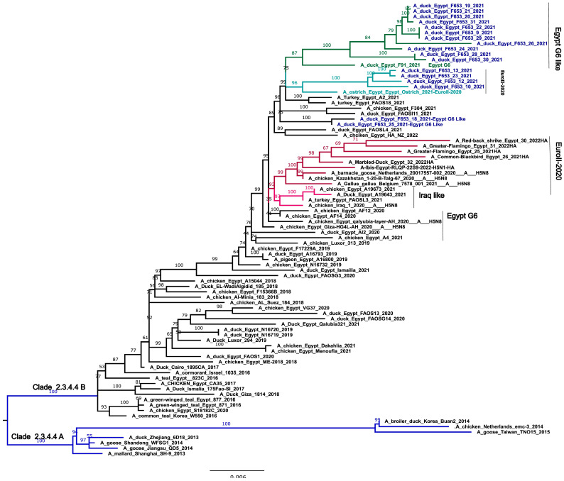
HA gene analysis revealed two distinct genetic groups (I and II), with group II further subdivided into G1-II (evolved from EuroII-2020) and G2-II (from Egypt Genotype 6), suggesting a mix of local and imported viral strains.
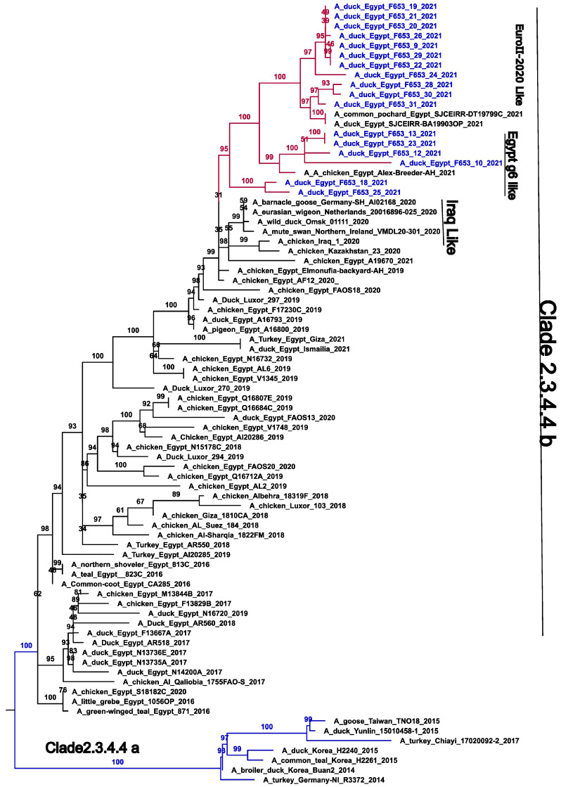
However, in the NA gene, genetic group I is subdivided into three subgroups with a different evolutionary origin: G1 (I and II), which evolved from Egypt Genotype 6, and G1-III, which evolved from the EuroII-2020 subclade. The long branch length between the genetic group and its ancestors (EuroII-2020 subclade) suggests that these genetic groups may have been circulating undetected in wild or domestic birds, thus indicating potential local transmission prior to detection. These contrasting genetic patterns highlight the complexity and potential reassortment events between the EuroII-2020 subclade and Genotype 6 in Egypt during 2019–2021 (Figure 2 and Figure 3).
Figure 2.
Phylogenetic trees of the nucleotide sequences of the HA gene segments of the avian influenza H5 subtype. Maximum-likelihood calculations were performed using the IQ-TREE v 2.1.3 software with the best fit model according to the Bayesian criterion (TIM + F + G4). Egyptian HPAI H5N8 viruses used in this study are colored in blue. Blue branches may indicate clade 2.3.4.4A, Red branches may indicate EuroII-2020 subclade, light green indicate viruses originated from EuroII-2020 subclade and deep green indicate viruses originated from Egypt G6.
Figure 3.
Phylogenetic trees of the nucleotide sequences of the NA gene segments of the avian influenza H5 subtype. Maximum-likelihood calculations were performed using the IQ-TREE v 2.1.3 software with the best fit model according to the Bayesian criterion (TIM+F+G4). Egyptian HPAI H5N8 viruses used in this study are colored in blue. Blue branches may indicate clade 2.3.4.4A.
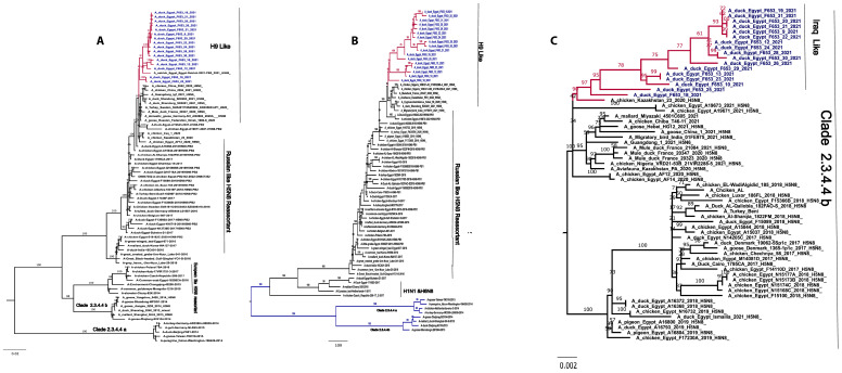
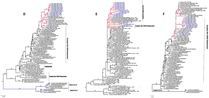
The PB2 and PB1 gene sequences of the isolates showed a nucleotide-sequence homology ranging from 98% to 100% and 97% to 99%, respectively. In the case of PB2 and PB1, they are related to H9-like viruses (Figure 4).
Figure 4.
Phylogenetic tree of internal genes. The PB1 and PB2 genes of all Egyptian strains cluster in H9-like viruses and PA genes are related to A/chicken/Kazakhstan/23/20209 Iraqi-like viruses (A) Phylogenetic tree of the pb2 gene. (B) Phylogenetic tree of the PB1 gene. (C) Phylogenetic tree of the PA gene. Egyptian HPAI H5N8 viruses used in this study are colored in blue. The NP related to European-like H5N8 reassortant 2016 and European-like H5N8 reassortant 2020, M related to Russian-like H5N8 reassortant 2016 and Russian reassortant H5N5 2021, and NS closely related to Russian reassortant-like H5N8 2016. (D) Phylogenetic tree of the NP gene. (E) Phylogenetic tree of the M gene. (F) Phylogenetic tree of the NS gene. Egyptian HPAI H5N8 viruses used in this study are colored in blue.
The PA gene sequences of the isolates showed 98-100% nucleotide homology and formed distinct genetic groups originating from chicken/Kazakhstan/23/2020 H5N8 (Iraqi-like viruses). The long horizontal branch lengths observed between these PA gene groups and their closest relatives suggest that the virus may have been circulating undetected in wild or domestic birds before it was introduced into Egypt (Figure 4).
The M gene sequences of the isolates showed 98-100% nucleotide homology, and phylogenetic analysis revealed three distinct genetic groups (I, II and III) within clade 2.3.4.4b. Each group evolved independently from different strains, including EuroII-2020 strains, Russian reassortant H5N5 2021-like, shelduck/Kalmykia/1814-1/2021 (H5N5), and A/Dalmatian pelican/Astrakhan/417-1/2021 (H5N5). Group I, considered an outgroup, mainly originated from Egyptian endemic Russian-like H5N8 reassortant strains. The NS gene sequences of the isolates showed 97-100% nucleotide homology and formed distinct genetic groups originating from Russian reassortant-like H5N8 2016 (Figure 4).
The NP gene sequences of the isolates showed 98-100% nucleotide homology, forming three distinct genetic groups (I, II and III). Group I is associated with European H5N8 reassortant strains from 2016, while groups II and III are linked to European H5N8 reassortant strains 2021 (Figure 4).
BLAST searches on the NCBI platform for all eight genes revealed that our H5N8 viruses exhibit close genetic relationships with strains from China, India, Kazakhstan, and Iraq, sharing over 98% nucleotide-sequence similarity (Table 3 and Table 4).
Table 3.
Nucleotide-sequence homology comparison of the gene segments of HPAI H5N8 viruses from Egypt with available sequences in NCBI platform.
| Accession No. | Closest Relative | Identity (%) | |
|---|---|---|---|
| PB2 | EPI1902241 | A/chicken/Egypt/F17230B/2019 (H5N8) | 99% |
| ON862701.1 | A/duck/Shandong/SD0263/2021 (H5N6) | 98.6% | |
| EPI1814302 | A/goose/Omsk/30006/2020 (A/H5N8) segment 1 (PB2) | 98% | |
| EPI1811685 | A/goose/Russian Federation/Kurgan/1345-25/2020 (A/H5N8) segment 1 (PB2) | 98% | |
| ON329039.1 | A/chicken/China/2042/2020 (H9N2) | 98.6% | |
| PB1 | MT261707.1 | A/duck/Egypt/A16793/2019 (H5N8) | 98.7% |
| EPI2162079 | A/chicken/Kazakhstan/23/2020 (H5N8) | 98% | |
| MW505413.1 | A/Cygnus columbianus/Hubei/56/2020 (H5N8) | 98.77% | |
| ON862694.1 | A/duck/Shandong/SD0261/2021 (H5N6) | 98.64% | |
| EPI1811626 | A/chicken/Iraq/1/2020 (H5N8) segment 2 (PB1) | 99% | |
| PA | OP740951.1 | A/Aviafauna/Kazakhstan/PA/2020 (H5N8) | 99% |
| EPI1881024 | A/bean goose/Hubei/BQ11/2020 (A/H5N8) segment 3 (PA) | 99% | |
| EPI2162080 | A/chicken/Kazakhstan/23/2020 (A/H5N8) segment 3 (PA) | 99% | |
| HA | MW137842.1 | A/Aviafauna/Kazakhstan/HA/2020 (H5N8) | 98.10% |
| OP740959.1 | A/chicken/Egypt/F17229A/2019 (H5N8) | 98.30% | |
| OQ711838.1 | A/Migratory bird/India/01FE975/2021 (H5N8) | 98.10% | |
| EPI1942903 | A/Anser_Brachyrhynchus_Anser_Anser/Belgium/13846/2020 (A/H5N8) segment 4 (HA) | 98% | |
| NP | MT261706.1 | A/duck/Egypt/A16793/2019 (H5N8) | 99.27% |
| OL354983.1 | A/duck/Egypt/A19643/2021 (H5N8) | 98.93% | |
| ON943055.1 | A/chicken/Kazakhstan/23/2020 (H5N8) | 98.80% | |
| EPI1811629 | A/chicken/Iraq/1/2020(H5N8) segment 5 (NP) | 98% | |
| NA | MW137803.1 | A/chicken/Egypt/F17230B/2019 (H5N8) | 99.15% |
| MZ882181.1 | A/goose/China/21FU005/2020 (H5N8) | 99.08% | |
| EPI1920391 | A/white-tailed eagle/Germany-SH/AI02170/2020 (H5N8) | 99% | |
| EPI1860070 | A/anser_anser/Spain/297-1_21VIR1230-5/2021 (H5N8) | 99% | |
| EPI1919630 | A/barnacle goose/Germany-SH/AI02190/2020 (H5N8) | 97% | |
| MP | OP597600.1 | Shelduck/Kalmykia/1814-1/2021 (H5N5) | 98.78% |
| OL353691.1 | A/chicken/Egypt/A19670/2021 (H5N8) | 98.78% | |
| OP597560.1 | A/Dalmatian pelican/Astrakhan/417-1/2021 (H5N5) | 98.67% | |
| EPI1920856 | A/domestic goose/Germany-MV/AI02558/2021 (H5N8) | 99% | |
| EPI1848412 | A/common buzzard/Sweden/SVA210212SZ0284/KN000390/2021 | 99% | |
| NS | MW505387.1 | A/Cygnus columbianus/Hubei/51/2020 (H5N8) | 98.21% |
| EPI1913525 | A/chicken/Egypt/AF12/2020 (H5N8) segment 8 (NS) | 98% | |
| EPI1927723 | A/pigeon/Kazakhstan/15-20-B-Talg-5/2020 (H5N8) | 99% | |
| EPI2152264 | A/brown-headed gull/Tibet/N38/2021 (H5N8) segment 8 (NS) | 98% | |
| OR048707.1 | A/mallard duck/Japan/KU-d89/2021 (H5N8) | 98.09% | |
| MW137800.1 | A/chicken/Egypt/F17230B/2019 (H5N8) | 98.21% |
Table 4.
Evolutionary ancestors and subgroups of HPAI H5N8 viruses of the present study.
| Gene | Evolutionary Ancestor | Subgroup | Subtype |
|---|---|---|---|
| HA | Egypt G6 | Egypt G6 | HPAI |
| Local Endemic EuroII-2020 subclade Egy/ostrich/2021 |
EuroII-2020 | ||
| Egypt G6 A/duck/Egypt/F91/2021 | Egypt G6-like | ||
| NA | Egypt G6 PA/chicken/Egypt/Elmonufia-backyard-AH/2019 | Egypt G6-like | HPAI |
| Egypt G6 A/chicken/Egypt/Alex-Breeder-AH/2021 | Egypt G6-like | ||
| Local Endemic EuroII-2020 Egy/ostrich/2021 |
EuroII-2020 | ||
| M | A/chicken/Egypt/AF12/2020 (H5N8) | Russian-like H5N8 reassortant 2016 | |
| A/chicken/Kazakhstan/23/2020 | EuroII-2020 | HPAI | |
| A/shelduck/Kalmykia/18141/2021 (H5N5) A/Dalmatian pelican/Astrakhan/417-1/2021 (H5N5) A/pelican/Dagestan/397-1/2021 (H5N5 |
Russian reassortant H5N5 2021 | ||
| NP | A/chicken/Egypt/AF12/2020 (H5N8) A/chicken/Egypt/AF14/2020 (H5N8) |
European-like H5N8 reassortant 2016 | LPAI |
| A/chicken/Egypt/A19670/2021 | European-like H5N8 reassortant 2020 | ||
| PA | A/chicken/Kazakhstan/23/2020 A/chicken/Iraq/1/2020 |
Iraqi-like virus | HPAI |
| PB2 | Eurasian LPAI A/duck/Shandong/SD0261/2021 (H5N6) A/chicken/China/2042/2020 (H9N2) Egy/ostrich/2021 H5N8 |
H9-like | LPAI |
| PB1 | Eurasian LPAI A/duck/Shandong/SD0261/2021 (H5N6) |
H9-like | LPAI |
| NS | A/chicken/Egypt/A19670/2021 | Russian-like H5N8 reassortant 2016 and Russian-like H5N8 reassortant 2020 (Russian-like H5N8 reassortant 2016-like) | HPAI |
4. Discussion
Since 2016, H5N8 viruses in clade 2.3.4.4.b have been spreading across Egypt [17,18,19,20,21,22,23,24,25,26,27,28,29,30,31,32,33,34,35,36,37,38,39,40,41,42,43,44,45,46,47,48,49,50,51,52,53,54,55,56,57,58,59,60,61,62,63,64,65,66,67,68,69,70,71,72,73,74,75,76,77,78]. Previous reports indicated that the major genotypes of H5N8 avian influenza viruses (IAVs) in Egypt can be divided into six distinct genotypes [17,51]. Recent studies have also reported the introduction of the EuroII-2020 subclade, particularly found in ostriches [18].
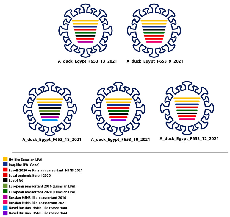
In this study, molecular and phylogenetic analyses of H5N8 strains found in Egypt revealed a significant link to waterfowl-associated strains from Europe. Our findings identified two distinct types of low-pathogenic avian influenza (LPAI) viruses in the region. The first type includes locally endemic reassortant viruses, such as the H5N8 reassortant 2016 and Russian-like H5N8 reassortant 2016, derived from European sources. These viruses have established themselves in the local population since 2016, demonstrating persistence and integration with existing endemic viruses, highlighting their adaptation and stability within the local environment. The second type comprises recently introduced viruses from Eurasia, such as H9-like strains, representing new genetic variants that were not previously detected locally, indicating ongoing viral introductions and growing genetic diversity (Figure 5).
Figure 5.
Representation of the newly identified genotypes of H5N8 viruses in poultry in Egypt. The newly identified genotypes of H5N8 viruses in poultry in Egypt are thought to be a result of genetic contributions from various sources, including Iraqi-like viruses (Kazakhstan/PA/2020 (H5N8), EuroII-2020 subclade, Eurasian LPAI, and Egypt G6. Notably, the NS gene differs in A/duck/Egypt/F653/10/2021, A/duck/Egypt/F653/12/2021, and A/duck/Egypt/F653/13/2021, while the other segments exhibit a high degree of similarity. See also Table 4.
The evolution of the EuroII-2020 and endemic Genotype 6 strains has led to the emergence of new genotypes resembling these original strains. Furthermore, reassortment events have occurred between these newly evolved genotypes, the recently introduced Eurasian viruses—including H9-like strains—and the endemic Russian-like and European-like reassortants from 2016. Afterward, Egypt witnessed the emergence of five distinct genotypes of the H5N8 avian influenza virus, displaying a similar genome constellation where the internal genes (NP, PB1, PB2) were derived from Eurasian low-pathogenic avian influenza viruses (LPIAVs). In contrast, the M, NS, and PA genes were sourced from highly pathogenic avian influenza (HPAI) viruses. However, a recent study has identified a new genome constellation where the majority of genes are from LPIAVs, with exceptions for the HA, NA, and NS genes. This shift reflects an evolving genetic landscape in the H5N8 virus [77].
The whole-genome analyses of HPAI H5N8 viruses indeed revealed several key mutations (T156A, S123P, D183N, and S223R) that could influence the virus’s adaptation, persistence, and virulence. These mutations were found to be associated with viruses from the 2.3.4 clade, which contained particular residues (T156A, S123P, D183N, and S223R) and exhibited transient persistence before implementing a compensatory mechanism that entailed a modification in the NA protein. This suggests that while these HA mutations initially provided some advantage, the viruses required additional changes in the NA protein to achieve stable adaptation and persistence [78]. In our study, we identified novel alterations in the amino acid sequences of the neuraminidase hemadsorption site which could impact the enzyme’s catalytic efficiency. The hemadsorption site is essential for NA function and plays a key role in interacting with HA receptor-binding specificity. Novel mutations in NA, combined with changes in HA, may be crucial for the emergence of pandemic influenza viruses. This underscores the importance of NA in viral evolution and adaptation [79].
Recent whole-genome analyses of the HPAI H5N8 viruses have revealed molecular mutations that could potentially increase their zoonotic potential. The H5 protein of all H5N8 viruses described here carries the mutations S133A, D94N, S154N, T156A, and V182N (H5 numbering). In addition, the H5 protein of the H5N8 virus A/duck/Egypt/F653/12/2021 features K189R/T mutations. These mutations in the H5 protein have been shown to increase binding to alpha2,6-SA. The substitution K189T in the HA protein, which is transported by Egyptian strains, can be utilized to evaluate the potential pandemic risk throughout surveillance of emerging viruses [38]. Furthermore, it is important to note that the S133A mutation characterizes the HA gene of all H5 viruses identified in Europe since early 2021 [34,80]. Moreover, the substitution T156A significantly modifies the antigenic characteristics of the 2.3.4 H5 viruses. Notably, T156A is consistently detected in airborne transmission-capable mutants adapted to ferrets. While most H5N1 viruses typically have a glycosylation site at positions 154–156, the T156A mutation leads to the loss of this glycosylation site. The loss of this glycosylation site is associated with an increased ability of the virus to infect mammals, suggesting that the loss of this glycosylation site enhances the virus’s adaptability and virulence in mammalian hosts [81]. In this investigation, it was determined that the HA protein of all our H5N8 Egyptian viruses contains Q222 and G224 residues (H5 numbering). These residues signal a preference for avian-like receptors, which reduces the likelihood of human-to-human transmission [82]. Additionally, the presence of these residues contributes to an increased proinflammatory response [34].
The N30D, I43M, and T215A substitutions in the M gene, which are noted in all isolates, have been associated with increased virulence in animal models [42]. The CCHH zinc-finger motif within helix 9 of the M1 protein is believed to play a crucial role in regulating virulence, particularly in mice [83].
Notably, there are mammalian adaptive markers present in the PB2 protein of thirteen H5N8 viruses, including L89V, G309D, T339K, R477G, I495V, K627E, and A676T. The L89V substitution in the PB2 protein is located in the vicinity of residues 51–259, which is implicated in interactions with PB1, and residues 1–515, which is the region accountable for binding to heat shock protein 90 (HSP90) [84]. Furthermore, substitutions G309D, T339K, R477G, and I495V have been identified in the region where HSP90 binds. By facilitating interactions between polymerase subunits or between the polymerase and host factors, these substitutions increase polymerase activity and, consequently, mammalian virulence [85].
It is noteworthy that in publicly available sequences, a combination of these six mutations along with 627E in PB2 was detected in 58 out of 340 avian influenza viruses (IAVs) responsible for human infections, including H5N1, H7N7, and H9N2. Moreover, a substantial proportion (67.5%, 13,074 out of 19,338) of influenza A viruses (IAVs) isolated from avian hosts possess this combination of mutations in PB2. Indeed, prior reports have highlighted that numerous low-pathogenic avian influenza viruses (LPIAVs) isolated from wild birds, which exhibit a combination of six specific mutations, can replicate in the BALB/c mouse model without the need for prior adaptation [86]. In accordance with our study, the substitutions L89V, G309D, and T339K were noted in Egypt H5N1 viruses recently isolated [87]. Moreover, the K497R substitution in the PA protein of these seventeen H5N8 viruses is particularly significant due to its pivotal role in facilitating the adaptation of avian viruses to mammalian hosts. Furthermore, it is worth noting that the pathogenicity and host specificity of influenza viruses can be directly influenced by the K615R substitution in the PA gene [85]. Also, in the PB1 protein, the D3V and D622G substitutions are associated with increased viral replication and enhanced polymerase activity, both in mammalian and avian cell lines. These mutations also correlate with elevated polymerase activity and heightened virulence in rodents. Additionally, the P598L mutation in PB1 is known to further augment polymerase activity in mammalian cells and rodents [46]. This modification has been documented to enhance the polymerase activity of a weakened human influenza virus containing the PB2 K627E mutation, which is typically involved in host adaptation and virulence enhancement in mammals. These mutations contribute to the virus’s ability to replicate more efficiently and potentially increase its pathogenicity across species [46].
Surprisingly, the NS1 protein in all Egyptian strains of these study lacks a PDZ motif and contains residues F103 and M106 within the cleavage and polyadenylation specificity factor (CPSF30) binding site [88]. These residues, which are frequently found in human isolates, inhibit the production of beta interferon (IFN-β) mRNA and stabilize CPSF30 binding [89].
Similar to a previous study in Egypt, numerous mutations were found in different viral proteins, and mammalian adaptations and virulence-related characteristics were observed in current Egyptian H5N8. These mutations were detected in PB2 (588V, 598T, and 504V), PA (127V, 550L, and 672L), PB1 (13P), NP (398Q), NS2 (31I), and NS1 (42S and 189N). The mutations identified in key viral proteins, especially in PB2 and PA, suggest an increased ability of the virus to adapt to mammalian hosts, potentially posing a higher risk for human infections. Despite these genetic characteristics, no proof of human infection with these viruses was found [17].
In conclusion, domestic ducks play a significant role as intermediaries in the transmission of avian influenza, bridging migratory waterfowl and terrestrial poultry. As asymptomatic carriers, they can spread the influenza A virus (IAV) to other species, underscoring the need for improved surveillance and management strategies.
To effectively control avian influenza in Egypt, it is crucial to enhance monitoring of (LPIAVs) in domestic ducks and poultry. Targeting specific strains that drive virus evolution and reinforcing biosecurity measures on farms and in live-poultry markets are essential steps.
Education of poultry farmers and workers on best practices, alongside collaboration with international organizations, will facilitate the exchange of information and strategies. Investment in research to understand virus reassortment and the development of new vaccines and preventive measures will be vital for managing outbreaks and mitigating risks.
A comprehensive strategy must include ongoing monitoring of genetic changes, evaluation of virological attributes, and strengthened surveillance initiatives. Updating vaccine seeds to address antigenic mismatches with circulating strains is recommended. Consistent vaccination efforts, particularly in backyard ducks, are necessary to overcome challenges related to vaccination failures. Adherence to rigorous biosafety and biosecurity measures is critical to safeguard poultry health and prevent future outbreaks of HPAI.
Acknowledgments
The authors would like to express their gratitude to all the veterinarians and farmers who contributed to this study. Special thanks are extended to our colleagues, Enas Hamed, Dina Arafa, and Wessam Hasan Mady at the Animal Health Research Institute, Egypt, for their invaluable guidance and support throughout the research process.
Supplementary Materials
The following supporting information can be downloaded at: https://www.mdpi.com/article/10.3390/v16111655/s1, Table S1. AIVs derived from ducks in Egypt between 2020/2021 and 2021/2022; Table S2. Summary of the genotyping of Egyptian AIV H5N8 genes analyzed in the current study with their accession numbers in GenBank; Table S3. Amino acid substitutions in the hemadsorbing sites and stalk region; Table S4. Comparison between Glycosylation sites of Neuraminidase (NA) of Egyptian viruses, the Egypt- ostrich-2021 and subclades of2.3.4.4 H5N8 b; Table S5. Amino acid differences between the three-reassortant H5N8 viruses and current study viruses; Table S6. Analysis of virulence determinants in the viral PB2, PB1, PA, NP, M2, NS1, and NS2 proteins in comparison with A/chicken/Egypt/NZ/2022 [65,90,91,92,93,94,95,96,97,98,99,100,101]. Table S7. Analysis of host range genetic determinants in the PB2, PB1, PA, NP, M1, M2, NS1, and NS2 proteins in H5N8 viruses in comparison with A/chicken/Egypt/NZ/2022 [58,88,90,92,93,94,102,103,104,105,106,107,108,109,110,111,112,113,114,115].
Author Contributions
N.S., M.E., I.K., M.O., A.M.K., S.O. and K.O.: Conceived and designed the study; N.S.: samples collection and data curation; N.S.: cDNA transport to Japan; N.S.: reviewing and editing; N.S.: Methodology and software; B.Y.A. and M.A.S.: supervision, reviewing, and editing. All authors have read and agreed to the published version of the manuscript.
Institutional Review Board Statement
The birds used in our study were freshly dead specimens obtained from farms and backyards, specifically those that had succumbed to avian influenza infection. As these birds were not euthanized for the study, but rather were collected after their natural demise, ethical review and approval were not required according to local legislation and institutional guidelines.
Informed Consent Statement
Not applicable.
Data Availability Statement
Data are available upon reasonable request from the corresponding authors.
Conflicts of Interest
The authors declare no conflicts of interest.
Funding Statement
The Researchers would like to thank the Deanship of Graduate Studies and Scientific Research at Qassim University for financial support (QU-APC-2024-9/1).
Footnotes
Disclaimer/Publisher’s Note: The statements, opinions and data contained in all publications are solely those of the individual author(s) and contributor(s) and not of MDPI and/or the editor(s). MDPI and/or the editor(s) disclaim responsibility for any injury to people or property resulting from any ideas, methods, instructions or products referred to in the content.
References
- 1.Swayne D., Suarez D.L. Highly pathogenic avian influenza. Rev. Sci. Tech. 2000;19:463–475. doi: 10.20506/rst.19.2.1230. [DOI] [PubMed] [Google Scholar]
- 2.Samji T. Influenza A: Understanding the viral life cycle. Yale J. Biol. Med. 2009;82:153. [PMC free article] [PubMed] [Google Scholar]
- 3.Abdelwhab E.M., Abdel-Moneim A.S. Recent Advances in Animal Virology. Springer; Singapore: 2019. Orthomyxoviruses; pp. 351–378. [Google Scholar]
- 4.Bhalerao U., Mavi A.K., Manglic S., Sakshi, Chowdhury S., Kumar U., Rohil V. An Updated Review on Influenza Viruses. In Emerging Human Viral Diseases. Volume I. Springer; Singapore: 2023. pp. 71–106. [Google Scholar]
- 5.Alexander D.J. A review of avian influenza in different bird species. Vet. Microbiol. 2000;74:3–13. doi: 10.1016/S0378-1135(00)00160-7. [DOI] [PubMed] [Google Scholar]
- 6.Torrontegui Vega O. Ph.D. Thesis. Universidad del País Vasco-Euskal Herriko Unibertsitatea; Eibar, Spain: 2017. Pathogen Dynamics in Wild Bird Species: Circulation of Avian Influenza Viruses in Natural vs. Anthropic Ecosystems and Concurrent Infections with Other Agents in Waterbirds. [Google Scholar]
- 7.de Franca M.S. Ph.D. Thesis. University of Georgia; Athens, Georgia: 2013. Evaluation of Host and Viral Factors Involved in the Infectivity, Pathogenesis and Transmission of Avian Influenza Viruses in Wild Birds. [Google Scholar]
- 8.Lee Y.-J., Kang H.-M., Lee E.-K., Song B.-M., Jeong J., Kwon Y.-K., Kim H.-R., Lee K.-J., Hong M.-S., Jang I.J.E. Novel reassortant influenza A (H5N8) viruses, South Korea, 2014. Emerg. Infect. Dis. 2014;20:1087. doi: 10.3201/eid2006.140233. [DOI] [PMC free article] [PubMed] [Google Scholar]
- 9.Lee D.-H., Bahl J., Torchetti M.K., Killian M.L., Ip H.S., DeLiberto T.J., Swayne D.E.J.E. Highly pathogenic avian influenza viruses and generation of novel reassortants, United States, 2014–2015. Emerg. Infect. Dis. 2016;22:1283. doi: 10.3201/eid2207.160048. [DOI] [PMC free article] [PubMed] [Google Scholar]
- 10.Lee D.-H., Sharshov K., Swayne D.E., Kurskaya O., Sobolev I., Kabilov M., Alekseev A., Irza V., Shestopalov A. Novel reassortant clade 2.3. 4.4 avian influenza A (H5N8) virus in wild aquatic birds, Russia, 2016. Emerg. Infect. Dis. 2017;23:359. doi: 10.3201/eid2302.161252. [DOI] [PMC free article] [PubMed] [Google Scholar]
- 11.Selim A.A., Erfan A.M., Hagag N., Zanaty A., Samir A.-H., Samy M., Abdelhalim A., Arafa A.-S.A., Soliman M.A., Shaheen M.J.E. Highly pathogenic avian influenza virus (H5N8) clade 2.3. 4.4 infection in migratory birds, Egypt. Emerg. Infect. Dis. 2017;23:1048. doi: 10.3201/eid2306.162056. [DOI] [PMC free article] [PubMed] [Google Scholar]
- 12.European Food Safety Authority. European Centre for Disease Prevention and Control. European Union Reference Laboratory for Avian Influenza. Adlhoch C., Fusaro A., Gonzales J.L., Kuiken T., Marangon S., Niqueux É., Staubach C., et al. Avian influenza overview February–May 2021. EFSA J. 2021;19:e06951. doi: 10.2903/j.efsa.2021.6951. [DOI] [PMC free article] [PubMed] [Google Scholar]
- 13.Alkie T.N., Cox S., Embury-Hyatt C., Stevens B., Pople N., Pybus M.J., Xu W., Hisanaga T., Suderman M., Koziuk J. Characterization of neurotropic HPAI H5N1 viruses with novel genome constellations and mammalian adaptive mutations in free-living mesocarnivores in Canada. Emerg. Microbes Infect. 2023;12:2186608. doi: 10.1080/22221751.2023.2186608. [DOI] [PMC free article] [PubMed] [Google Scholar]
- 14.Postel A., King J., Kaiser F.K., Kennedy J., Lombardo M.S., Reineking W., de le Roi M., Harder T., Pohlmann A., Gerlach T., et al. Infections with highly pathogenic avian influenza A virus (HPAIV) H5N8 in harbor seals at the German North Sea coast, 2021. Emerg. Microbes Infect. 2022;11:725–729. doi: 10.1080/22221751.2022.2043726. [DOI] [PMC free article] [PubMed] [Google Scholar]
- 15.Lewis N.S., Banyard A.C., Whittard E., Karibayev T., Al Kafagi T., Chvala I., Byrne A., Meruyert S., King J., Harder T., et al. Infections, Emergence and spread of novel H5N8, H5N5 and H5N1 clade 2.3. 4.4 highly pathogenic avian influenza in 2020. Emerg. Microbes Infect. 2021;10:148–151. doi: 10.1080/22221751.2021.1872355. [DOI] [PMC free article] [PubMed] [Google Scholar]
- 16.Naguib M.M., Verhagen J.H., Samy A., Eriksson P., Fife M., Lundkvist Å., Ellström P., Järhult J.D. Avian influenza viruses at the wild–domestic bird interface in Egypt. Infect. Ecol. Epidemiol. 2019;9:1575687. doi: 10.1080/20008686.2019.1575687. [DOI] [PMC free article] [PubMed] [Google Scholar]
- 17.Kandeil A., Moatasim Y., El Taweel A., El Sayes M., Rubrum A., Jeevan T., McKenzie P.P., Webby R.J., Ali M.A., Kayali G., et al. Genetic and antigenic characteristics of highly pathogenic avian influenza A(H5N8) viruses circulating in domestic poultry in Egypt, 2017–2021. Microorganisms. 2022;10:595. doi: 10.3390/microorganisms10030595. [DOI] [PMC free article] [PubMed] [Google Scholar]
- 18.Elsayed H.S., Adel A., Alshaya D.S., Safhi F.A., Elmasry D.M., Selim K., Erfan A.A., Eid S., Selim S., El-Saadony M.T. First isolation of influenza a subtype H5N8 in ostrich: Pathological and genetic characterization. Poult. Sci. 2022;101:102156. doi: 10.1016/j.psj.2022.102156. [DOI] [PMC free article] [PubMed] [Google Scholar]
- 19.Mosaad Z., Elhusseiny M.H., Zanaty A., Fathy M.M., Hagag N.M., Mady W.H., Said D., Elsayed M.M., Erfan A.M., Rabie N.J.P. Emergence of Highly Pathogenic Avian Influenza A Virus (H5N1) of Clade 2.3. 4.4 b in Egypt, 2021–2022. Pathogens. 2023;12:90. doi: 10.3390/pathogens12010090. [DOI] [PMC free article] [PubMed] [Google Scholar]
- 20.Kandeil A., Kayed A., Moatasim Y., Aboulhoda B.E., El Taweel A.N., Kutkat O., El Sayes M., Gomaa M., El-Shesheny R., Webby R.J.M.P. Molecular identification and virological characteristics of highly pathogenic avian influenza A/H5N5 virus in wild birds in Egypt. Microb. Pathog. 2023;174:105928. doi: 10.1016/j.micpath.2022.105928. [DOI] [PubMed] [Google Scholar]
- 21.Ali A.A., Kotb G., Soliman A., Swede K.S. Isolation and Identification of The Highly Pathogenic Avian Influenza H5N8 Virus Isolated From Commercial Layer Chickens in Al-Sharkia Province in Egypt During 2019–2021. Zagazig Vet. J. 2024;52:117–129. doi: 10.21608/zvjz.2024.268382.1234. [DOI] [Google Scholar]
- 22.Chokkakula S., Oh S., Choi W.-S., Kim C.I., Jeong J.H., Kim B.K., Park J.-H., Min S.C., Kim E.-G., Baek Y.H. Mammalian adaptation risk in HPAI H5N8: A comprehensive model bridging experimental data with mathematical insights. Emerg. Microbes Infect. 2024;13:2339949. doi: 10.1080/22221751.2024.2339949. [DOI] [PMC free article] [PubMed] [Google Scholar]
- 23.Domingo E. Introduction to Virus Origins and Their Role in Biological Evolution. Virus Popul. 2020:1–33. doi: 10.1016/B978-0-12-816331-3.00001-5. [DOI] [Google Scholar]
- 24.Oie A. Manual of Diagnostic Tests and Vaccines for Terrestrial Animals. Office International des Epizooties; Paris, France: 2015. pp. 1092–1106. [Google Scholar]
- 25.Khalil A.M., Hatai H., Fujimoto Y., Kojima I., Okajima M., Esaki M., Kinoshita K., Ozawa M. A lethal case of natural infection with the H5N8 highly pathogenic avian influenza virus of clade 2.3. 4.4 in a mandarin duck. Zoonotic Dis. 2022;2:32–36. doi: 10.3390/zoonoticdis2010004. [DOI] [Google Scholar]
- 26.Okuya K., Esaki M., Tokorozaki K., Hasegawa T., Ozawa M. Isolation and genetic characterization of multiple genotypes of both H5 and H7 avian influenza viruses from environmental water in the Izumi plain, Kagoshima prefecture, Japan during the 2021/22 winter season. Comp. Immunol. Microbiol. Infect. Dis. 2024;109:102182. doi: 10.1016/j.cimid.2024.102182. [DOI] [PubMed] [Google Scholar]
- 27.Hoffmann E., Stech J., Guan Y., Webster R., Perez D. Universal primer set for the full-length amplification of all influenza A viruses. Arch. Virol. 2001;146:2275–2289. doi: 10.1007/s007050170002. [DOI] [PubMed] [Google Scholar]
- 28.Suttie A., Deng Y.-M., Greenhill A.R., Dussart P., Horwood P.F., Karlsson E.A.J.V.G. Inventory of molecular markers affecting biological characteristics of avian influenza A viruses. Virus Genes. 2019;55:739–768. doi: 10.1007/s11262-019-01700-z. [DOI] [PMC free article] [PubMed] [Google Scholar]
- 29.Stevens J., Blixt O., Tumpey T.M., Taubenberger J.K., Paulson J.C., Wilson I.A. Structure and receptor specificity of the hemagglutinin from an H5N1 influenza virus. Science. 2006;312:404–410. doi: 10.1126/science.1124513. [DOI] [PubMed] [Google Scholar]
- 30.Nguyen L.-T., Schmidt H.A., von Haeseler A., Minh B.Q. IQ-TREE: A fast and effective stochastic algorithm for estimating maximum-likelihood phylogenies. Mol. Biol. Evol. 2015;32:268–274. doi: 10.1093/molbev/msu300. [DOI] [PMC free article] [PubMed] [Google Scholar]
- 31.Hoang D.T., Chernomor O., von Haeseler A., Minh B.Q., Vinh L.S. UFBoot2: Improving the ultrafast bootstrap approximation. Mol. Biol. Evol. 2018;35:518–522. doi: 10.1093/molbev/msx281. [DOI] [PMC free article] [PubMed] [Google Scholar]
- 32.Tscherne D.M., García-Sastre A. Virulence determinants of pandemic influenza viruses. J. Clin. Investig. 2011;121:6–13. doi: 10.1172/JCI44947. [DOI] [PMC free article] [PubMed] [Google Scholar]
- 33.Su Y., Yang H.-Y., Zhang B.-J., Jia H.-L., Tien P. Analysis of a point mutation in H5N1 avian influenza virus hemagglutinin in relation to virus entry into live mammalian cells. Arch. Virol. 2008;153:2253–2261. doi: 10.1007/s00705-008-0255-y. [DOI] [PubMed] [Google Scholar]
- 34.Yang Z.-Y., Wei C.-J., Kong W.-P., Wu L., Xu L., Smith D.F., Nabel G.J.J.S. Immunization by avian H5 influenza hemagglutinin mutants with altered receptor binding specificity. Science. 2007;317:825–828. doi: 10.1126/science.1135165. [DOI] [PMC free article] [PubMed] [Google Scholar]
- 35.Wang W., Lu B., Zhou H., Suguitan Jr A.L., Cheng X., Subbarao K., Kemble G., Jin H. Glycosylation at 158N of the hemagglutinin protein and receptor binding specificity synergistically affect the antigenicity and immunogenicity of a live attenuated H5N1 A/Vietnam/1203/2004 vaccine virus in ferrets. J. Virol. 2010;84:6570–6577. doi: 10.1128/JVI.00221-10. [DOI] [PMC free article] [PubMed] [Google Scholar]
- 36.Gao Y., Zhang Y., Shinya K., Deng G., Jiang Y., Li Z., Guan Y., Tian G., Li Y., Shi J.J.P. Identification of amino acids in HA and PB2 critical for the transmission of H5N1 avian influenza viruses in a mammalian host. PLoS Pathog. 2009;5:e1000709. doi: 10.1371/journal.ppat.1000709. [DOI] [PMC free article] [PubMed] [Google Scholar]
- 37.Lu X., Qi J., Shi Y., Wang M., Smith D.F., Heimburg-Molinaro J., Zhang Y., Paulson J.C., Xiao H., Gao G.F. Structure and receptor binding specificity of hemagglutinin H13 from avian influenza A virus H13N6. J. Virol. 2013;87:9077–9085. doi: 10.1128/JVI.00235-13. [DOI] [PMC free article] [PubMed] [Google Scholar]
- 38.Peng W., Bouwman K.M., McBride R., Grant O.C., Woods R.J., Verheije M.H., Paulson J.C., de Vries R.P. Enhanced human-type receptor binding by ferret-transmissible H5N1 with a K193T mutation. J. Virol. 2018;92:e02016-17. doi: 10.1128/JVI.02016-17. [DOI] [PMC free article] [PubMed] [Google Scholar]
- 39.Wessels U., Abdelwhab E.M., Veits J., Hoffmann D., Mamerow S., Stech O., Hellert J., Beer M., Mettenleiter T.C., Stech J. A dual motif in the hemagglutinin of H5N1 goose/guangdong-like highly pathogenic avian influenza virus strains is conserved from their early evolution and increases both membrane fusion pH and virulence. J. Virol. 2018;92:10–1128. doi: 10.1128/JVI.00778-18. [DOI] [PMC free article] [PubMed] [Google Scholar]
- 40.Bosch F., Garten W., Klenk H.-D., Rott R.J.V. Proteolytic cleavage of influenza virus hemagglutinins: Primary structure of the connecting peptide between HA1 and HA2 determines proteolytic cleavability and pathogenicity of Avian influenza viruses. Virology. 1981;113:725–735. doi: 10.1016/0042-6822(81)90201-4. [DOI] [PubMed] [Google Scholar]
- 41.Hurt A.C., Selleck P., Komadina N., Shaw R., Brown L., Barr I.G. Susceptibility of highly pathogenic A (H5N1) avian influenza viruses to the neuraminidase inhibitors and adamantanes. Antivir. Res. 2007;73:228–231. doi: 10.1016/j.antiviral.2006.10.004. [DOI] [PubMed] [Google Scholar]
- 42.Fan S., Deng G., Song J., Tian G., Suo Y., Jiang Y., Guan Y., Bu Z., Kawaoka Y., Chen H. Two amino acid residues in the matrix protein M1 contribute to the virulence difference of H5N1 avian influenza viruses in mice. Virology. 2009;384:28–32. doi: 10.1016/j.virol.2008.11.044. [DOI] [PubMed] [Google Scholar]
- 43.Gao W., Zu Z., Liu J., Song J., Wang X., Wang C., Liu L., Tong Q., Wang M., Sun H., et al. Prevailing I292V PB2 mutation in avian influenza H9N2 virus increases viral polymerase function and attenuates IFN-β induction in human cells. J. Gen. Virol. 2019;100:1273–1281. doi: 10.1099/jgv.0.001294. [DOI] [PMC free article] [PubMed] [Google Scholar]
- 44.Hu M., Yuan S., Zhang K., Singh K., Ma Q., Zhou J., Chu H., Zheng B.-J. PB2 substitutions V598T/I increase the virulence of H7N9 influenza A virus in mammals. Virology. 2017;501:92–101. doi: 10.1016/j.virol.2016.11.008. [DOI] [PubMed] [Google Scholar]
- 45.Xiao C., Ma W., Sun N., Huang L., Li Y., Zeng Z., Wen Y., Zhang Z., Li H., Li Q., et al. PB2-588 V promotes the mammalian adaptation of H10N8, H7N9 and H9N2 avian influenza viruses. Sci. Rep. 2016;6:19474. doi: 10.1038/srep19474. [DOI] [PMC free article] [PubMed] [Google Scholar]
- 46.Centers for Disease Control and Prevention . H5N1 Genetic Changes Inventory: A Tool for Influenza Surveillance and Preparedness. CDC; Atlanta, GA, USA: 2012. [Google Scholar]
- 47.Sun H., Cui P., Song Y., Qi Y., Li X., Qi W., Xu C., Jiao P., Liao M. PB2 segment promotes high-pathogenicity of H5N1 avian influenza viruses in mice. Front. Microbiol. 2015;6:73. doi: 10.3389/fmicb.2015.00073. [DOI] [PMC free article] [PubMed] [Google Scholar]
- 48.Li J., Ishaq M., Prudence M., Xi X., Hu T., Liu Q., Guo D. Single mutation at the amino acid position 627 of PB2 that leads to increased virulence of an H5N1 avian influenza virus during adaptation in mice can be compensated by multiple mutations at other sites of PB2. Virus Res. 2009;144:123–129. doi: 10.1016/j.virusres.2009.04.008. [DOI] [PubMed] [Google Scholar]
- 49.Elgendy E.M., Arai Y., Kawashita N., Daidoji T., Takagi T., Ibrahim M.S., Nakaya T., Watanabe Y. Identification of polymerase gene mutations that affect viral replication in H5N1 influenza viruses isolated from pigeons. J. Gen. Virol. 2017;98:6–17. doi: 10.1099/jgv.0.000674. [DOI] [PubMed] [Google Scholar]
- 50.Feng X., Wang Z., Shi J., Deng G., Kong H., Tao S., Li C., Liu L., Guan Y., Chen H. Glycine at position 622 in PB1 contributes to the virulence of H5N1 avian influenza virus in mice. J. Virol. 2016;90:1872–1879. doi: 10.1128/JVI.02387-15. [DOI] [PMC free article] [PubMed] [Google Scholar]
- 51.Abdel-Ghany A., El Tawee A., Moatasim Y., Ata N., Adel A., El-Deeb A., Kandeil A., Ali M., Hussein H. Prevalence of two distinct genotypes of highly pathogenic avian influenza A/h5N8 viruses in backyard waterfowls in upper egypt during 2018. Adv. Anim. Vet. Sci. 2023;11:820–831. doi: 10.17582/journal.aavs/2023/11.5.820.831. [DOI] [Google Scholar]
- 52.Kamal R.P., Alymova I.V., York I.A. Evolution and virulence of influenza A virus protein PB1-F2. Int. J. Mol. Sci. 2017;19:96. doi: 10.3390/ijms19010096. [DOI] [PMC free article] [PubMed] [Google Scholar]
- 53.Chen C.-J., Chen G.-W., Wang C.-H., Huang C.-H., Wang Y.-C., Shih S.-R. Differential localization and function of PB1-F2 derived from different strains of influenza A virus. J. Virol. 2010;84:10051–10062. doi: 10.1128/JVI.00592-10. [DOI] [PMC free article] [PubMed] [Google Scholar]
- 54.McAuley J.L., Zhang K., McCullers J. The effects of influenza A virus PB1-F2 protein on polymerase activity are strain specific and do not impact pathogenesis. J. Virol. 2010;84:558–564. doi: 10.1128/JVI.01785-09. [DOI] [PMC free article] [PubMed] [Google Scholar]
- 55.Lee J., Henningson J., Ma J., Duff M., Lang Y., Li Y., Li Y., Nagy A., Sunwoo S., Bawa B., et al. Effects of PB1-F2 on the pathogenicity of H1N1 swine influenza virus in mice and pigs. J. Gen. Virol. 2017;98:31–42. doi: 10.1099/jgv.0.000695. [DOI] [PMC free article] [PubMed] [Google Scholar]
- 56.Yamada H., Chounan R., Higashi Y., Kurihara N., Kido H. Mitochondrial targeting sequence of the influenza A virus PB1-F2 protein and its function in mitochondria. FEBS Lett. 2004;578:331–336. doi: 10.1016/j.febslet.2004.11.017. [DOI] [PubMed] [Google Scholar]
- 57.Zell R., Krumbholz A., Eitner A., Krieg R., Halbhuber K.-J., Wutzler P. Prevalence of PB1-F2 of influenza A viruses. J. Gen. Virol. 2007;88:536–546. doi: 10.1099/vir.0.82378-0. [DOI] [PubMed] [Google Scholar]
- 58.Finkelstein D.B., Mukatira S., Mehta P.K., Obenauer J.C., Su X., Webster R.G., Naeve C.W. Persistent host markers in pandemic and H5N1 influenza viruses. J. Virol. 2007;81:10292–10299. doi: 10.1128/JVI.00921-07. [DOI] [PMC free article] [PubMed] [Google Scholar]
- 59.Yamayoshi S., Yamada S., Fukuyama S., Murakami S., Zhao D., Uraki R., Watanabe T., Tomita Y., Macken C., Neumann G.J. Virulence-affecting amino acid changes in the PA protein of H7N9 influenza A viruses. J. Virol. 2014;88:3127–3134. doi: 10.1128/JVI.03155-13. [DOI] [PMC free article] [PubMed] [Google Scholar]
- 60.DesRochers B.L., Chen R.E., Gounder A.P., Pinto A.K., Bricker T., Linton C.N., Rogers C.D., Williams G.D., Webby R.J., Boon A.C.J.V. Residues in the PB2 and PA genes contribute to the pathogenicity of avian H7N3 influenza A virus in DBA/2 mice. Virology. 2016;494:89–99. doi: 10.1016/j.virol.2016.04.013. [DOI] [PMC free article] [PubMed] [Google Scholar]
- 61.Song J., Xu J., Shi J., Li Y., Chen H. Synergistic effect of S224P and N383D substitutions in the PA of H5N1 avian influenza virus contributes to mammalian adaptation. Sci. Rep. 2015;5:10510. doi: 10.1038/srep10510. [DOI] [PMC free article] [PubMed] [Google Scholar]
- 62.Yamayoshi S., Kiso M., Yasuhara A., Ito M., Shu Y., Kawaoka Y. Enhanced replication of highly pathogenic influenza A (H7N9) virus in humans. Emerg. Infect. Dis. 2018;24:746. doi: 10.3201/eid2404.171509. [DOI] [PMC free article] [PubMed] [Google Scholar]
- 63.Tada T., Suzuki K., Sakurai Y., Kubo M., Okada H., Itoh T., Tsukamoto K. Emergence of avian influenza viruses with enhanced transcription activity by a single amino acid substitution in the nucleoprotein during replication in chicken brains. J. Virol. 2011;85:10354–10363. doi: 10.1128/JVI.00605-11. [DOI] [PMC free article] [PubMed] [Google Scholar]
- 64.Wasilenko J.L., Sarmento L., Pantin-Jackwood M.J. A single substitution in amino acid 184 of the NP protein alters the replication and pathogenicity of H5N1 avian influenza viruses in chickens. Arch. Virol. 2009;154:969–979. doi: 10.1007/s00705-009-0399-4. [DOI] [PubMed] [Google Scholar]
- 65.Jiao P., Tian G., Li Y., Deng G., Jiang Y., Liu C., Liu W., Bu Z., Kawaoka Y., Chen H. A single-amino-acid substitution in the NS1 protein changes the pathogenicity of H5N1 avian influenza viruses in mice. J. Virol. 2008;82:1146–1154. doi: 10.1128/JVI.01698-07. [DOI] [PMC free article] [PubMed] [Google Scholar]
- 66.Ayllon J., Domingues P., Rajsbaum R., Miorin L., Schmolke M., Hale B.G., García-Sastre A. A single amino acid substitution in the novel H7N9 influenza A virus NS1 protein increases CPSF30 binding and virulence. J. Virol. 2014;88:12146–12151. doi: 10.1128/JVI.01567-14. [DOI] [PMC free article] [PubMed] [Google Scholar]
- 67.Li J., Zhang K., Chen Q., Zhang X., Sun Y., Bi Y., Zhang S., Gu J., Li J., Liu D., et al. Three amino acid substitutions in the NS1 protein change the virus replication of H5N1 influenza virus in human cells. Virology. 2018;519:64–73. doi: 10.1016/j.virol.2018.04.004. [DOI] [PubMed] [Google Scholar]
- 68.Li Z., Jiang Y., Jiao P., Wang A., Zhao F., Tian G., Wang X., Yu K., Bu Z., Chen H. NS1 gene contributes to the virulence of H5N1 avian influenza viruses. J. Virol. 2006;80:11115–11123. doi: 10.1128/JVI.00993-06. [DOI] [PMC free article] [PubMed] [Google Scholar]
- 69.Kuo R.-L., Krug R.M. Influenza a virus polymerase is an integral component of the CPSF30-NS1A protein complex in infected cells. J. Virol. 2009;83:1611–1616. doi: 10.1128/JVI.01491-08. [DOI] [PMC free article] [PubMed] [Google Scholar]
- 70.Allen J.E., Gardner S.N., Vitalis E.A., Slezak T.R. Conserved amino acid markers from past influenza pandemic strains. BMC Microbiol. 2009;9:77. doi: 10.1186/1471-2180-9-77. [DOI] [PMC free article] [PubMed] [Google Scholar]
- 71.Chen S., Quan K., Wang D., Du Y., Qin T., Peng D., Liu X. Truncation or deglycosylation of the neuraminidase stalk enhances the pathogenicity of the H5N1 subtype avian influenza virus in mallard ducks. Front. Microbiol. 2020;11:583588. doi: 10.3389/fmicb.2020.583588. [DOI] [PMC free article] [PubMed] [Google Scholar]
- 72.Park S., Il Kim J., Lee I., Bae J.-Y., Yoo K., Nam M., Kim J., Sook Park M., Song K.-J., Song J.-W. Adaptive mutations of neuraminidase stalk truncation and deglycosylation confer enhanced pathogenicity of influenza A viruses. Sci. Rep. 2017;7:10928. doi: 10.1038/s41598-017-11348-0. [DOI] [PMC free article] [PubMed] [Google Scholar]
- 73.Hassan K.E., Saad N., Abozeid H.H., Shany S., El-Kady M.F., Arafa A., El-Sawah A.A.A., Pfaff F., Hafez H.M., Beer M., et al. Genotyping and reassortment analysis of highly pathogenic avian influenza viruses H5N8 and H5N2 from Egypt reveals successive annual replacement of genotypes. Infect. Genet. Evol. 2020;84:104375. doi: 10.1016/j.meegid.2020.104375. [DOI] [PubMed] [Google Scholar]
- 74.Salaheldin A.H., Elbestawy A.R., Abdelkader A.M., Sultan H.A., Ibrahim A.A., Abd El-Hamid H.S., Abdelwhab E.M. Isolation of Genetically Diverse H5N8 Avian Influenza Viruses in Poultry in Egypt, 2019-2021. Viruses. 2022;14:1431. doi: 10.3390/v14071431. [DOI] [PMC free article] [PubMed] [Google Scholar]
- 75.Yehia N., Erfan A.M., Adel A., El-Tayeb A., Hassan W.M.M., Samy A., Abd El-Hack M.E., El-Saadony M.T., El-Tarabily K.A., Ahmed K.A. Pathogenicity of three genetically distinct and highly pathogenic Egyptian H5N8 avian influenza viruses in chickens. Poult. Sci. 2022;101:101662. doi: 10.1016/j.psj.2021.101662. [DOI] [PMC free article] [PubMed] [Google Scholar]
- 76.Hassan K.E., El-Kady M.F., EL-Sawah A.A., Luttermann C., Parvin R., Shany S., Beer M., Harder T. Respiratory disease due to mixed viral infections in poultry flocks in Egypt between 2017 and 2018: Upsurge of highly pathogenic avian influenza virus subtype H5N8 since 2018. Transbound. Emerg. Dis. 2021;68:21–36. doi: 10.1111/tbed.13281. [DOI] [PubMed] [Google Scholar]
- 77.Yehia N., Rabie N., Adel A., Mossad Z., Nagshabandi M.K., Alharbi M.T., El-Saadony M.T., El-Tarabily K.A., Erfan A. Differential replication characteristic of reassortant avian influenza A viruses H5N8 clade 2.3. 4.4 b in Madin-Darby canine kidney cell. Poult. Sci. 2023;102:102685. doi: 10.1016/j.psj.2023.102685. [DOI] [PMC free article] [PubMed] [Google Scholar]
- 78.Antigua K.J.C., Baek Y.H., Choi W.S., Jeong J.H., Kim E.H., Oh S., Yoon S.W., Kim C., Kim E.G., Choi S.Y., et al. Multiple HA substitutions in highly pathogenic avian influenza H5Nx viruses contributed to the change in the NA subtype preference. Virulence. 2022;13:990–1004. doi: 10.1080/21505594.2022.2082672. [DOI] [PMC free article] [PubMed] [Google Scholar]
- 79.Uhlendorff J., Matrosovich T., Klenk H.-D., Matrosovich M. Functional significance of the hemadsorption activity of influenza virus neuraminidase and its alteration in pandemic viruses. Arch. Virol. 2009;154:945–957. doi: 10.1007/s00705-009-0393-x. [DOI] [PMC free article] [PubMed] [Google Scholar]
- 80.Herfst S., Schrauwen E.J.A., Linster M., Chutinimitkul S., de Wit E., Munster V.J., Sorrell E.M., Bestebroer T.M., Burke D.F., Smith D.J., et al. Airborne Transmission of Influenza A/H5N1 Virus Between Ferrets. Science. 2012;336:1534–1541. doi: 10.1126/science.1213362. [DOI] [PMC free article] [PubMed] [Google Scholar]
- 81.Poucke S.V., Doedt J., Baumann J., Qiu Y., Matrosovich T., Klenk H.-D., Reeth K.V., Matrosovich M. Role of Substitutions in the Hemagglutinin in the Emergence of the 1968 Pandemic Influenza Virus. J. Virol. 2015;89:12211–12216. doi: 10.1128/JVI.01292-15. [DOI] [PMC free article] [PubMed] [Google Scholar]
- 82.EFSA (European Food Safety Authority) ECDC (European Centre for Disease Prevention and Control) EURL (European Reference Laboratory for Avian Influenza) Adlhoch C., Fusaro A., Gonzales J.L., Kuiken T., Marangon S., Niqueux É., Staubach C., et al. Avian influenza overview June–September 2022. EFSA J. 2022;20:e07597. doi: 10.2903/j.efsa.2022.7597. [DOI] [PMC free article] [PubMed] [Google Scholar]
- 83.Hui E.K.-W., Smee D.F., Wong M.-H., Nayak D.P. Mutations in influenza virus M1 CCHH, the putative zinc finger motif, cause attenuation in mice and protect mice against lethal influenza virus infection. J. Virol. 2006;80:5697–5707. doi: 10.1128/JVI.02729-05. [DOI] [PMC free article] [PubMed] [Google Scholar]
- 84.Momose F., Naito T., Yano K., Sugimoto S., Morikawa Y., Nagata K. Identification of Hsp90 as a stimulatory host factor involved in influenza virus RNA synthesis. J. Biol. Chem. 2002;277:45306–45314. doi: 10.1074/jbc.M206822200. [DOI] [PubMed] [Google Scholar]
- 85.Guo F., Luo T., Pu Z., Xiang D., Shen X., Irwin D.M., Liao M., Shen Y. Increasing the potential ability of human infections in H5N6 avian influenza A viruses. J. Infect. 2018;77:349–356. doi: 10.1016/j.jinf.2018.07.015. [DOI] [PubMed] [Google Scholar]
- 86.Lee Y.-N., Lee D.-H., Cheon S.-H., Park Y.-R., Baek Y.-G., Si Y.-J., Kye S.-J., Lee E.-K., Heo G.-B., Bae Y.-C., et al. Genetic characteristics and pathogenesis of H5 low pathogenic avian influenza viruses from wild birds and domestic ducks in South Korea. Sci. Rep. 2020;10:12151. doi: 10.1038/s41598-020-68720-w. [DOI] [PMC free article] [PubMed] [Google Scholar]
- 87.El-Shesheny R., Moatasim Y., Mahmoud S.H., Song Y., El Taweel A., Gomaa M., Kamel M.N., Sayes M.E., Kandeil A., Lam T.T.Y., et al. Highly Pathogenic Avian Influenza A(H5N1) Virus Clade 2.3.4.4b in Wild Birds and Live Bird Markets, Egypt. Pathogens. 2023;12:36. doi: 10.3390/pathogens12010036. [DOI] [PMC free article] [PubMed] [Google Scholar]
- 88.Xu C., Hu W.-B., Xu K., He Y.-X., Wang T.-Y., Chen Z., Li T.-X., Liu J.-H., Buchy P., Sun B. Amino acids 473V and 598P of PB1 from an avian-origin influenza A virus contribute to polymerase activity, especially in mammalian cells. J. Gen. Virol. 2012;93:531–540. doi: 10.1099/vir.0.036434-0. [DOI] [PubMed] [Google Scholar]
- 89.Spesock A., Malur M., Hossain M.J., Chen L.-M., Njaa B.L., Davis C.T., Lipatov A.S., York I.A., Krug R.M., Donis R.O. The virulence of 1997 H5N1 influenza viruses in the mouse model is increased by correcting a defect in their NS1 proteins. J. Virol. 2011;85:7048–7058. doi: 10.1128/JVI.00417-11. [DOI] [PMC free article] [PubMed] [Google Scholar]
- 90.Wang J., Sun Y., Xu Q., Tan Y., Pu J., Yang H., Brown E.G., Liu J. Mouse-adapted H9N2 influenza A virus PB2 protein M147L and E627K mutations are critical for high virulence. PLoS ONE. 2012;7:e40752. doi: 10.1371/journal.pone.0040752. [DOI] [PMC free article] [PubMed] [Google Scholar]
- 91.Rolling T., Koerner I., Zimmermann P., Holz K., Haller O., Staeheli P., Kochs G. Adaptive mutations resulting in enhanced polymerase activity contribute to high virulence of influenza A virus in mice. J. Virol. 2009;83:6673–6680. doi: 10.1128/JVI.00212-09. [DOI] [PMC free article] [PubMed] [Google Scholar]
- 92.Teng Q., Zhang X., Xu D., Zhou J., Dai X., Chen Z., Li Z. Characterization of an H3N2 canine influenza virus isolated from Tibetan mastiffs in China. Vet. Microbiol. 2013;162:345–352. doi: 10.1016/j.vetmic.2012.10.006. [DOI] [PubMed] [Google Scholar]
- 93.Mok C.K.P., Lee H.H.Y., Lestra M., Nicholls J.M., Chan M.C.W., Sia S.F., Zhu H., Poon L.L.M., Guan Y., Peiris J.S.M. Amino acid substitutions in polymerase basic protein 2 gene contribute to the pathogenicity of the novel A/H7N9 influenza virus in mammalian hosts. J. Virol. 2014;88:3568–3576. doi: 10.1128/JVI.02740-13. [DOI] [PMC free article] [PubMed] [Google Scholar]
- 94.Chen G.-W., Chang S.-C., Mok C.-K., Lo Y.-L., Kung Y.-N., Huang J.-H., Shih Y.-H., Wang J.-Y., Chiang C., Chen C.-J., et al. Genomic Signatures of Human versus Avian Influenza A Viruses. Emerg. Infect. Dis. 2006;12:1353–1360. doi: 10.3201/eid1209.060276. [DOI] [PMC free article] [PubMed] [Google Scholar]
- 95.Lee M., Deng M., Lin Y., Chang C., Shieh H.K., Shiau J., Huang C. Characterization of an H5N1 avian influenza virus from Taiwan. Vet. Microbiol. 2007;124:193–201. doi: 10.1016/j.vetmic.2007.04.021. [DOI] [PubMed] [Google Scholar]
- 96.Lycett S.J., Ward M.J., Lewis F.I., Poon A.F.Y., Pond S.L.K., Brown A.J.L. Detection of Mammalian Virulence Determinants in Highly Pathogenic Avian Influenza H5N1 Viruses: Multivariate Analysis of Published Data. J. Virol. 2009;83:9901–9910. doi: 10.1128/JVI.00608-09. [DOI] [PMC free article] [PubMed] [Google Scholar]
- 97.Li Z., Chen H., Jiao P., Deng G., Tian G., Li Y., Hoffmann E., Webster R.G., Matsuoka Y., Yu K. Molecular Basis of Replication of Duck H5N1 Influenza Viruses in a Mammalian Mouse Model. J. Virol. 2005;79:12058–12064. doi: 10.1128/JVI.79.18.12058-12064.2005. [DOI] [PMC free article] [PubMed] [Google Scholar]
- 98.Otte A., Sauter M., Daxer M., McHardy A., Klingel K., Gabriel G. Adaptive mutations that occurred during circulation in humans of H1N1 influenza virus in the 2009 pandemic enhance virulence in mice. J. Virol. 2015;89:7329–7337. doi: 10.1128/JVI.00665-15. [DOI] [PMC free article] [PubMed] [Google Scholar]
- 99.Chen L., Wang C., Luo J., Li M., Liu H., Zhao N., Huang J., Zhu X., Ma G., Yuan G., et al. Amino Acid Substitution K470R in the Nucleoprotein Increases the Virulence of H5N1 Influenza A Virus in Mammals. Front. Microbiol. 2017;8:1308. doi: 10.3389/fmicb.2017.01308. [DOI] [PMC free article] [PubMed] [Google Scholar]
- 100.Dankar S.K., Wang S., Ping J., Forbes N.E., Keleta L., Li Y., Brown E.G. Influenza A virus NS1 gene mutations F103L and M106I increase replication and virulence. Viro. J. 2011;8:1–13. doi: 10.1186/1743-422X-8-13. [DOI] [PMC free article] [PubMed] [Google Scholar]
- 101.Subbarao K., Shaw M.W. Molecular aspects of avian influenza (H5N1) viruses isolated from humans. Rev. Med. Virol. 2000;10:337–348. doi: 10.1002/1099-1654(200009/10)10:5<337::AID-RMV292>3.0.CO;2-V. [DOI] [PubMed] [Google Scholar]
- 102.Shaw M., Cooper L., Xu X., Thompson W., Krauss S., Guan Y., Zhou N., Klimov A., Cox N., Webster R. Molecular changes associated with the transmission of avian influenza a H5N1 and H9N2 viruses to humans. J Med Virol. 2002;66:107–114. doi: 10.1002/jmv.2118. [DOI] [PubMed] [Google Scholar]
- 103.Guilligay D., Tarendeau F., Resa-Infante P., Coloma R., Crepin T., Sehr P., Lewis J., Ruigrok R.W., Ortin J., Hart D.J. The structural basis for cap binding by influenza virus polymerase subunit PB2. Nat. Struct. Mol. Biol. 2008;15:500–506. doi: 10.1038/nsmb.1421. [DOI] [PubMed] [Google Scholar]
- 104.Kuzuhara T., Kise D., Yoshida H., Horita T., Murazaki Y., Nishimura A., Echigo N., Utsunomiya H., Tsuge H. Structural basis of the influenza A virus RNA polymerase PB2 RNA-binding domain containing the pathogenicity-determinant lysine 627 residue. J. Biol. Chem. 2009;284:6855–6860. doi: 10.1074/jbc.C800224200. [DOI] [PMC free article] [PubMed] [Google Scholar]
- 105.Liu Q., Lu L., Sun Z., Chen G.-W., Wen Y., Jiang S. Genomic signature and protein sequence analysis of a novel influenza A (H7N9) virus that causes an outbreak in humans in China. Microbes Infect. 2013;15:432–439. doi: 10.1016/j.micinf.2013.04.004. [DOI] [PubMed] [Google Scholar]
- 106.Koçer Z.A., Fan Y., Huether R., Obenauer J., Webby R.J., Zhang J., Webster R.G., Wu G. Survival analysis of infected mice reveals pathogenic variations in the genome of avian H1N1 viruses. Sci. Rep. 2014;4:1–11. doi: 10.1038/srep07455. [DOI] [PMC free article] [PubMed] [Google Scholar]
- 107.Taubenberger J.K., Reid A.H., Lourens R.M., Wang R., Jin G., Fanning T.G. Characterization of the 1918 influenza virus polymerase genes. Nature. 2005;437:889–893. doi: 10.1038/nature04230. [DOI] [PubMed] [Google Scholar]
- 108.Wanitchang A., Jengarn J., Jongkaewwattana A. The N terminus of PA polymerase of swine-origin influenza virus H1N1 determines its compatibility with PB2 and PB1 subunits through a strain-specific amino acid serine 186. Virus Res. 2011;155:325–333. doi: 10.1016/j.virusres.2010.10.032. [DOI] [PubMed] [Google Scholar]
- 109.Wang D., Tang G., Huang Y., Yu C., Li S., Zhuang L., Fu L., Wang S., Li N., Li X. A returning migrant worker with avian influenza A (H7N9) virus infection in Guizhou, China: A case report. J. Med. Case Rep. 2015;9:1–6. doi: 10.1186/s13256-015-0580-1. [DOI] [PMC free article] [PubMed] [Google Scholar]
- 110.Brown E., Liu H., Kit L.C., Baird S., Nesrallah M. Pattern of mutation in the genome of influenza A virus on adaptation to increased virulence in the mouse lung: Identification of functional themes. Proc. Natl. Acad. Sci. USA. 2001;98:6883–6888. doi: 10.1073/pnas.111165798. [DOI] [PMC free article] [PubMed] [Google Scholar]
- 111.Yamaji R., Yamada S., Le M.Q., Ito M., Sakai-Tagawa Y., Kawaoka Y. Mammalian adaptive mutations of the PA protein of highly pathogenic avian H5N1 influenza virus. J. Virol. 2015;89:4117–4125. doi: 10.1128/JVI.03532-14. [DOI] [PMC free article] [PubMed] [Google Scholar]
- 112.Lipatov A., Yen H.-L., Salomon R., Ozaki H., Hoffmann E., Webster R. The role of the N-terminal caspase cleavage site in the nucleoprotein of influenza A virus in vitro and in vivo. Arch. Virol. 2008;153:427–434. doi: 10.1007/s00705-007-0003-8. [DOI] [PubMed] [Google Scholar]
- 113.Katz J.M., Lu X., Tumpey T.M., Smith C.B., Shaw M.W., Subbarao K. Molecular correlates of influenza A H5N1 virus pathogenesis in mice. J. Virol. 2000;74:10807–10810. doi: 10.1128/JVI.74.22.10807-10810.2000. [DOI] [PMC free article] [PubMed] [Google Scholar]
- 114.Pan C., Jiang S. E14-F55 combination in M2 protein: A putative molecular determinant responsible for swine-origin influenza A virus transmission in humans. PLoS Curr. 2009;1:RRN1044. doi: 10.1371/currents.RRN1044. [DOI] [PMC free article] [PubMed] [Google Scholar]
- 115.Peralta B., Costes P., Lacroux C., Guérin J.-L., Volmer R. Species-specific contribution of the four C-terminal amino acids of influenza A virus NS1 protein to virulence. J. Virol. 2010;84:6733–6747. doi: 10.1128/JVI.02427-09. [DOI] [PMC free article] [PubMed] [Google Scholar]
Associated Data
This section collects any data citations, data availability statements, or supplementary materials included in this article.
Supplementary Materials
Data Availability Statement
Data are available upon reasonable request from the corresponding authors.


