Abstract
10X Visium, a popular Spatial transcriptomics (ST) method, faces limited adoption due to its high cost and restricted sample usage per slide. To address these issues, we propose Microarray Integrated Spatial Transcriptomics (MIST), combining conventional tissue microarray (TMA) with Visium, using laser-cutting and 3D printing to enhance slide throughput. Our design facilitates independent replication and customization in individual labs to suit specific experimental needs. We provide a step-by-step guide from designing TMAs to the library preparation step. We demonstrate MIST’s cost-effectiveness and technical benefits over Visium and GeoMx Nanostring. We also introduce ‘AnnotateMap’, a novel computational tool for efficient analysis of multiple ROIs processed through MIST.
Subject terms: Computational biology and bioinformatics, Cancer
Background
Spatial transcriptomics (ST) has potentially revolutionized the investigation of various disease pathologies, predominantly cancer 1. It allows the study of the positional mapping of cells and their gene expression in a histological tissue section at near single-cell resolution 2. ST helps delineate the geographical heterogeneity of tumors in terms of cell populations, gene expression signatures, neighboring cell relations, intercellular communication, etc. One of the majorly used spatial transcriptomics techniques is the Visium technology from 10X genomics.
The Visium spatial gene expression slides contain an array of barcoded oligo dT capture primers. Each slide has four capture areas in which the sequencing reaction takes place. Each capture area is surrounded by a fiducial frame and measures 6.5 ×6.5 mm; it contains 5,000 barcoded spots that are 55 µm in diameter, providing an average resolution of 1-10 cells 3. Each spot has millions of primers containing elements required for in situ cDNA synthesis, e.g., polyT-tail to capture mRNA. In addition to the PolyT-tail, the capture probes contain a spatial barcode unique for each spot and a unique molecular identifier (UMI) to identify each mRNA molecule. For FFPE tissues, LHS and RHS probes 4 are used to capture mRNA instead of oligo dT primers due to the degradative effect of formalin on RNA 5. A tissue section, either FFPE or FF of 5 µm or 10 µm thickness, respectively, is placed on the slide, it is first stained by either H&E (hematoxylin and eosin) staining or immunofluorescence staining, followed by synthesis of the spatial gene expression library.
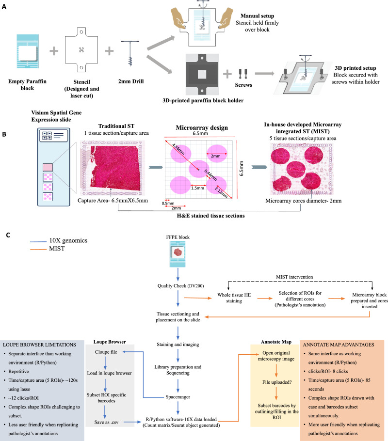
The 10X Visium assay poses 5 major challenges: i) Limited sample usage, ii) High cost per sample, iii) Variability among different slide batches, iv) Low level of replication, v) Barcode extraction for data analysis is closely linked with the loupe browser. “Low level of replication” in the context of Visium technology refers to the difficulty of performing multiple replicates of an experiment, primarily due to the high costs and the limited number of samples that can be processed per slide. Because only a few samples can be analyzed at a time, it becomes challenging to run multiple replicates under identical conditions. The loupe browser is a proprietary tool from 10X genomics that is not flexible to accommodate new features to assist with efficient analysis. The visium gene expression slide has four capture areas measuring 6.5 × 6.5 mm sq each. Due to the size limitation of the capture areas, the current protocol recommends using 1 tissue section per capture area. Therefore, only four tissue sections per slide from single or multiple tissue blocks can be used for the assay. 10X Visium is costly; the price of processing a single gene expression slide is approximately 11000 USD, excluding additional sequencing charges. Therefore, many researchers do not readily adopt this technology, especially in low-resource countries. There are two ways in which researchers reduce the cost of imaging-based assays—first, they try to fit in more number of samples in the same imaged area or frame, and second, they employ computational tools, specifically, machine learning to extrapolate high-resolution data from their low-resolution images6,7. To reduce this prohibitively high cost of spatial transcriptomics so that the technology can be ubiquitously used, we propose to overcome the restriction of limited sample usage and increase throughput per slide through the integration of conventional tissue microarray(TMA) with 10X Visium spatial transcriptomics, introducing a novel approach called MIST (Microarray Integrated Spatial Transcriptomics). We also introduce a novel computational tool named ‘AnnotateMap’ that addresses key challenges posed by the loupe browser (Fig. 1C).
Fig. 1. Overview of the MIST approach and its integration with AnnotateMap.
A, B Representative image of traditional spatial transcriptomics and our novel in-house developed microarray approach (MIST). C Flow chart depicting the steps of the Visium FFPE assay with MIST, accompanied by a comparative analysis of the 10X Genomics Loupe Browser and our proprietary tool, AnnotateMap. Figure 1 was created using Inkscape, STL and DXF design file images from Solidworks, and assembled in Microsoft PowerPoint.
Tissue microarray was a remarkable invention in pathology in the early 2000s and is still commonly used in hospitals. A tissue microarray uses a single slide to simultaneously assay >1000 minute tissues 8 subsampled from different patient biopsies. It facilitates low-cost multiplexed high throughput analysis of many patient samples and allows the assessment of molecular targets in a cohort under uniform standard conditions, thus removing any variations occurring due to a different slide batch. Tissue microarray enables judicial utilization of irreplaceable tissue resections. This approach leads to multi-fold cost reduction, making the aim of integrating ST into the clinical workflows more feasible.
MIST can facilitate the assay of samples from multiple patients or subsample multiple regions of interest (ROIs) from the same patient where ROIs are distantly located. Multiple sequential sections (replicates) from the same region of interest can also be processed at a reasonable cost. Additionally, it is worth noting that certain regions within the tissue might not offer pathologically interesting insights, such as the presence of a huge blood vessel traversing through a tumor sample, necrosis, etc. As a consequence, these areas might not yield substantial gene expression information. While such occurrences are not uncommon in biological tissues, they may not provide clinically significant information for the specific pathological investigation being conducted. MIST increases the flexibility of the sampling choice, therefore, by deploying MIST, researchers can assay the regions with the most relevant pathological features to interpret variations and capture maximum heterogeneity to ensure comprehensive conclusions. Building on these innovative approaches to multiplexing and tissue processing, Monasterio et al. employed a tissue rolling technique to maximize the use of the capture area, facilitating the analysis of the entire murine gastrointestinal tract 9. Le Naour et al. used an improved Swiss rolling technique that was first described in the 1960s to assay fresh frozen sections of the entire murine colon 10.
A substantial amount of tissue is available after surgical resection of entire tumors. However, when dealing with biopsies, the available tissue is often limited, consisting of very small areas. For instance, for metastatic patients, core needle biopsies (CNB) and fine needle aspirations (FNA) are the primary methods utilized in the diagnostic procedure11, where CNBs are cylindrical biopsies of ≤2 mm in diameter 12. MIST can be used to perform spatial assay of multiple ( ~ 20) CNBs in a single slide under identical reaction conditions, which would help prevent potential batch effects and ensure accurate analysis. Similarly, the spatial analysis of multiple FNAs (with a diameter of ≤1 mm) can be conducted using the fundamental principles of MIST, as elaborated in the ‘Discussion’ section.” MIST can also be employed in pilot studies to generate preliminary data generation for hypothesis building.
TMAs are officially unsupported by 10X Genomics, and they do not provide any guidance on TMA block generation, tissue sectioning, or placement onto gene expression slides. While 10X Genomics offers some basic information on TMAs, they recommend using a service provider for preparing TMA blocks, and their assessment of TMA compatibility is limited to the 11 mm Visium CytAssist Spatial Gene Expression Slides. There is no guidance from 10X on creating TMA blocks, troubleshooting, or placing TMA tissue sections for CytAssist slides 13.
Our research article addresses these significant gaps by providing an in-depth exploration of TMA for Spatial Transcriptomics. We offer a comprehensive examination of crucial aspects, such as troubleshooting, tissue type selection, sectioning methods, and the accurate placement of tissues on glass slides. Additionally, we discuss various applications and use cases for TMAs, highlighting their practical utility in research. A notable aspect of our work is the focus on customizing TMAs to suit specific study requirements, thereby offering solutions that are currently unavailable from 10X Genomics.
Standard Tissue Microarray (TMA) molds often come with fixed distances between wells, limiting your ability to optimize tissue packing. Although custom TMA designs are an option, they typically require ordering from third-party suppliers. Moreover, building a TMA using an automated tissue arrayer or commercially available rubber molds tends to be costly and time-consuming 14. In contrast, our technique is crafted for self-sufficient replication in individual labs. It offers the flexibility to customize and adapt the design according to specific experimental requirements, free from external dependencies. This approach is less resource-intensive and enables labs to efficiently tailor TMAs to their unique experimental needs in a short span of time.
Results
The proposed protocol consists of four parts: i)3D printing and preparation of the microarray block, ii) Image processing, library preparation, and sequencing, iii)Extraction of core-specific data using the Loupe browser or AnnotateMap, and iv) Data analysis.
10X Visium assay involves tissue permeabilization and probe release onto the gene expression slide, this step may cause diffusion of probes from their original location in the tissue. We 3D printed, optimized, and designed a microarray to accommodate standard circular tissue cores of 2 mm diameter from a biopsy, each with enough distance between them to prevent mRNA diffusion across different cores. Additionally, we ensured a minimum distance between the fiducial frame and the tissue cores to prevent overlap with the fiducial frames, as they help in the accurate overlay and alignment of the tissue image with the gene expression data. Keeping these crucial details in mind, we could successfully accommodate five tissue cores in the 6.5 × 6.5 mm capture area. Figure 1A and B show the HE-stained tissue with our in-house developed microarray approach (MIST). MIST has only been applied and tested with FFPE tissue samples. The details of the microarray design are mentioned in the Methods section. The flowchart illustrated in Fig. 1C outlines the limitations of the Loupe browser that our computational tool, AnnotateMap, is designed to address and overcome.
We have used FFPE tissue sections from resected surgery blocks of a single cancer patient. The whole section H&E staining was done, and 5 different regions of interest (ROIs) to be used for microarray were marked by an expert pathologist; check Methods for details. Figure 2 shows a preliminary analysis of the spatial data generated using the MIST approach.
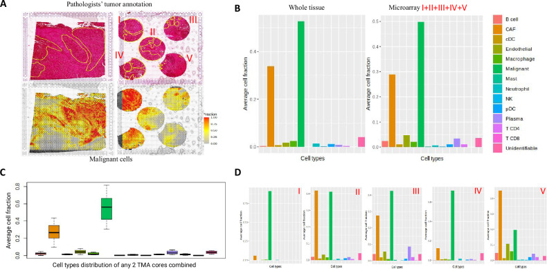
Fig. 2. Comparison of spatial cell distribution between whole tissue and microarray cores.
A Top left image shows HE stained image of the tissue section spanning the whole fiducial frame. The image in the top right displays a HE-stained image of the microarray derived from the entire tissue. Notably, the five cores in the microarray (labeled as I, II, III, IV, V) were precisely extracted from the identical region from which the whole tissue was initially sectioned. The bottom two images show malignant cell fraction in the two tissue designs. B Average cell fractions for different cell types for whole tissue and microarray. For microarray, average fractions from all the five cores are depicted together. C Box plot with average cell fraction from any two unique microarray cores combined. D Cell fraction from 5 different microarray cores plotted separately. The top two components of Fig. 2A were annotated and created using Leica ImageScope, while the bottom components of Fig. 2A, B, C, and D were created in R.
The major tumor areas annotated by an expert pathologist are shown in yellow contours in the top two HE-stained images of Fig. 2A. In the bottom two images, we have overlaid the malignant cell fraction onto the HE-stained image. The malignant cell fraction was annotated using the SpaCET tool, which identifies spots forming malignant cell clusters based on copy number variations (CNVs) and gene expression signatures. We observe that the majority of the areas annotated align with the pathologists’ annotation. The tissue area covered in the microarray design is ~60% of the whole tissue. Remarkably, we observed that all the cell types recorded in the whole tissue are captured in the microarray design as well. The bar plot in Fig. 2B depicts the average cell fraction across all spots for each cell type. Further, we wanted to check if the heterogeneity of a large tissue section spanning the entire capture area can be reflected in a few 2 mm cores. To check this, we separately plotted the average cell fraction of the five different cores (Fig. 2D) and observed that the cellular heterogeneity can be captured in less than 60% of the entire tissue. Each boxplot in Fig. 2C shows the distribution of the specific cell type across 10 different pairwise combinations of the five ROIs. For each combination, the mean of the cell fraction across the two ROIs is plotted. It can be observed that 2/5th of the 60% area of the whole tissue covered in the microarray is sufficient to capture maximum heterogeneity from a whole tissue section. This underscores the need for a judicious selection of Regions of Interest (ROIs) by an expert pathologist, ensuring that the chosen areas accurately reflect the intricate heterogeneity. Customizing the methodology to align with the research objectives is of utmost importance. Depending on the research query, homogenous or heterogenous samples can be selected and assayed. For instance, for studies focused on malignant cells, selecting cancer-specific areas becomes crucial.
AnnotateMap
In order to extract barcode information and efficiently analyze the data from multiple samples, we developed AnnotateMap that addresses the annotation challenges in LoupeBrowser. AnnotateMap is a user-friendly, open-source tool that integrates seamlessly with existing R/Python workflows for efficient barcode annotation from tissue images. It allows pathologists to draw boundaries around complex ROI shapes, replicating their intuitive workflow. AnnotateMap processes barcodes from a single capture area with 5 ROIs in approximately 85 (29.2% faster) seconds compared to loupe browser (120 seconds). While the 35-second improvement in processing time compared to Loupe Browser may seem modest, the true value of AnnotateMap lies beyond just speed improvements.
AnnotateMap overcomes key limitations of Loupe Browser and offers broader benefits for Spatial Transcriptomics analysis. As an open-source tool, AnnotateMap fosters collaboration and enables developers to contribute features tailored to the specific needs of the community. It offers advanced region extraction capabilities, such as contrast-based extraction of regions of interest (ROIs) into separate files, which significantly reduces manual processing time compared to the Loupe Browser. Additionally, AnnotateMap integrates seamlessly with existing R and Python workflows, streamlining data analysis pipelines by leveraging widely used bioinformatics tools. The customizable graphical user interface (GUI) further enhances its utility, allowing researchers to tailor the software to their specific needs and preferences.
By addressing these limitations of Loupe Browser, AnnotateMap offers a more powerful and flexible platform for Spatial Transcriptomics analysis. The potential for user-driven development and advanced features makes AnnotateMap a valuable tool for researchers in this field. Details on AnnotateMap can be found in the methods section.
Cost estimation
The Visium FFPE protocol uses three kits, namely: i) Visium Spatial Gene Expression Slide- 4 reactions (6.5 ×6.5 mm), ii) Visium FFPE Reagent Kit -small, iii) Visium Human Transcriptome Probe Kit - Small. The combined cost of the three kits is approximately ~11000 USD. Once the spatial transcriptomics library is prepared, it undergoes sequencing on the Illumina platform. It is important to note that this cost estimation does not include third-party items or library sequencing charges on the Illumina platform.
Since ST generates high-throughput data with a requirement of 25,000 read pairs/spot (for FFPE samples), an entire Illumina flow cell (P2 flow cell- 400 million reads: 100 cycles) is usually used for sequencing leading to a cost of ~3000 USD.
Overall, the data generation using ST can amount to around 14,000 USD per slide, which translates to approximately 3500 USD per capture area. However, we have made a promising advancement regarding cost optimization. Using MIST, we fit 5 tissue cores in one capture area, therefore the MIST method significantly reduces the expense of slide processing, bringing down the cost of single sample processing to 875 USD (1/5th of the original cost, i.e., 3500 USD) or at least 1750 USD if using 2 cores to capture maximum heterogeneity of a specific sample type.
Additionally, 10X Genomics has introduced an updated version of their Visium gene expression slides with an expanded 11 × 11 mm capture area, accommodating two reactions per slide. The entire slide pack is priced at approximately $10,400. By utilizing square packing, up to 16 circular tissue cores, each 2 mm in diameter, can be arranged within a single 11 × 11 mm capture area, thereby reducing the overall cost per sample to just $325. Notably, these larger capture area slides allow for the simultaneous processing of up to 32 samples, effectively minimizing the risk of batch effects. The cost per sample can be further reduced by using smaller tissue cores, such as 1 mm diameter cores, enabling even greater sample throughput. A key advantage of MIST is its ability to accommodate multiple cores of same or even varying sizes, tailored to specific experimental requirements. This flexibility in customization can be achieved directly within a personal lab setting, without external dependencies.
Comparison of MIST with TMA used in GeoMX nanostring
Another spatial transcriptomics technology, GeoMx DSP Nanostring, facilitates the use of tissue microarray(TMA), where multiple circular tissue cores of variable diameter (0.6-6 mm)15 can be fit in the center (35.3 × 14.1 mm)16 of a glass slide. For DSP nanostring, the region of interest is defined as the area in the tissue from which gene expression data is acquired; an ROI can be further subdivided into different AOIs (segments) with distinct morphologies to obtain separate gene expression profiles17. Nanostring recommends assaying ≥6 ROIs for a specific region from one patient tissue sample; if ROIs are assayed irrespective of the region type, then ≥24 ROIs are recommended per sample 18. The size range of the ROIs is 10um to 600um17 in diameter. For RNA analysis, although single-cell resolution (10um) AOI selection is possible, nanoString recommends capturing a minimum of 200 cells from each ROI or segment 18. Any AOI/ROI would give gene expression data as a mini-bulk of cells it encompasses. Hence, as the size of the AOI increases, the resolution decreases, and it would give an expression of all the cells composing the AOI bulk. To compare, MIST based on visium provides a uniform resolution of 50um. As per the GeoMx DSP experimental design guideline (Manual number: MAN-10108-01), the usual areas of illumination (AOIs) assayed per ROI are in tens. Such an assay design would not allow a robust understanding of the spatial arrangement inside a tissue; it would only provide information on a limited area (ROI) from the tissue region. It does not come close to the comprehensive analysis MIST provides regarding the complete spatial assay of the core. In our microarray design, the number of spots (analogous to AOIs) analyzed for a 2 mm core varies between 400 and 500.
According to personal communication, the cost of DSP Nanostring is 200 USD for an ROI/AOI of 200um diameter (including sequencing charges). In general, a larger AOI is likely to incur higher costs compared to smaller AOIs, as a larger AOI would require more reagents and higher sequencing depth. Irrespective of the area of the AOI, cost increases as more AOIs are analyzed. If the cost is translated to a 2 mm core with 50 AOIs, each having a diameter of 200 micrometers, the estimated cost would be approximately 10,000 USD. In contrast, MIST presents a considerably more economical option, costing ~300–800 USD for analyzing approximately 500 spots (analogous to AOIs), and provides a comprehensive and improved understanding of the spatial dynamics (Table 1).
Table 1.
Comparison between GeoMx DSP nanostring and MIST
| Comparison parameters | GeoMx DSP Nanostring | MIST (VISIUM) |
|---|---|---|
| Setup cost | Requires GeoMx digital spatial profiling instrument ($295,000) | Benchtop (no investment) |
| Single slide processing cost | 10,000 USD for one ROI assaying up to 50 AOIs of 200um diameter |
400–500 spots/ROI: ~300–800 USD. 20 such ROIs can be processed per slide *spots(50um diameter are analogous to AOIs) |
| Choice of sampling | Should be ideally homogenous for a specific ROI |
Size of spots is fixed- 50um Heterogenous sampling is ideal- captures maximum heterogeneity |
| ROI analysis e.g. 2 mm ROI | Less comprehensive; AOIs, maximum in tens are assayed | More comprehensive, entire 2 mm area is assayed |
| Resolution | Larger AOI- more cells-mini bulk of all the cells in the AOI | Fixed resolution corresponding to the spot size(50um) |
Discussion
MIST offers a practical solution to the cost and sample plexity challenges of spatial transcriptomics, making this innovative technique more accessible for comprehensive research in diverse clinical settings. There are tissues with a low cellular density, one such example pertains to the lung tissue. For lung samples, normal lung used as a control contains large alveolar spaces, which do not contribute to significant gene expression information on the gene expression slide as they are essentially empty spaces. To obtain sufficient gene expression data from normal lung samples for comparison with the diseased phenotype, multiple cores from the normal lung must be assayed. Similarly, in the case of high-density cellular regions such as lymph node, a small-sized ROI would capture the distinct cell types.
Cancer is a complex molecular disease. Spatial markers for cancer stage identification, drug efficiency, recovery, and relapse once discovered can be utilized to help understand the prognosis of multiple patients together on the same slide with MIST, which would be feasible in clinical applications. MIST can be employed to gain spatial information from different histology regions as lymph nodes, normal adjacent to the tumor (NAT), tumor, tumor-normal interface, inflammation, vascular inflammation etc. This technology allows for the processing of multiple Regions of Interest (ROIs) from diverse tumor profiles—ranging from cohesive tumor masses to disintegrated tumors or regions with scattered tumor cells—all within a single slide. Such an assay would be very beneficial in use cases where researchers want to explore the cancer evolution in single or multiple patients.
While generating spatial data for machine learning or deep learning applications, cost-efficiency is crucial. Here, a larger sample size takes precedence, ensuring an adequate number of samples for effective distribution into training, validation, and test sets. MIST facilitates the simultaneous processing of multiple samples under consistent conditions, effectively mitigating any potential batch effects. Moreover, a larger sample size not only offers a more accurate representation of disease pathology but also captures sufficient variation, mirroring the diversity within a population. This approach is particularly significant in clinical applications, where robust and diverse datasets are essential for developing and validating predictive models that can be used for diagnostics, prognostics, and personalized treatment planning.
Furthermore, MIST stands as a powerful tool to address specific research inquiries. For example, when focusing solely on investigating distinct inflammatory spatial patterns within a particular type of cancer, researchers can meticulously select and analyze multiple inflammatory ROIs, even if these regions are spatially distant. This capacity to selectively target and thoroughly examine specific regions adds a layer of precision and efficiency to cancer research endeavors. In summary, while the initial cost of the Visium FFPE protocol is exorbitant, leveraging MIST allows for substantial cost reduction in slide processing, making this innovative technique an affordable option with single-sample processing at approximately 300 to 800 USD per sample.
In the context of future research, firstly, we propose using MIST for visium slides with larger capture areas and more spots; 11 mm x 11 mm capture area and ~14,000 spots. Similarly, when paired with BGI STomics, which offers various capture area sizes (50 mm², 100 mm², 200 mm², 36 cm², and 174.24 cm²) 19, MIST can potentially enhance the number of samples processed in a single slide. Currently, we have used single-size cores for MIST, we envision an alternative microarray design that uses different sized cores to maximize the use of space within a capture area. In this specific design, we positioned the corner cores at 0.25 mm distance from the edge of the fiducial frame. We placed a 1 mm core between each pair of corner cores at 0.5 mm from each adjoining corner core. This strategic layout allowed us to incorporate a total of nine circular cores comprising two different diameters: five 2 mm cores and an additional four 1 mm cores. Researchers are encouraged to explore the feasibility of this design for their specific applications. For easy implementation, we have provided the required design files in the supplementary information (Supplementary File 1). Similarly, based on the research question, all 1 mm cores, as seen in fine needle aspirations, can also be used to design the laser cut stencil for MIST. This would further allow a more dense packing of the cores and increase the number of samples assayed in a single slide.
To summarize, Microarray Integrated Spatial Transcriptomics (MIST) allows flexible selection of diverse regions of interest (ROIs), allowing judicious use of precious tissue biopsies and also reduces the cost of Visium assay by fivefold. We describe a detailed protocol with critical troubleshooting steps to execute TMA integration with the Visium ST platform. This work presents the first experimental evidence at the user end with a detailed protocol that performs integration of tissue microarray with an ST platform in a customized manner. MIST democratizes access to spatial transcriptomics for a wider research audience. This approach not only makes spatial transcriptomics more accessible to a broader range of researchers but also allows for the processing of multiple samples under consistent conditions, thereby minimizing batch effects and enabling a comprehensive analysis of disease pathology as discussed in specific use-cases.
Methods
Microarray block preparation
We used a 1 cm thick acrylic sheet to laser cut a stencil of a larger size (2.5 × 2.5 cm) with microarray cores laser cut as per the proposed microarray design (Fig. 1A) (Supplementary File 1). Laser cutting technology ensures a precision of 0.1 mm, resulting in a highly accurate microarray design. The corner cores are positioned at a distance of 0.5 mm from the edge of the fiducial frames, with a separation of 1.5 mm between them. Additionally, the center core is situated at a distance of 0.44 mm from the corner cores (Fig. 1B). A larger stencil, compared to the size of the capture area of the slide, was used to give firm holding space while drilling the microarray block. A blank paraffin microarray block was prepared, and cylindrical cores were drilled out from it with the help of the stencil overlayed on the paraffin block, using a 2 mm drill through manual drilling (hand-operated). The stencil can also be printed with a high precision 3D-printer.
Improved microarray block preparation setup
The described protocol employs manual drilling by positioning the stencil firmly over the paraffin block. However, this manual approach may introduce errors, especially when dealing with smaller than 0.5 mm resolutions. To overcome this challenge, we have developed a custom-made apparatus that consists of a 3D-printed mold designed (Fig. 1B) to hold a standard FFPE block cassette. Users can place the paraffin block cassette into the mold. On top of this setup, the stencil is positioned, and the entire assembly is secured using screws. Subsequently, the microarray blocks can be created using either a 2 mm automated pen drill or manual drilling. The design files necessary for 3D printing the mold and laser cutting the stencil can be found in the supplementary information (Supplementary File 1 and Supplementary File 1). We trimmed off the extra wax from the microarray block to achieve a size of about 6.5 × 6.5 mm.
Tissue selection
This study uses formalin-fixed paraffin-embedded (FFPE) samples from a single cancer patient. The necessary ethical clearance was granted by the Institute Ethics Committee, All India Institute of Medical Sciences (AIIMS), New Delhi. RNA extraction was performed with 2-3 FFPE sections of 10um thickness using the Qiagen RNeasy FFPE kit (Catalogue number- 73504). The preliminary RNA quality assessment was done on the Bioanalyser to determine RNA fragment size, specifically, the DV200 value (percentage of fragments with ≥200 nucleotides). RNA quality assessment is essential to ensure the success of the downstream library preparation protocol as formalin has a degrative effect on RNA. It is recommended to consult the 10X Genomics user guide and our publication for comprehensive guidance.
Microarray tissue integration
Simultaneous with the microarray block preparation, tissue sections from whole tissue blocks were HE stained, and regions of interest (ROIs) were annotated by an expert pathologist. After a microarray block with hollow cylindrical cut-outs is prepared, tissue cores from the ROIs are extracted from the whole tissue FFPE block using a standard 2 mm cylindrical microarray punch. The cylindrical core is then snug fit into one of the cylindrical cut-outs of the prepared microarray block. Once all five microarray cores were embedded with tissue, the block was incubated at 37 °C on a hot plate for 1 hr, followed by incubation at 4 °C for 15–20 min. Post this, the block is trimmed on the microtome for any uneven surface. It is then again put at 4 °C for 5 min, the blocks are kept at 4 °C for long-term storage. We sectioned the microarray blocks 4days after being made, except for one block for which the whole tissue section and microarray section were used on the same slide (samples used in the analysis for this paper). The storage at 4 °C aids in allowing the cores to settle effectively within the paraffin block. The optimum temperature and the duration of incubation for microarray block preparation can vary depending on the properties of the paraffin wax. To validate the accurate embedding of ROIs, HE staining was done on sections from the microarray blocks on a plain glass slide and examined by a pathologist.
There are four capture areas (6.5 × 6.5 mm) in the Visium spatial gene expression slide. For the 1st capture area, we performed the spatial assay for a whole section to span the entire capture area of the Visium slide. For the 2nd capture area, once the whole tissue was sectioned and placed onto the slide, an FFPE microarray block was prepared with tissue cores from the same region as the whole tissue. This was done by marking the region on the whole tissue HE-stained slide from which the tissue was sectioned. The marked slide was then overlaid on the tissue to extract 5 ROIs from the same region. The remaining two capture areas (3rd and 4th) were used with the microarray approach from different regions of interest (ROIs), their results are not included in this study.
In the case of whole tissue and microarray sections taken from the same FFPE block (Fig. 1A), we adopted a slightly modified microarray block preparation protocol such that the embedded cores are properly settled within the wax block in a shorter time frame. After the TMA block was prepared, it was incubated for 1 h at 37 °C, followed by 20 min at -20°C. Importantly, the standard visium protocol doesn’t specify if a time delay during subsequent tissue placement on the slide would affect downstream analysis. Due to the study requirements in our protocol, a time gap of 1 hour and 20 minutes was introduced between the first two tissue placements. Through our study, we demonstrate that having such a time gap is safe and does not adversely impact the analysis.
Tissue sectioning and placement
While tissue sectioning, it is important to ensure an even block surface. If the block protrudes from the upper end in the microtome block holder, there’s a risk of cores dislodging during sectioning. It is important to note down the orientation of the TMA block when placed in the microtome block holder, to further match the orientation of the tissue section floated in the water bath and when placed onto the 10X Genomics gene expression slide.
Sectioning Tissue Microarrays (TMAs) presents unique challenges due to the incorporation of cores from diverse Formalin-Fixed, Paraffin-Embedded (FFPE) blocks. It’s critical to ensure that all tissues undergo uniform processing during fixation and that a consistent type of wax is used for all the FFPE blocks contributing to the microarray cores. This uniformity is key to achieving consistent sectioning quality. To further enhance the sectioning process, it’s advisable to group tissues with similar properties within the same TMA, which helps in maintaining uniform sectioning parameters and improving overall results.
When placing tissue on the 10X gene expression slide, start with the topmost capture area, then proceed to the others. Practice section placement on non-charged plain glass slides with manually drawn capture areas using dummy TMA blocks. Align the top edge of the fiducial frame with any edge of the tissue section floating in the water bath, using a brush for directional assistance, and carefully transfer the section onto the slide. Remember to note the specific edge from which the section is placed on the slide for consistent orientation.
MIST assay troubleshooting: solutions for challenges
Microarray block preparation for MIST can present several challenges. Preventing tissue loss while sectioning is crucial, especially while handling blocks with calcified tissue, which can occur due to excess alcohol usage during FFPE block preparation. Calcified tissues become hardened and brittle, making it difficult to section them into thin layers. To mitigate this issue, one must exercise caution and consider either avoiding using such tissue blocks altogether or hydrating them in ice-cold water for 10–15 min depending upon the extent of hydration. Hemorrhage commonly occurs during surgical procedures, it may also happen when resected tissue is pressed firmly by forceps prior to fixation 20. Tissue sections with clotting or hemorrhage tend to wash off during staining.
In some instances, the microarray core punched from the whole tissue block may not be uniform or cylindrical due to uneven dimensions of the resected tissue while fixing in paraffin, leading to a loss in the sectioned tissue area. If a significant tissue area is lost, one may punch out the intact cores and reinsert them in another wax microarray block.
If the tissue section exceeds the capture area’s size and risks overlapping with the fiducial frames during the experiment, it can be managed effectively. Place the section on a non-charged, plain glass slide, where 6.5 × 6.5 mm squares are manually marked on the back. This allows for quick trimming of any excess wax. Afterwards, the section can be refloated and then accurately transferred onto the 10X Visium slide. Alternatively, if time permits, extra wax can be trimmed off the wax block.
Additionally, while preparing the microarray block, it is important to drill the paraffin block to the bottom to ensure proper embedding of the tissue cores. Improper drilling will lead to protruding tissue cores, which are at risk of expelling out from the paraffin block during sectioning. This can also happen due to uneven surfaces of the microarray block. In such cases too, another core from a similar pathological annotation may be embedded along with the existing intact cores into a new wax block to avoid loss of space in the Visium gene expression slide. It is advisable to practice microarray block preparation and tissue section placement using dummy blocks before proceeding to the actual experiment slide.
Library preparation
The FFPE tissue sections placed on the Visium slide were HE stained and imaged using the Leica Aperio Versa 8 microscope at 20X magnification. Library preparation was done in accordance with the 10X genomics protocol for Visium FFPE samples. After library preparation, we used Qubit to measure the library concentration and Tapestation to estimate the size of the fragments. The library concentrations for the four capture areas were 18.2 ng/µl, 12.2 ng/µl,16.2 ng/µl, and 14.7 ng/µl. The fragment sizes of the libraries from these areas matched the recommended size by 10X genomics, with peak sizes of 259 bp, 270 bp, 269 bp, and 256 bp, respectively.
Data preprocessing
The HE-stained FFPE tissue sections were captured at 20X magnification using the Leica Aperio Versa 8 microscope, which generates images in the scn format. The size of the scn files for the entire 6.5 × 6.5 mm capture area is huge, ranging between 1GB and 2.5 GB. In order to make these images compatible with the 10X genomics Spaceranger software, we preprocessed the images using FIJI. Spaceranger requires images in either tiff or jpeg format. To start, we integrated the Bio-Formats plugin into FIJI. The first step is to open the image using the Bio-Formats plugin using the Bio-Formats Importer option. Upon selecting the scn image, a pop-up window titled ‘Bio-Formats Import Options’ will appear, where only the following four options have to be selected: Hyperstack, Group files with similar names, Use virtual stack, and Default color mode, click OK. Following this, another window named ‘Bio-Formats File Stitching’ will open, keep the default parameters selected. In the subsequent ‘Bio-Formats Series Options’ window, only select the images corresponding to the image of interest, click OK. Once the image opens, the final step is to save the image as either a JPEG or tiff file format.
Post image preprocessing, we performed demultiplexing of Illumina’s base call files (BCLs) into FASTQs by running ‘spaceranger mkfastq’ on Linux with the designated sample indices. Subsequently, ‘spaceranger count’ was run to generate Spaceranger outputs, which were used for further data analysis.
Data analysis
Extraction of SpotIDs for each ROI
In order to process the different sample cores from the TMA, extraction of core-specific barcodes is required. Loupe browser (version 6.3.0) from 10X Genomics can be used to extract the spot IDs corresponding to each ROI in the microarray design cloupe file was loaded on the loupe browser (version 6.3.0) using the ‘Open Loupe File’ option.
Once the cloupe file is loaded, select the ‘lasso’ option from the top menu, and select an ROI. A pop-up window appears, type the category and cluster name. We have used ‘ST’ as category name for reference. Subsequently, select the spots from all the ROIs using the paintbrush or lasso and save it with a unique cluster name. The recommended approach is to label the Region of Interest (ROI) with the name of the nearest fiducial frame corner, extending from any existing designation, such as triangle, circle, hourglass, or 7-point hexagon. This would prevent any potential confusion that may arise due to image rotation.
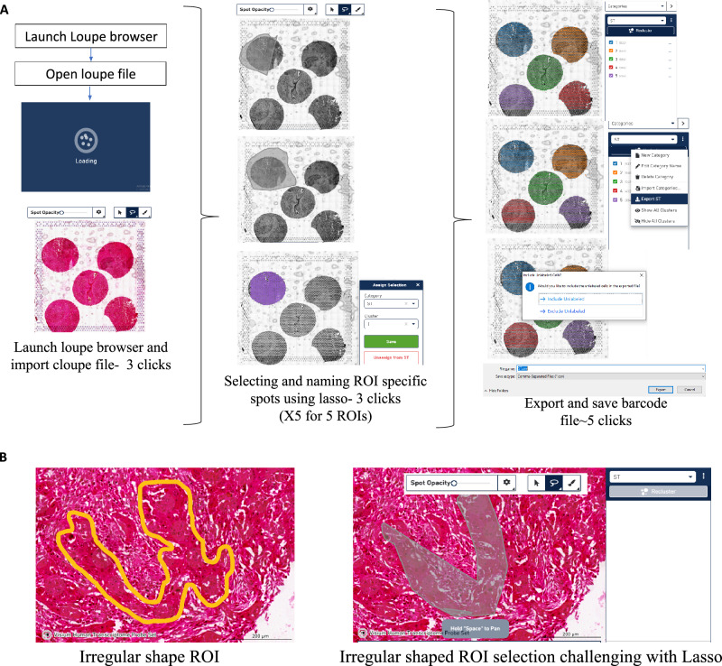
The final step is to export the barcodes in a CSV format using the ‘Export Sample-C’ option. The CSV file is then imported to the working environment- R/python for further analysis. The exported barcodes or SpotIDs can be used to subset ROI-specific barcodes from either the Seurat object or the count matrix of a particular capture area. See Fig. 3A.
Fig. 3. Selecting ROI-specific barcodes in the Loupe browser.
A Steps followed in the Loupe browser to subset ROI/core-specific barcodes. B Challenge depicted when attempting to select an area with a non-uniform boundary using the lasso or paint tool. The figure is generated with the help of Loupe browser software.
Overcoming challenges of the Loupe Browser and extraction of core-specific data using AnnotateMap
While the Loupe Browser offers a user-friendly interface, it has certain limitations. The previously outlined steps (Fig. 3A) must be performed iteratively to extract barcodes for each Region of Interest (ROI). Time taken per capture area (5 ROIs) for subsetting barcodes using the ‘lasso’ is ~2 minutes after sufficient experience with the loupe browser. To put this into context, processing a whole slide would take 8 minutes, and for 10 such slides (common in Spatial Transcriptomics studies), it amounts to 80 minutes. This is further compounded when dealing with huge datasets making the process cumbersome and time-intensive. Given that the Loupe Browser operates as a separate interface, transitioning from R or Python to the Loupe Browser solely for barcode extraction presents additional inefficiencies. Additionally, the need to organize data in two separate interfaces poses a limitation. Having a unified platform for all operations would be highly convenient, particularly when dealing with large spatial datasets. An additional drawback of the Loupe Browser is the limitation in selecting ROIs with non-uniform or complex boundaries using the lasso/paint option (Fig. 3B). Manually replicating the pathologists’ annotations again in the Loupe Browser to extract barcodes for further genomic analysis is not very user friendly.
To overcome these challenges, we have developed an efficient and streamlined tool named AnnotateMap. It seamlessly integrates with users’ R/Python analysis workflows, offering a singular interface. Here, barcodes from the 5 ROIs from a capture area can be subsetted by drawing and filling in a boundary around the region of interest, the tool essentially takes 85 seconds to process barcodes from a single capture area with 5 ROIs. Our tool simplifies the process by allowing users to draw boundaries around complex irregular shaped ROIs, making replication of pathologists annotations more user friendly. This also aligns with the intuitive approach typically followed by pathologists.
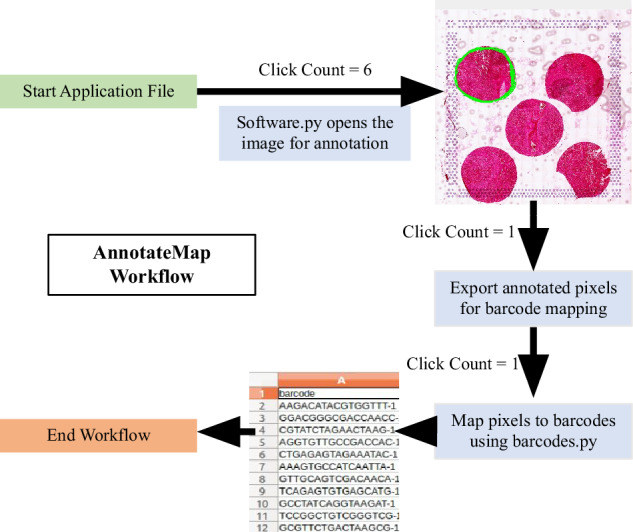
The integrated Python scripts provide a comprehensive workflow for annotating tissue images and extracting relevant barcode information (Fig. 4A). The software.py script utilizes OpenCV to enable interactive annotation of tissue images, offering a resizable display for annotating regions of interest (ROIs) through the drawing of contours. These contours, represented by coordinates, are saved in a CSV file (contours.csv). Subsequently, barcodes.py processes the annotated coordinates from contours.csv and matches them with tissue positional data stored in tissue_positions.csv. It employs a KDTree-based nearest neighbor search to identify the closest tissue elements to each contour point and retrieves their associated barcodes. After removing duplicate barcodes, the script generates a new CSV file (barcodes.csv) containing the unique barcodes corresponding to the annotated tissue regions. This integrated workflow allows efficient annotation and subsequent extraction of specific tissue barcodes.
Fig. 4. Working of AnnotateMap. More details can be found in our GitHub repository.

Figure was created using our in-house developed AnnotateMap tool and was assembled using Microsoft Powerpoint.
Annotation of malignant and nonmalignant cell types
Spatial data was normalized using RC normalization on the Seurat object prior to analysis using the SpaCET tool. The Seurat object was converted to the SpaCET object for analysis. 10X Genomics Visium platform offers approximately 1–12 cell resolution in a 50 µm diameter spot size. In this case, cell type annotation is challenging since multiple cells might contribute to gene expression read out from a single spot. To accommodate this complexity, we used the SpaCET tool21, which first identifies malignant cells using both copy number alterations and malignant gene expression signatures. Next, it deconvolves the non-malignant cell fraction and adjusts different cell densities in a single spot by employing linear regression, with gene expression profiles of individual cell types identified from various single-cell RNA sequencing (scRNAseq) datasets. The cell fraction in a sample (Fig. 2B, D) was calculated by averaging the specific cell fraction across all spots in that sample.
Supplementary information
Acknowledgements
We would like to acknowledge Abhilash Dasari for his input on the microarray design. Pallavi Gupta and Triveni Shelke for their help in the wet lab experiments. Rajan, for this help in beta testing the working of AnnotateMap. Prakrithi and Nirmal Singh Mahar for their inputs in the analysis discussions. We also acknowledge and thank the Department of Biotechnology, Government of India, for their support through the Ramalingaswami Fellowship (ST/HRD/35/02/200).
Author contributions
J. designed, optimized, and prepared the microarray apparatus using 3D printing and laser cutting. She contributed to make the microarray blocks, performed the library preparation and sequencing for the Spatial transcriptomics experiment. She performed the data analysis, guided with AnnotateMap, and wrote the manuscript. P.S. is the lead developer of AnnotateMap and contributed to writing the AnnotateMap section. K.Y. contributed to the preparation of microarray blocks, tissue sectioning, and placement on the gene expression slide. S.D. helped with designing the custom microarray design. S.M. contributed to designing the modified version of the microarray setup using 3D printing. S.K. performed the surgery and gave the samples for the study. D.J. was the leading pathologist for the study and contributed to selecting the regions of interest. P.S.M. involved in ethical clearances, patient sampling and helped in the study design. I.G. conceptualized the study, guided the data analysis and writing of the manuscript.
Data availability
Data related to this study is available on reasonable request. Interested researchers may contact the corresponding author to gain access.
Code availability
Source code for AnnotateMap is available at: https://github.com/Riyu44/MicroarrayProcessor. The underlying code for this study is not publicly available but may be made available to qualified researchers on reasonable request from the corresponding author.
Competing interests
All authors have reviewed potential conflicts of interest and declare that there are no competing interests related to the manuscript. This declaration is made to maintain transparency and uphold the integrity of our research.
Footnotes
Publisher’s note Springer Nature remains neutral with regard to jurisdictional claims in published maps and institutional affiliations.
Supplementary information
The online version contains supplementary material available at 10.1038/s41540-024-00462-1.
References
- 1.Method of the Year 2020. spatially resolved transcriptomics. Nat. Methods18, 1 (2021). [DOI] [PubMed] [Google Scholar]
- 2.Moses, L. & Pachter, L. Museum of spatial transcriptomics. Nat. Methods19, 534–546 (2022). [DOI] [PubMed] [Google Scholar]
- 3.Visium spatial tissue optimization reagents kits user guide - official 10x genomics support. In: 10x Genomics [Internet]. [cited 11 Aug 2024]. Available: https://www.10xgenomics.com/support/spatial-gene-expression-fresh-frozen/documentation/steps/tissue-optimization/visium-spatial-tissue-optimization-reagents-kits-user-guide.
- 4.User guides - official 10x genomics support. In: 10x Genomics [Internet]. [cited 11 Aug 2024]. Available: https://www.10xgenomics.com/support/user-guides/spatial-gene-expression-ffpe?menu%5BproductNames%5D=Spatial%20Gene%20Expression%20for%20FFPE.
- 5.Jones, W. et al. Deleterious effects of formalin-fixation and delays to fixation on RNA and miRNA-Seq profiles. Sci. Rep.9, 6980 (2019). [DOI] [PMC free article] [PubMed] [Google Scholar]
- 6.He, B. et al. Integrating spatial gene expression and breast tumour morphology via deep learning. Nat. Biomed. Eng.4, 827–834 (2020). [DOI] [PubMed] [Google Scholar]
- 7.Xie R. et al. Spatially Resolved Gene Expression Prediction from H&E Histology Images via Bi-modal Contrastive Learning. arXiv [cs.CV]. 2023. Available: http://arxiv.org/abs/2306.01859.
- 8.Jawhar, N. M. T. Tissue Microarray: A rapidly evolving diagnostic and research tool. Ann. Saudi Med29, 123–127 (2009). [DOI] [PMC free article] [PubMed] [Google Scholar]
- 9.Monasterio, G. et al. A versatile tissue-rolling technique for spatial-omics analyses of the entire murine gastrointestinal tract. Nat. Protoc.10.1038/s41596-024-01001-2 (2024). [DOI] [PubMed] [Google Scholar]
- 10.Le Naour, J. et al. Improved Swiss-rolling method for histological analyses of colon tissue. MethodsX9, 101630 (2022). [DOI] [PMC free article] [PubMed] [Google Scholar]
- 11.Elsheikh, T. M. & Silverman, J. F. Fine needle aspiration and core needle biopsy of metastatic malignancy of unknown primary site. Mod. Pathol.32, 58–70 (2019). [DOI] [PubMed] [Google Scholar]
- 12.VanderLaan, P. A. Fine-needle aspiration and core needle biopsy: An update on 2 common minimally invasive tissue sampling modalities. Cancer Cytopathol.124, 862–870 (2016). [DOI] [PubMed] [Google Scholar]
- 13.Are Tissue Microarrays (TMAs) compatible with the Visium CytAssist Spatial Gene Expression for FFPE assay? In: 10X Genomics [Internet]. [cited 11 Aug 2024]. Available: https://kb.10xgenomics.com/hc/en-us/articles/13704485511565-Are-Tissue-Microarrays-TMAs-compatible-with-the-Visium-CytAssist-Spatial-Gene-Expression-for-FFPE-assay.
- 14.Srinath, S., Kendole, R. K., Gopinath, P., Krishnappa, S. & Vishwanath, S. K. Economic methods used in fabrication of tissue microarray: A pilot study. J. Oral. Maxillofac. Pathol.20, 86–90 (2016). [DOI] [PMC free article] [PubMed] [Google Scholar]
- 15.Hernandez, S. et al. Challenges and Opportunities for Immunoprofiling Using a Spatial High-Plex Technology: The NanoString GeoMx® Digital Spatial Profiler. Front Oncol.12, 890410 (2022). [DOI] [PMC free article] [PubMed] [Google Scholar]
- 16.GeoMx Manuals & Guides. In: NanoString [Internet]. NanoString Technologies, Inc.; 24 Jan 2021 [cited 1 Dec 2023]. Available: https://nanostring.com/resources/geomx-manuals-guides/.
- 17.Deshpande N. GeoMx DSP: What’s in an ROI? In: NanoString [Internet]. NanoString Technologies, Inc.; 21 Mar 2023 [cited 16 Oct 2023]. Available: https://nanostring.com/blog/geomx-dsp-whats-in-an-roi/.
- 18.Spatial profiling of COVID-19 samples: Experimental designs to capture regional heterogeneity with the GeoMx DSP. In: NanoString University [Internet]. [cited 6 Dec 2023]. Available: https://university.nanostring.com/spatial-profiling-of-covid-19-samples-experimental-designs-to-capture-regional-heterogeneity-with-th/876246.
- 19.Transcriptomics Solution - STOmics. [cited 11 Aug 2024]. Available: https://en.stomics.tech/products/stereo-seq-transcriptomics-solution/list.html.
- 20.Bindhu, P., Krishnapillai, R., Thomas, P. & Jayanthi, P. Facts in artifacts. J. Oral. Maxillofac. Pathol.17, 397–401 (2013). [DOI] [PMC free article] [PubMed] [Google Scholar]
- 21.Ru, B., Huang, J., Zhang, Y., Aldape, K. & Jiang, P. Estimation of cell lineages in tumors from spatial transcriptomics data. Nat. Commun.14, 568 (2023). [DOI] [PMC free article] [PubMed] [Google Scholar]
Associated Data
This section collects any data citations, data availability statements, or supplementary materials included in this article.
Supplementary Materials
Data Availability Statement
Data related to this study is available on reasonable request. Interested researchers may contact the corresponding author to gain access.
Source code for AnnotateMap is available at: https://github.com/Riyu44/MicroarrayProcessor. The underlying code for this study is not publicly available but may be made available to qualified researchers on reasonable request from the corresponding author.


