Abstract
The precise assessment of hemothorax risk resulting from a rib fracture is not feasible. CT images, patient characteristics, and clinical experience are utilized in daily practice to assess risk intuitively. This study aimed to identify specific markers on CT images that can predict the risk of hemothorax. The study was retrospectively conducted between May 2021 and December 2023 at three different centers. Patients diagnosed with hemothorax at the initial assessment or during follow-up were identified among those being followed for rib fractures. An investigation was carried out to examine the relationship between the number of rib fractures, displacement status, and the location of the fracture on the rib arch with the risk of hemothorax. Of the 273 patients included in the study, 201 (73.6%) were male. The mean age was 53.9 ± 17.27 (19–93) years. Lateral (p = 0.029) and posterior (p < 0.001) location of the fracture and displacement of at least one fracture (p = 0.003) were associated with an increased risk. There was a significant correlation between the number of rib fractures and the risk of hemothorax (p < 0.001). The optimal cut-off for the number of rib fractures associated with a high risk of hemothorax was determined to be 4. Anatomical characteristics of a rib fracture can be useful to assess the risk of hemothorax practically in patients with thoracic trauma especially in emergency rooms. Patients with four or more rib fractures, at least one displaced rib fracture, and lateral and posterior rib fractures should be followed more carefully for hemothorax.
Keywords: Displacement, Number of fractures, Fracture location, Hemothorax, Rib fracture
Subject terms: Anatomy, Risk factors, Respiratory tract diseases, Trauma
Introduction
Rib fracture is a common pathologic finding after blunt thoracic trauma1. Although most cases can be treated with basic medical care, some cases may develop vascular injury, parenchymal laceration, and hemopneumothorax that require significant intervention2. Due to the varying clinical course, it is essential to accurately identify the risk factors, especially for hemothorax, a potentially life-threatening condition.
Approximately 33% of blunt trauma patients develop hemothorax following a rib fracture. Hemothorax typically originates from damage to intercostal or parenchymal vascular structures caused by a rib fracture3. Although this connection is well known, there are few studies on the risk of hemothorax caused by local factors associated with rib fractures4,5. It is still uncertain whether a single displaced rib fracture or consecutive non-displaced fractures pose a greater risk, and the preferred method is to evaluate each patient individually.
This study focused on identifying the independent factors that contribute to an increased risk of hemothorax in patients who experience rib fractures following blunt thoracic trauma. The goal is to offer a practical method for identifying patients at high risk for hemothorax by analyzing tomography images in busy work environments like emergency departments.
Methods
The study was a multicenter retrospective investigation conducted by the thoracic surgery departments of three training and research hospitals. It was initiated with the decision of the hospital ethics committee dated 29.05.2024 and numbered 2024/285. We retrospectively recorded the data of patients who were observed in the thoracic surgery clinic for thoracic trauma from May 2021 to December 2023. The study involved patients who were admitted to the emergency department following blunt thoracic trauma. All patients underwent thoracic tomography upon admission and were diagnosed with a rib fracture. All patients were hospitalized in the thoracic surgery inpatient clinic and followed up. Excluded from the study were patients who had radiologic evidence of injury affecting multiple systems upon admission, who had no thoracic tomography, patients with various types of bone fractures (such as those involving the thoracic vertebrae) that could potentially result in hemothorax, patients with indications of significant damage to large blood vessels, patients who presented with penetrating injuries, patients who were under the age of 18. Following the initial screening, the following patients were excluded from the study in order to create an isolated group:
33 patients with bilateral rib fractures,
17 patients with fractures in different regions of the same rib,
17 patients taking medication that could potentially disrupt the balance of bleeding-coagulation.
15 patients with sternum fracture,
8 patients who were admitted more than 24 h following the trauma,
6 patients who could potentially have pleural effusion based on their clinical history.
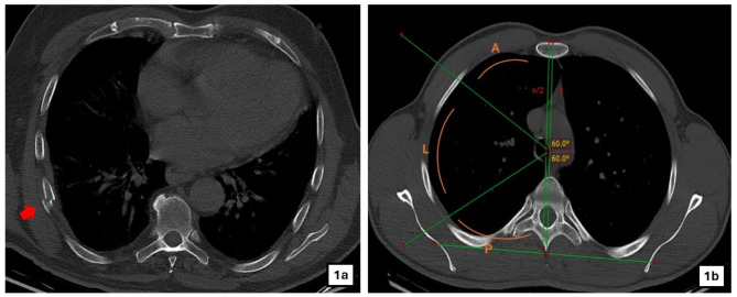
A study was conducted on 273 patients to analyze the relationship between age, gender, trauma mechanism, number of rib fractures, displacement of the fracture (displaced-nondisplaced), location of the fracture on the rib arch (anterior-lateral-posterior), and the occurrence of hemothorax. The trauma mechanisms were documented based on the patient’s statement. The fall mechanism, which has the potential to disrupt the standardization of trauma energy, was divided into two distinct classifications. A simple fall was defined as a fall that occurs from the patient’s current level. Falls from heights were categorized as traumas occurring at a vertical distance between 1 and 3 m. Since the study exclusively focused on patients who were observed in the inpatient ward and not in the intensive care unit, there is a lack of data regarding patients who suffered trauma exceeding a distance of 3 m. Accidents that people have while in a car are defined as in-vehicle traffic accidents. The term out-of-vehicle traffic accident is used when a vehicle hits a pedestrian. All radiologic evaluations were performed by a single thoracic surgeon with 10 years of experience. The term “displaced fracture” is used to describe a situation where more than 50% of the rib’s thickness is moved out of its normal position (Fig. 1a). A modification was made to the previously used method in the literature to precisely determine the location of the rib fracture6. An imaginary line was drawn from the back rib plane to the centerline of the sternum. The thorax was divided into three equal parts, each separated by 60-degree angles, starting from the midpoint of the line. The areas were classified as anterior, lateral, and posterior according to their respective positions (Fig. 1b).
Fig. 1.
(a) Displaced fracture of a rib in the right side of the chest (b) The method to define the localization of the rib fracture (x: anterior posterior distance of the thorax, x/2: midpoint of the anterior posterior distance of the thorax, A: area defining the anterior rib fracture when the hemithorax is divided into three equal parts, L: lateral area, P: posterior area).
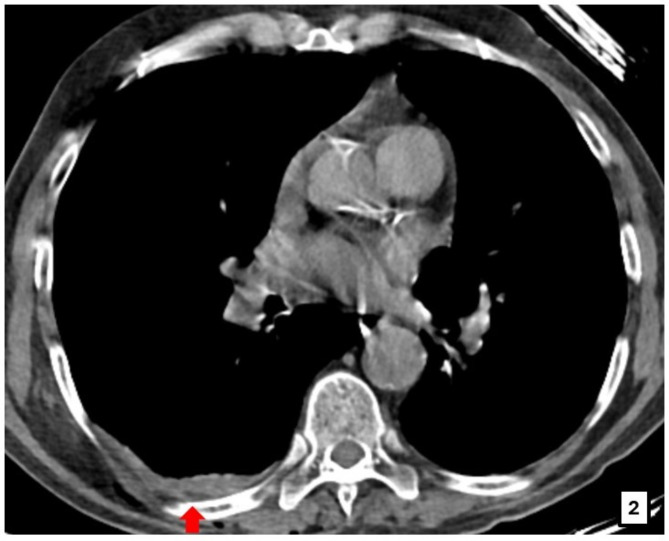
All measurable fluids that could be associated with the trauma mechanism, side and observed on the side of the rib fracture were considered as hemothorax (Fig. 2). Patients who did not initially present with hemothorax but later developed a blunt costophrenic sinus and a decrease in hemogram during follow-up were also categorized as patients with hemothorax. An investigation was conducted to analyze the relationship between local factors associated with rib fractures and the likelihood of hemothorax.
Fig. 2.
Hemothorax less than 10 mm due to the rib fracture in the right hemithorax.
Ethical consideration
The ethical research board committee of Izmir Democracy University Buca Seyfi Demirsoy Education and Research Hospital approved the research (REF. 2024/285). All study participants provided informed consent to use their medical and surgical data in this study. This study was carried out in accordance with the Helsinki Declaration’s contents.
Statistical analysis
In descriptive statistics, number and percentage values for categorical variables and mean ± standard deviation, min-max values for numerical variables were presented. The normality assumption of the quantitative data was assessed with the Shapiro-Wilk test. Comparison of numerical variables with normal distribution in two groups was performed with Independent sample t-test. The comparison of categorical variables between two groups was analyzed by Pearson Chi-square test. In these univariate tests, variables with a p value of <0.10 and considered to be clinically important were included in the Multiple Logistic Regression model and analyzed whether they were risk factors for hemothorax. Odds ratio and 95% confidence intervals were calculated for each risk factor. The diagnostic performance of the number of rib fractures for the risk of hemothorax was evaluated by Receiver Operator Characteristic (ROC) curve. In addition, the AUC (Area Under the Curve) value, which reflects the ability of the test to discriminate between individuals with and without hemothorax diagnosis, was used. In the ROC analysis, the optimal cut-off value that maximizes the sensitivity and specificity of the test was calculated with the Youden index. IBM SPSS Statistics 25.0 (IBM SPSS Statistics for Windows, Version 25.0. Armonk, NY: IBM Corp.) package program was used to analyze the data obtained from the study. The significance level was set as 0.05 in all analyzes.
Results
The study included a total of 273 patients, with 201 (73.6%) being male. The mean age was 53.9 ± 17.27 (19–93) years. The predominant mechanism of trauma observed was a simple fall, accounting for 99 cases (36.3%). The next most common causes were falls from high places, which accounted for 54 cases (19.8%), in vehicle traffic accidents, which accounted for 31 cases (11.4%), and descending stairs in a tumbling fashion, which accounted for 30 cases (11%). The occurrence of injuries to the right and left hemithorax following trauma was almost equal, with an incidence of 50.2% and 49.8% respectively. The median number of rib fractures was 3 (1–10). Patients most presented with 3 rib fractures (27.5%), followed by two (21.6%), four (16.8%) and one (12.1%), respectively. A total of 169 patients (61.9%) had at least one displaced rib fracture. The rib fracture was most frequently located posteriorly, accounting for 129 cases (47.3%). There were 102 cases (37.4%) with measurable hemothorax following trauma.
After analyzing the demographic characteristics of the patients, it was determined that age did not have a significant impact on the risk of hemothorax (p = 0.296). The prevalence of hemothorax was significantly higher in male patients, accounting for 82.4% of cases (p = 0.012). The mechanism of trauma was not identified as a prognostic factor for the risk of hemothorax (p = 0.173) (Table 1).
Table 1.
Univariate analyses and significance values on the development of hemothorax.
| Patient characteristics | Hemothorax (n/mean) | Significance | ||
|---|---|---|---|---|
| None | Exist | General | P*** | |
| Age** | 53.1 ± 17.4(19–93) | 55.3 ± 17(19–87) | 53.9 ± 17.3 (19–93) | 0.296 |
| Gender | ||||
| Male | 117 | 84 | 201 | 0.012 |
| Female | 54 | 18 | 72 | |
| Mechanism | ||||
| Simple fall | 63 | 36 | 99 | 0.173 |
| Fall from height | 31 | 23 | 54 | |
| IVTA* | 22 | 9 | 31 | |
| Stairway fall | 23 | 7 | 30 | |
| Assault | 11 | 12 | 23 | |
| OVTA* | 8 | 11 | 19 | |
| Others | 13 | 4 | 17 | |
| Side | ||||
| Left | 89 | 47 | 136 | 0.340 |
| Right | 82 | 55 | 137 | |
| Number of rib fracture** | 3 ± 1.4 (1–8) | 4 ± 2 (1–10) | 3.4 ± 1.8 (1–10) | < 0.001 |
| Displaced rib fracture | ||||
| None | 84 | 20 | 104 | <0.001 |
| Exist | 87 | 82 | 169 | |
| Location of the fracture on the rib | ||||
| Anterior | 44 | 9 | 53 | 0.001 |
| Lateral | 59 | 32 | 91 | |
| Posterior | 68 | 61 | 129 | |
Statistically significant p-values are highlighted in bolditalics.
* IVTA: In-vehicle traffic accident, OVTA: Out-vehicle traffic accident ** Numerical data are presented as mean, categorical data are presented as number (n). ***Variables with significant p value were included in multiple regression analysis.
When examining the impact of rib fracture characteristics on hemothorax, it was observed that patients with hemothorax had a mean of 4 ± 2 (1–10) rib fractures, while patients without hemothorax had a mean of 3 ± 1.4 (1–8) rib fractures. An increased number of rib fractures was determined to be a significant contributing factor in terms of the risk of hemothorax (p < 0.001). Among the patients diagnosed with hemothorax, a displaced rib fracture was observed in 80.4% of cases. Based on this finding, the occurrence of a displaced rib fracture was determined to be a significant risk factor for hemothorax (p < 0.001). When the location of the rib fracture was evaluated, it was found that the most common location of the fracture was posterior with a rate of 59.8% in patients with hemothorax (p < 0.001) (Table 1).
A multiple logistic regression model was constructed by including gender, number of rib fractures, location of the fracture on the rib arch, and displacement status, all of which were determined to have statistical significance in the univariate analyses. Based on this model, the hemothorax was significantly affected by the number of rib fractures, the displacement of the fractures, and the location of the fracture on the rib. According to the regression analysis, each additional rib fracture was associated with a 38% higher risk of hemothorax (p < 0.001). There was a 2.7-fold higher risk of hemothorax (p = 0.003) when at least one rib fracture was displaced. The risk of hemothorax was 2.7 times higher for lateral rib fractures and 5.8 times higher for posterior rib fractures compared to anterior rib fractures (p = 0.029, p < 0.001, respectively). Table 2 presents a summary of the data from the multiple regression analysis. The most effective cut-off for determining the presence of hemothorax based on the number of rib fractures was found to be 4, with a sensitivity of 55.0% and specificity of 71.0%. The area under the curve (AUC) value, which represents the accuracy of the diagnostic test, was calculated to be 0.65 (95% CI: 0.59; 0.72).
Table 2.
Results of the multiple regression analysis.
| OR | 95% CI for OR | p | ||
|---|---|---|---|---|
| Lower | Upper | |||
| Gender | 1.698 | 0.881 | 3.271 | 0.114 |
| Number of rib fracture | 1.383 | 1.161 | 1.647 | < 0.001 |
| Displacement status | 2.650 | 1.407 | 4.992 | 0.003 |
| Lateral rib fracture | 2.734 | 1.112 | 6.724 | 0.029 |
| Posterior rib fracture | 5.830 | 2.434 | 13.965 | <0.001 |
Statistically significant p-values are highlighted in bolditalics.
Discussion
Traumatic hemothorax is a commonly encountered and potentially fatal condition in the practice of thoracic surgery. It can occur as a result of penetrating trauma or blunt trauma. While injuries like vertebral fracture and pulmonary parenchymal laceration can contribute to the development of hemothorax following blunt thoracic trauma, the primary issue in the majority of cases is rib fracture and its associated secondary pathologies3,7–9. According to Yu et al.3, 33% of patients who experienced blunt thoracic trauma and had rib fractures developed hemothorax. Put simply, while most patients can complete treatment without any hemothorax being detected, around 30% of patients will experience a hemothorax that requires further monitoring. Currently, identifying the patients who are at greater risk to develop hemothorax will help direct the treatment.
It is a plausible theory that increasing the number of broken ribs increases the risk of hemothorax. Increasing the number of broken ribs may also lead to damage to more intercostal structures and damage to more parenchymal structures. Furthermore, ribs have the potential to cause bleeding independently, without causing harm to nearby structures. These theories have served as a source of motivation for numerous studies, leading to the inclusion of rib fractures in trauma scoring systems10–12. Our study provided concrete evidence of this relationship, demonstrating that each additional rib fracture increased the risk of hemothorax by 38%. Furthermore, the analysis revealed that the cut-off value for rib fractures was determined to be 4. While this analysis does not specify the appropriate setting for follow-up care (outpatient, inpatient, or intensive care unit) for a patient with four rib fractures, it does suggest that patients with four or more rib fractures require more vigilant monitoring.
The second specific data identified in our study was the correlation between displaced rib fractures and the occurrence of hemothorax. Typically, the term displaced rib fracture is used to refer to a serious fracture that indicates a higher risk of fatal injuries and complications like hemothorax. Although these significant characteristics exist, there are numerous definitions for displaced rib fracture in the literature13. Chien CY (10) et al. have defined a displaced rib fracture as a fracture line that is displaced by a minimum of half the thickness of the fractured rib. Chapman BC et al.14 coined the term “severe displaced rib fracture” to describe a condition where the rib is displaced by a distance greater than its width, with no connection between the proximal and distal ends. While there may be varying definitions for displaced rib fracture, it is generally agreed upon that it signifies a significant trauma. Our study clearly demonstrated a concrete relationship, showing that the presence of at least one displaced rib fracture increased the risk of hemothorax by a factor of 2.7.
In our study, we clearly demonstrated the correlation between the location of rib fractures and the occurrence of hemothorax. While rib fractures often cause damage to intercostal vascular structures and nearby organs, there are also instances where they lead to significant injury to large blood vessels. Aortic injury has been reported in both the early and late periods, particularly in rib fractures located towards the back15,16. Our study excluded cases with major vascular structure injuries, as we focused specifically on investigating the characteristics of rib fractures. Our study aligns with the existing literature in demonstrating that anterior rib fractures have the lowest risk of hemothorax when considering the clinical reflection of anatomical features. Lateral rib fractures were identified as having the greatest risk. That study focused exclusively on the midline ribs and established a distinct categorization for the upper and lower ribs17. In our study, we classified the data into anterior, lateral, and posterior categories, without considering any differences in levels. Based on the data, it was found that lateral rib fractures were 2.7 times more likely to result in hemothorax compared to anterior rib fractures (p = 0.029). Similarly, posterior rib fractures were 5.8 times more likely to result in hemothorax compared to anterior rib fractures (p < 0.001). The fact that intercostal arteries originate from the aorta and gradually become relatively thinner in size throughout their course may support the finding that posterior fractures have a higher bleeding tendency compared to anterior fractures.
The occurrence of hemothorax has been linked to the nature and intensity of trauma in numerous studies documented in the literature. High-energy traumas are expected to cause more extensive tissue damage and an increased risk of hemothorax compared to traumas like simple falls. Our study found no statistically significant correlation between the mechanism of trauma and the occurrence of hemothorax. This difference may be explained by the exclusion criteria. Our study aimed to thoroughly investigate the correlation between rib fracture and hemothorax. Consequently, patients with other factors contributing to the development of hemothorax, such as vertebral fracture or diaphragmatic injury, were deliberately excluded from the study. As a secondary consequence of these exclusion criteria, multi-systemic trauma patients were also excluded from the study and only patients with minor injuries from high-energy trauma patients such as vehicular traffic accidents could be included in the study. While initially seen as a disadvantage, this feature is actually crucial for standardizing studies. The study’s retrospective nature poses a drawback in terms of data collection. The inclusion of 3 different centers in our study enhances the data’s security and the variety of cases. However, the implementation of rigorous standardization resulted in the exclusion of some major trauma cases. Our study suggests that the risk of hemothorax is influenced by the local characteristics of the rib fracture, regardless of the severity of trauma. We believe that our study may be a step towards providing a practical view on the risk of hemothorax.
Conclusions
Our study concluded that there is a significant increase in the risk of hemothorax associated with more rib fractures and specifically more posterior rib fractures. This risk remains consistent regardless of factors such as trauma mechanism, age, and gender. We discovered that the displacement of even a single rib fracture significantly increased the risk of hemothorax. There is currently insufficient data in thoracic surgery practice to determine which patients should be prioritized for follow-up. According to our results, we recommend that physicians evaluating trauma patients should follow up more closely and carefully patients with posterior rib fractures, at least one displaced rib fracture and more than four rib fractures within their capabilities and by utilizing their clinical experience.
Author contributions
All authors contributed to the study conception and design. S.A: Writing – original draft, Visualization, Software, Project administration, Methodology, Investigation, Formal analysis, Data curation, Conceptualization. S.K.A: Writing – review & editing, Methodology, Project administration. BG: Data curation. SGG: Data curation. OK: Writing – review & editing, Supervision. OFD: Conceptualization, Formal analysis, MethodologyAll authors commented on previous versions of the manuscript. All authors read and approved the final manuscript.
Data availability
The datasets generated during and/or analyzed during the current study are available from the corresponding author on reasonable request.
Declarations
Competing interests
The authors declare no competing interests.
Institutional review board statement
Izmir Democracy University Buca Seyfi Demirsoy Education and Research Hospital Clinical Research Ethics. Committee were approved for this study (approval number 2024/285). All methods were performed in accordance with the relevant guidelines and regulations.
Informed consent
All study participants provided informed consent to use their medical and surgical data in this study.
Footnotes
Publisher’s note
Springer Nature remains neutral with regard to jurisdictional claims in published maps and institutional affiliations.
References
- 1.Kani, K. K., Mulcahy, H., Porrino, J. A. & Chew, F. S. Thoracic cage injuries. Eur. J. Radiol.11010.1016/j.ejrad.2018.12.003 (2019). 225 – 32. [DOI] [PubMed]
- 2.Flores-Funes, D. et al. Is the number of rib fractures a risk factor for delayed complications? A case–control study. Eur. J. Trauma. Emerg. Surg.46, 435–440. 10.1007/s00068-018-1012-x (2020). [DOI] [PubMed] [Google Scholar]
- 3.Yu, H., Isaacson, A. J. & Burke, C. T. Management of traumatic Hemothorax, retained Hemothorax, and other thoracic collections. Curr. Trauma. Rep.3, 181–189. 10.1007/s40719-017-0101-3 (2017). [Google Scholar]
- 4.Liman, S. T., Kuzucu, A., Tastepe, A. I., Ulasan, G. N. & Topcu, S. Chest injury due to blunt trauma. Eur. J. Cardiothorac. Surg.23 (3), 374–378. 10.1016/s1010-7940(02)00813-8 (2003). [DOI] [PubMed] [Google Scholar]
- 5.Dogrul, B. N., Kiliccalan, I., Asci, E. S. & Peker, S. C. Blunt trauma related chest wall and pulmonary injuries: An overview. Chin. J. Traumatol.23 (03), 125–138. 10.1016/j.cjtee.2020.04.003 (2020). [DOI] [PMC free article] [PubMed] [Google Scholar]
- 6.Liebsch, C. et al. Patterns of serial rib fractures after blunt chest trauma: An analysis of 380 cases. PLoS One. 14 (12), e0224105. 10.1371/journal.pone.0224105 (2019). [DOI] [PMC free article] [PubMed] [Google Scholar]
- 7.Miller, D. L. & Mansour, K. A. Blunt traumatic lung injuries. Thorac. Surg. Clin.17 (1), 57–61. 10.1016/j.thorsurg.2007.03.017 (2007). [DOI] [PubMed] [Google Scholar]
- 8.Talbot, B. S. et al. Traumatic Rib Injury: Patterns, imaging pitfalls, complications, and treatment. Radiographics. 37 (2), 628–651. 10.1148/rg.2017160100 (2017). [DOI] [PubMed] [Google Scholar]
- 9.Simon, B. J., Chu, Q., Emhoff, T. A., Fiallo, V. M. & Lee, K. F. Delayed hemothorax after blunt thoracic trauma: An uncommon entity with significant morbidity. J. Trauma.45 (4), 673–676. 10.1097/00005373-199810000-00005 (1998). [DOI] [PubMed] [Google Scholar]
- 10.Chien, C. Y. et al. The number of displaced rib fractures is more predictive for complications in chest trauma patients. Scand. J. Trauma. Resusc. Emerg. Med.25 (1), 19. 10.1186/s13049-017-0368-y (2017). [DOI] [PMC free article] [PubMed] [Google Scholar]
- 11.Ziegler, D. W. & Agarwal, N. N. The morbidity and mortality of rib fractures. J. Trauma.37 (6), 975–979. 10.1097/00005373-199412000-00018 (1994). [DOI] [PubMed] [Google Scholar]
- 12.Chen, J., Jeremitsky, E., Philp, F., Fry, W. & Smith, R. S. A chest trauma scoring system to predict outcomes. Surgery. 156 (4), 988–993. 10.1016/j.surg.2014.06.045 (2014). [DOI] [PubMed] [Google Scholar]
- 13.Kavurmacı, Ö. et al. We need a common definition and treatment algorithm for displaced rib fracture. Curr. Thorac. Surg.7 (2), 74–81. 10.26663/cts.2022.012 (2022). [Google Scholar]
- 14.Chapman, B. C. et al. RibScore: a novel radiographic score based on fracture pattern that predicts pneumonia, respiratory failure, and tracheostomy. J. Trauma. Acute Care Surg.80 (1), 95–101. 10.1097/TA.0000000000000867 (2016). [DOI] [PubMed] [Google Scholar]
- 15.Boyles, A. D., Taylor, B. C. & Ferrel, J. R. Posterior rib fractures as a cause of delayed aortic injury: A case series and literature review. Injury. 44 (5), 43–45. 10.1016/j.injury.2013.03.011 (2013). [Google Scholar]
- 16.Yanagawa, Y., Kaneko, N., Hagiwara, A., Kimura, T. & Isoda, S. Delayed sudden cardiac arrest induced by aortic injury with a posterior fracture of the left rib. Gen. Thorac. Cardiovasc. Surg.56, 91–92. 10.1007/s11748-007-0186-7 (2008). [DOI] [PubMed] [Google Scholar]
- 17.Pines, G., Gotler, Y., Lazar, L. O. & Lin, G. Clinical significance of rib fractures’ anatomical patterns. Injury. 51 (8), 1812–1816. 10.1016/j.injury.2020.05.023 (2020). [DOI] [PubMed] [Google Scholar]
Associated Data
This section collects any data citations, data availability statements, or supplementary materials included in this article.
Data Availability Statement
The datasets generated during and/or analyzed during the current study are available from the corresponding author on reasonable request.


