Abstract
Infectious bursal disease viruses (IBDVs), belonging to the family Birnaviridae, exhibit a wide range of immunosuppressive potential, pathogenicity, and virulence for chickens. The genomic segment A encodes all the structural (VP2, VP4, and VP3) and nonstructural proteins, whereas segment B encodes the viral RNA-dependent RNA polymerase (VP1). To identify the molecular determinants for the virulence, pathogenic phenotype, and cell tropism of IBDV, we prepared full-length cDNA clones of a virulent strain, Irwin Moulthrop (IM), and constructed several chimeric cDNA clones of segments A and B between the attenuated vaccine strain (D78) and the virulent IM or GLS variant strain. Using the cRNA-based reverse-genetics system developed for IBDV, we generated five chimeric viruses after transfection by electroporation procedures in Vero or chicken embryo fibroblast (CEF) cells, one of which was recovered after propagation in embryonated eggs. To evaluate the characteristics of the recovered viruses in vivo, we inoculated 3-week-old chickens with D78, IM, GLS, or chimeric viruses and analyzed their bursae for pathological lesions 3 days postinfection. Viruses in which VP4, VP4-VP3, and VP1 coding sequences of the virulent strain IM were substituted for the corresponding region in the vaccine strain failed to induce hemorrhagic lesions in the bursa. In contrast, viruses in which the VP2 coding region of the vaccine strain was replaced with the variant GLS or virulent IM strain caused rapid bursal atrophy or hemorrhagic lesions in the bursa, as seen with the variant or classical virulent strain, respectively. These results show that the virulence and pathogenic-phenotype markers of IBDV reside in VP2. Moreover, one of the chimeric viruses containing VP2 sequences of the virulent strain could not be recovered in Vero or CEF cells but was recovered in embryonated eggs, suggesting that VP2 contains the determinants for cell tropism. Similarly, one of the chimeric viruses containing the VP1 segment of the virulent strain could not be recovered in Vero cells but was recovered in CEF cells, suggesting that VP1 contains the determinants for cell-specific replication in Vero cells. By comparing the deduced amino acid sequences of the D78 and IM strains and their reactivities with monoclonal antibody 21, which binds specifically to virulent IBDV, the putative amino acids involved in virulence and cell tropism were identified. Our results indicate that residues Gln at position 253 (Gln253), Asp279, and Ala284 of VP2 are involved in the virulence, cell tropism, and pathogenic phenotype of virulent IBDV.
Infectious bursal disease virus (IBDV) belongs to the genus Avibirnavirus and is a member of the family Birnaviridae (1, 9, 10). The genome of IBDV consists of two segments of double-stranded RNA, which are packaged in a nonenveloped icosahedral shell 60 nm in diameter. The larger segment, A, is 3,261 nucleotides long, and it encodes a 110-kDa precursor protein in a single large open reading frame (ORF), which is cleaved by autoproteolysis to yield mature VP2, VP3, and VP4 proteins (1, 12). VP2 and VP3 are the major structural proteins of the virion, whereas VP4 is a minor protein involved in the processing of the precursor protein (3, 12, 13, 28). VP2 is the major host-protective immunogen of IBDV and contains the determinants responsible for causing antigenic variation (6, 11, 40). VP3 is a group-specific antigen and forms a complex with VP1, which may have an essential role for the morphogenesis of IBDV particles (5, 19). Segment A also encodes a 17-kDa nonstructural (NS) protein from a small ORF which precedes and partly overlaps the large ORF (35). This NS protein is detected only in IBDV-infected cells, and it is not required for viral replication but plays an important role in pathogenesis (24, 43). The smaller segment, B, is 2,827 nucleotides long, and it encodes VP1, a 97-kDa protein having RNA-dependent RNA polymerase activity (36). This protein is covalently linked to the 5′ ends of the genomic RNA segments (34).
IBDV infects the precursors of antibody-producing B cells in the bursa of Fabricius (BF), which can cause severe immunosuppression and mortality in young chickens (2, 15). Viruses of serotype I are pathogenic to chickens, whereas serotype II viruses are avirulent for chickens (21). IBDV isolates of serotype I display a wide range of immunosuppressive potential, pathogenicity, and virulence for chickens. Classic IBDV strains isolated from the United States in the early 1960s, such as the Edgar, 2512, and Irwin Moulthrop (IM) strains, induce hemorrhagic lesions accompanied by near-total B-cell follicle depletion and cause between 30 and 60% mortality in light-breed chickens. In the late 1980s, Delaware and GLS variant viruses, which cause rapid atrophy of the bursa without the accompanying inflammation, hemorrhage, or mortality caused by the earlier classical strains, were isolated from the Delmarva Peninsula (32, 33). In the mid 1990s, “very virulent” strains of IBDV which cause >70% mortality in chickens emerged in several European and Asian countries (6, 18, 37). To distinguish the very virulent strains from the classic vaccine strains, a monoclonal antibody (MAb) was generated against the virulent IM strain (22). This MAb 21 recognizes all the very virulent IBDV strains tested to date, but it does not react if these viruses are adapted in tissue culture (22, 41).
Earlier studies have shown that very virulent strains of IBDV lose their virulence potential after serial passage in non-B lymphoid chicken cells (42). Comparison of the deduced amino acid sequences of the very virulent (OKYM) and attenuated (OKYMT) strains showed specific amino acid substitutions within the hypervariable region of the VP2 protein. However, due to the lack of a reverse-genetics system that can generate virulent IBDV, it was difficult to pinpoint the amino acids involved in virulence and cell tropism. By carrying out site-directed mutagenesis of residues 279 and 284 in VP2, Lim and coworkers demonstrated that very virulent IBDV could be adapted to chicken embryo fibroblast (CEF) cell culture (17). Similarly, Mundt reported that residues 253 and 284 of the VP2 protein of the variant virus are necessary for tissue culture infectivity (23). However, none of these viruses were tested in chickens to verify the role of these residues in IBDV virulence and pathogenicity. In a recent study, Boot and coworkers rescued a very virulent IBDV, using a fowlpox-based reverse-genetics system, and demonstrated that VP2 is not the sole determinant of the very virulent phenotype (4). However, except for VP2, the possible role of viral proteins in virulence, cell tropism, and the pathogenic phenotype has not yet been determined.
Therefore, in order to identify the viral protein(s) of IBDV that is involved in virulence, cell tropism, and the pathogenic phenotype, we constructed chimeric clones between the attenuated vaccine strain D78 and either the virulent (IM) or the variant (GLS) strain by exchanging VP2-, VP4-, VP4 and -3, or VP1-encoding cDNA fragments. Using the cRNA-based reverse-genetics system for IBDV, we recovered five chimeric viruses, including the virulent one, which contains the epitope recognized by MAb 21 (virulence marker). In this report, we describe the characteristics of these recovered viruses in vitro and in vivo and identify the protein(s) and putative amino acid residues involved in virulence, cell tropism, and the pathogenic phenotype.
MATERIALS AND METHODS
Cells, viruses, and hybridomas.
Vero cells were maintained in M199 medium supplemented with 5% fetal bovine serum (FBS) at 37°C in a humidified 5% CO2 incubator and were used for propagation of the virus and transfection experiments. Primary CEF cells were prepared from 10-day-old embryonated eggs (SPAFAS, Inc., Storrs, Conn.) as described previously (25). Secondary CEF cells were maintained in a growth medium consisting of M199-F10 (50%-50% [vol/vol]) and 5% FBS and were used for transfection, virus titration, immunofluorescence, and plaque assays. Virus stocks were established by serial passage of the recombinant viruses in the cell cultures, except one (recombinant IMVP2 [rIMVP2]), which was propagated in embryonated eggs. The D78 vaccine strain and the recovered chimeric viruses were titrated in secondary CEF cells as described previously (25), whereas rIMVP2 was titrated in eggs. The virulent IM and GLS strains of IBDV were obtained from bursae of infected specific-pathogen-free chickens and purified as described previously (25). A panel of MAbs, prepared against various strains of IBDV, was used to characterize IBDV antigens by antigen capture–enzyme-linked immunosorbent assay (AC-ELISA), as described previously (33, 39). All classical strains of IBDV reacted with MAbs B69, R63, and B29, whereas MAb 57 recognized only the GLS variant strain. In addition, MAb 21 prepared against the IM strain was used, which reacted only with the highly virulent IBDV strains (22).
Construction of full-length cDNA clones.
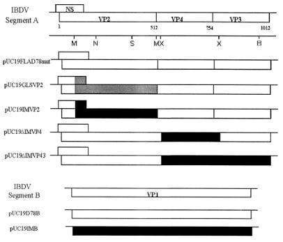
All manipulations of DNAs were performed according to standard protocols (27). Construction of a full-length cDNA clone of IBDV genome segment A of strain D78 (with sequence tags), pUC19FLAD78mut, has been described previously (25). It encodes all of the structural proteins (VP2, VP4, and VP3), as well as the NS protein (Fig. 1). The cloning of segment A of IBDV strain IM was achieved by generating four overlapping cDNA clones. First, the VP2 region of IBDV segment A was cloned by reverse transcription (RT)-PCR, using the oligonucleotide primers IBDVP2R (5′-CCAATTGCATGGGCTAGG-3′; binding to nucleotide positions 1534 to 1551) and BamBV (5′-ACGATCGCAGCGATGACAAACCTG-3′; binding to nucleotide positions 119 to 141). This fragment was cloned into a pCR2.1 vector (Invitrogen) to produce pCRIMVP2. The 5′ end of IBDV segment A was cloned by RT-PCR, using the oligonucleotide primers 5′IR (5′-CACAGTCAAAATGTAGGTCGA-3′; binding to nucleotide positions 256 to 277) and A5′D78 (25). Similarly, the 3′ end of IBDV segment A was cloned by RT-PCR, using the oligonucleotide primers A3′D78 (25) and 3′IF (5′-CAGATGAAAGATCTGCTTG-3′; binding to nucleotide positions 3011 to 3031). Both of these fragments were cloned into pCR2.1 to obtain the plasmids pCRIM5′ and pCRIM3′, which were used for sequence analysis. The remaining portion of IM segment A, which encodes VP4 and VP3, was cloned by RT-PCR, using primers IBDVP2F (5′-GCTTCAAAGACATAATCCGG-3′; binding to nucleotide positions 1458 to 1477) and BglR (5′-CAAGAGCAGATCTTTCATCTG-3′; binding to nucleotide positions 3011 to 3031). The resulting fragment was then cloned in pCR2.1 to obtain pCRIMVP43. Plasmids pUC19IMVP2 and pUC19GLSVP2 were prepared by replacing an MfeI fragment in plasmid pUC19FLAD78mut with the respective MfeI fragments derived from plasmids pCRIMVP2 and pGLS-5, as shown in Fig. 1. The cloning of genomic segment A of IBDV strain GLS and the construction of plasmid pGLS-5 have been described previously (39).
FIG. 1.
Schematic presentation of various cDNA constructs of IM, D78, and GLS strains in order to generate plus-sense RNA transcripts using T7 RNA polymerase. A map of the IBDV genome segments A and B, with its coding capacity, is shown at the top (drawn to scale). The open boxes depict the coding regions of the D78 strain, whereas the solid and shaded boxes represent the coding regions of the IM and GLS strains, respectively. Selected restriction sites, important for the construction of chimeric cDNA clones of segment A, are shown. B, BglII; M, MfeI, N, NdeI; S, SpeI; X, XhoI. The use of restriction enzymes to generate these chimeric clones did not alter the reading frame of the polyprotein and hence did not cause extensions or deletions of the VP2 (residues 1 to 512), VP4 (residues 513 to 754), or VP3 (residues 755 to 1012) proteins. All of the constructs contain a T7 polymerase promoter sequence at the 5′ end.
A modified pUC19 vector was constructed by digesting the vector with both NdeI and NarI and then religating it to form pUC19Δ. Next, a full-length D78 clone was constructed using this modified pUC19Δ vector. The 3′ end of the full-length D78 clone was modified by PCR to produce a plasmid, pUC19ΔD78F, that contained a KpnI site at the end instead of an EcoRI site. This plasmid was used as a backbone to construct the additional clones described below.
A full-length clone of IM segment A was constructed in several steps. First, plasmid pUC19IMVP2 was digested with SpeI and BglII to release a 1,530-bp fragment that was further digested with MfeI. This SpeI-MfeI fragment was then combined with the MfeI-BglII fragment of pCRIMVP43 and cloned back into the pUC19IMVP2 vector to obtain pUC19IMVP243. An RsrII-BglII fragment from this plasmid was obtained by digestion with the RsrII and BglII enzymes. This fragment was then combined with the BglII-BsrGI fragment of plasmid pCRIM3′NC and ligated into the RsrII-BsrGI-digested pUC19ΔD78F vector. This created a full-length clone, pUC19ΔIMA, which contains all of the coding sequence of IM segment A but lacks the two nucleotide changes in the 5′ noncoding region of wild-type IM. This construct is not shown in Fig. 1. To evaluate the role of VP4 in virulence, plasmid pUC19ΔD78F was digested with the XhoI enzyme to release the VP4 coding fragment, which was then replaced with the XhoI fragment, derived from pCRIMVP43 (without altering the reading frame), to create plasmid pUC19ΔIMVP4. In this chimera, residues at positions 517, 588, and 702 of D78 were replaced with residues of the IM strain (Table 1). Similarly, to prove that the determinants of virulence, cell tropism, and pathogenicity do not reside in VP4 and VP3, we constructed clone pUC19ΔIMVP43 by digesting plasmid pUC19ΔIMA with MfeI and replacing this fragment with the MfeI portion of pUC19ΔD78F (Fig. 1). As a result of this chimera, two additional residues at positions 923 and 981 of the D78 VP3 protein were replaced with residues of the IM strain (Table 1).
TABLE 1.
Location of amino acid changes in VP2, VP4, VP3, and VP1 proteins of IBDV strains D78 and IM
| Protein | Amino acid change (position) | ||||||
|---|---|---|---|---|---|---|---|
| D78 VP2 | Gly (76) | Val (242) | His (253) | Thr (270) | Asn (279) | Thr (284) | Arg (330) |
| IM VP2 | Ser | Ile | Gln | Ala | Asp | Ala | Ser |
| D78 VP4 | Tyr (517) | Glu (588) | Arg (702) | ||||
| IM VP4 | His | Lys | Lys | ||||
| D78 VP3 | Arg (923) | Leu (981) | |||||
| IM VP3 | Lys | Pro | |||||
| D78 VP1 | Thr (13) | Arg (115) | Gly (147) | Glu (515) | Leu (546) | Glu (653) | (879) |
| IM VP1 | Lys | Gly | Asp | Asp | Pro | Lys | Gln |
To construct a cDNA clone of segment B of the virulent IBDV strain IM, two primer pairs (B5′-D78 and B5-IPD78, and B3′-D78 and B3-IPD78) were synthesized and used for RT-PCR amplification. The sequences of the primers were identical to the one used for the construction of the segment B cDNA clone of strain D78 (25, 43). Using genomic double-stranded RNA as a template, cDNA fragments were synthesized and amplified by using a kit according to the supplier's protocol (Perkin-Elmer). The amplified fragments were cloned between the EcoRI site of a pCR2.1 vector to obtain plasmids pCRIMB5′ and pCRIMB3′. To construct a full-length clone of segment B, the 5′-end fragment of IBDV (from plasmid pCRIMB5′) was first subcloned between the EcoRI and PstI sites of the pUC19 vector to obtain pUC19IMB5′. Then the 3′-end fragment of IBDV (from plasmid pCRIMB3′) was inserted between the BglII and PstI sites of plasmid pUC19IMB5′ to obtain a full-length plasmid, pUC19IMB, which encodes the VP1 protein (Fig. 1).
DNA from the above-mentioned plasmids was sequenced by the dideoxy chain termination method (30), using an automated DNA sequencer (Applied Biosystem), and the sequence data were analyzed using PC/Gene (Intelligenetics) software. The integrity of the full-length constructs was tested by an in vitro transcription and translation coupled reticulocyte lysate system using T7 RNA polymerase (Promega Corp.). The resulting labeled products were separated on a sodium dodecyl sulfate–12.5% polyacrylamide gel and visualized by autoradiography.
Transcription and transfection of synthetic RNAs.
To generate synthetic transcripts of segments A and B, plasmids of segments A and B were linearized with the BsrGI and PstI enzymes, respectively, and treated as described previously (25). The linearized DNA was used to produce in vitro transcripts with the T7 mMessage mMachine kit (Ambion) according to the manufacturer's instructions. Briefly, approximately 3 μg of linearized DNA template was added to the transcription reaction mixture (20 μl) containing 40 mM Tris-HCl (pH 7.9); 10 mM NaCl; 6 mM MgCl2; 2 mM spermidine; 0.5 mM (each) ATP, CTP, and UTP; 0.1 mM GTP; 0.25 mM cap analog [m7G(5′)ppp(5′)G]; 120 U of RNasin; and 150 U of T7 RNA polymerase and incubated at 37°C for 1 h. Equimolar amounts of RNA transcripts of segments A and B (≈8 μg each) were directly used to transfect cells by the electroporation technique.
Electroporation of Vero and CEF cells was carried out in accordance with the protocol supplied by the manufacturer (Bio-Rad, Hercules, Calif.). Approximately 2 × 10⁶ cells and 9 μl of RNA transcripts from each segment were placed in a cuvette with a 0.4-cm gap and incubated on ice for 10 min prior to electroporation. Electroporation was done using a Genepulser set at 200 V with a capacitance of 960 μF. After the shock, the cuvettes were incubated on ice for 5 min, and then the cells were placed in medium supplemented with 10% FBS in a six-well plate. The plate was incubated at 37°C in a humidified 5% CO2 incubator. For all of the transfections, except the one performed with pUC19IMVP2, the medium was changed after 16 h. After 4 days, the cells were freeze-thawed three times, and the supernatant was passed into a T-25 flask containing confluent CEF or Vero cells with 5% FBS-supplemented medium. The monolayer was examined daily for cytopathic effect. Following electroporation of CEF cells with transcripts derived from pUC19IMVP2, the medium was changed after 6 h and replaced with 1 ml of 10% FBS-supplemented medium. After 24 h, the cells were freeze-thawed twice, and the supernatant was inoculated into the chorioallantoic membranes (CAMs) of 11-day-old embryos, as described below. After 6 days, the embryos were examined for lesions, and the CAM was collected for further propagation of virus.
Inoculation of CAM.
Eleven-day-old embryos were used for the CAM inoculation. Briefly, a hole was punched at the air cell as well as in the side of the egg, where the vein structure is well developed. The air was removed from the air cell with a syringe. The supernatant (100 to 500 μl) was dropped onto the CAM with a syringe through the hole punched in the side of the egg. Both holes were sealed, and the embryo was incubated for 6 days. The embryo was examined daily for survival by candling. Death of the embryo is often a sign of virus infection. After 6 days, the embryo was examined for other signs of viral infection, such as lesions on the CAM, lesions on the embryo, or stunting of the embryo's growth.
Characterization of recovered IBDV.
To determine the specificity of the recovered viruses, CEF cells were infected with the transfectant viruses and the infected cells were analyzed by immunofluorescence assay with rabbit anti-IBDV polyclonal serum, as described previously (25). To examine viral structural proteins expressed by recovered chimeric viruses, the viruses (including the one propagated in the CAM) were purified by sucrose gradient centrifugation and analyzed by Western blotting, as described previously (26, 39). To further characterize the recovered virus, RT-PCR was performed on the chimeric virus with the appropriate primer pair, used to produce the original clone. The resulting PCR product was directly sequenced as described above using one of the primers of the primer pair.
Growth curve of chimeric IBDV.
To analyze the growth characteristics of IBDV, confluent secondary CEF cells (in T-25 flasks) were infected with one of the recovered virus stocks (generated after five passages in Vero or CEF cells) at a multiplicity of infection of 0.1. Infected cell cultures were harvested at different time intervals, and the titer of infectious progeny was determined by plaque assay on CEF cells as described previously (25). For rIMVP2, which does not propagate in tissue culture, a 50% egg infectious dose (EID50) was used to determine the virus titer. Serial dilutions of the virus were made and then inoculated into CAMs as described previously. For each dilution, the number of embryos dead or showing lesions was determined. Then, and EID50 was determined using the Reed-Muench formula.
Chicken inoculation.
Three-week-old specific-pathogen-free chickens were obtained (SPAFAS, Inc.) and housed in isolators. Prior to inoculation, the chickens were bled, and their sera were tested by ELISA to ensure that they were negative for IBDV-specific antibodies. Eight groups of chickens were inoculated by the ocular route with either culture medium or wild-type IM, D78, rIMB, rIMVP43, rIMVP4, GLSVP2, or rIMVP2 virus. For viruses that were able to replicate in cell culture, a dose of 1,000 PFU was administered to each group of chickens, whereas a dose of 1,000 EID50 was given to chickens in the IM and rIMVP2 groups. After 3 days, any surviving chickens were humanely killed and the bursae were excised and bisected. One hemisection was used for RT-PCR assay and AC-ELISA, while the other was fixed and sectioned for histopathological examination.
Identification of recovered viruses by RT-PCR and AC-ELISA.
In order to determine whether the nucleotide sequence in the recovered viruses is of chimeric origin, total nucleic acids from IBDV-infected CEF cells, CAMs, or bursal homogenates were isolated and analyzed by RT-PCR, as described above. Segment-specific primers were used for RT of genomic RNA. Following RT, the reaction products were amplified by PCR with the desired segment-specific primer, and the amplified products were either directly sequenced or cloned into the pCR2.1 vector and then sequenced as described above. Generally, the nucleotide sequence in chimeric viruses was determined twice, once after recovery in vitro and again after in vivo studies of bursal homogenates. The recovered viruses were further characterized with a panel of IBDV-specific MAbs in an AC-ELISA, as previously described (40).
Histopathological studies.
The bursa tissues were fixed by immersion in 10% neutral buffered formalin. The ratio of fixative to bursa exceeded 10:1. Seven days later, a cross sectional portion of each bursa was processed through paraffin, stained with hematoxylin and eosin, and examined with a light microscope. The severity of the lesion was graded on a scale of 1 to 5, based on the extent of the lymphocyte necrosis, follicular depletion, and atrophy.
Nucleotide sequence accession numbers.
The complete nucleotide sequences of IBDV genome segments A and B of the IM strain have been deposited in the GenBank database under accession no. AY029166 and AY029165, respectively.
RESULTS
Sequence analysis of segments A and B of virulent IBDV strain IM.
We determined the complete nucleotide sequences of IM IBDV genome segments A and B, including the 5′- and 3′-terminal sequences. These segments are 3,261 and 2,827 bp long, respectively, which is identical to the sizes of the segments of strain D78. Comparison of the 5′- and 3′-terminal sequences of both IM segments with those of the D78 strain showed nucleotide substitutions of A→G and T→C at positions 69 and 80 in segment A and G→A and A→G at positions 59 and 69 in segment B, respectively. Comparison of the deduced amino acid sequences of IM segments A and B with those of the D78 strain showed 98.82 and 99.32% identity at the amino acid level, respectively. There are a total of 12 amino acid substitutions in segment A between IM and D78, of which seven are located in VP2, three in VP4, and two in the VP3 region of the polyprotein (Table 1). Furthermore, there are a total of seven amino acid changes in segment B between IM and D78, including an additional amino acid residue at the C terminus of VP1 in the IM strain (Table 1). This suggests that the classical strain IM is closely related to the D78 vaccine strain. A phylogenetic analysis, based on the deduced amino acid sequences of segments A and B of very virulent or virulent strains of IBDV, revealed two distinct groups. The first group consisted of classical virulent strains, namely, IM, STC, Edgar, and 52/70, that were isolated in the early 1960s and 1970s and caused about 30% mortality. The second group comprised all very virulent strains, such as UK661 (United Kingdom), OKYM (Japan), and HK46 (Hong Kong), that were isolated in the late 1980s and early 1990s and caused ≥70% mortality (data not shown).
Construction of full-length and chimeric cDNA clones.
To identify the molecular determinants of the virulence, pathogenic phenotype, and cell tropism of IBDV, we constructed chimeric cDNA clones between the attenuated vaccine strain D78 and the virulent strain IM or the variant GLS strain. Figure 1 shows the construction of these clones, in which VP2, VP4, and VP4-VP3 coding sequences of IM or GLS were substituted in the D78 backbone. The chimeric nature of these clones was confirmed by DNA sequence analysis. In addition, we also constructed a full-length cDNA clone of IM segment B to determine whether the RNA polymerase would play a role in virulence or cell tropism. The clones were constructed with a pUC19 vector, and all contained a T7 promoter sequence preceding the 5′ noncoding regions of the full-length cDNA clones. The functionality of all these clones was tested by in vitro transcription-coupled translation reactions, which yielded protein products that comigrated with the marker IBDV proteins after fractionation on a sodium dodecyl sulfate–12.5% polyacrylamide gel and autoradiography (data not shown).
Transfection and recovery of chimeric viruses.
To identify the protein(s) responsible for the cell tropism, virulence, and pathogenic phenotype of IBDV, we transfected Vero cells by an electroporation procedure with combined plus-sense transcripts derived from various plasmids, as shown in Table 2. From six transfection experiments, we recovered three chimeric viruses (rGLSVP2, rIMVP4, and rIMVP3) and the parental D78 virus, rD78, containing the tagged sequences. No other viruses could be recovered in Vero cells, even after five attempts, when the cRNA transcripts comprised the coding regions of the IM VP1 (pUC19IMB) or VP2 (pUC19IMVP2) protein. Therefore, CEF and Vero cells were transfected in parallel with the transcripts derived from the plasmid pairs (i) pUC19IMB and pUC19FLAD78mut, (ii) pUC19IMVP2 and pUC19D78B, and (iii) pUC19D78B and pUC19FLAD78mut. From these transfections (done in duplicate), the rIMB virus was recovered only in CEF cells, as shown in Table 2, whereas the parental rD78 virus was recovered in both Vero and CEF cells, as expected. To determine the presence of infectious virus in transfected Vero cells, the cells were freeze-thawed twice, and the supernatants were used to infect CEF cells. No virus could be recovered even after four passages in CEF cells. This result suggests that VP1 contains the determinants for cell-specific replication in Vero cells. Furthermore, the rIMVP2 virus (containing the IM VP2 protein) could not be recovered in CEF cells (after at least five attempts), suggesting that VP2 contains the molecular determinants for cell tropism. Since we could not recover the rIMVP2 virus, we examined the transient expression of IBDV-specific proteins in CEF cells. Twenty-four hours posttransfection, we detected the presence of viral proteins by immunofluorescence assay, using anti-IBDV polyclonal serum (data not shown). Therefore, to assess the presence of infectious virus in transfected CEF cells, the cells were freeze-thawed twice, and the supernatants were used for CAM inoculation of 11-day-old embryonated eggs. After 6 days, the embryos showed signs of viral infection, and the recovered rIMVP2 virus was propagated in the CAMs for further characterization. These results demonstrate that the rIMVP2 virus is able to replicate in CEF cells after transfection but cannot be propagated further in CEF cells. Since parental IM or rIMVP2 viruses are able to grow only in B lymphoid cells and not in CEF cells, it suggests that the major determinants of B-lymphocyte cell receptors reside in VP2.
TABLE 2.
Generation of chimeric IBDVs after transfection with transcripts derived from cloned cDNA of segments A and B in Vero cells, CEF cells, and CAM
| Plasmids used for transcription | Transfectiona
|
Recombinant virus | ||
|---|---|---|---|---|
| Vero | CEF | CAM | ||
| pUC19D78B | + | + | ND | rD78 |
| pUC19FLAD78mut | ||||
| pUC19D78B | + | + | ND | rGLSVP2 |
| pUC19GLSVP2 | ||||
| pUC19D78B | + | + | ND | rIMVP4 |
| pUC19ΔIMVP4 | ||||
| pUC19D78B | + | + | ND | rIMVP43 |
| pUC19ΔIMVP43 | ||||
| pUC19IMB | − | + | ND | rIMB |
| pUC19FLAD78mut | ||||
| pUC19D78B | − | − | +b | rIMVP2 |
| pUC19IMVP2 | ||||
+, virus recovered; −, no virus recovered; ND, not done.
Recovery of virus after transfection in CEF cells and passage in CAM.
Characterization of chimeric viruses.
To verify whether the recovered viruses were of recombinant origin, the genomic RNAs were isolated from these viruses and analyzed by RT-PCR using primer pairs specific for segment A or B. Sequence analysis of the cloned PCR product confirmed the expected nucleotide sequences in the VP2, VP4, or VP3 genes of the various chimeric viruses. However, when the VP1 gene of the recovered rIMB virus was sequenced, it showed reversion of two amino acid residues to the parental D78 segment B sequence. The residues that reverted were Gly→Arg and Lys→Glu at amino acid positions 115 and 653, respectively (Table 1). This result indicates that VP1 may play a role in cell-specific replication, as we were unable to recover this virus in Vero cells but could recover it in CEF cells after substitution of the above-mentioned residues. After four passages of the rIMB virus in CEF cells, the virus could replicate in Vero cells.
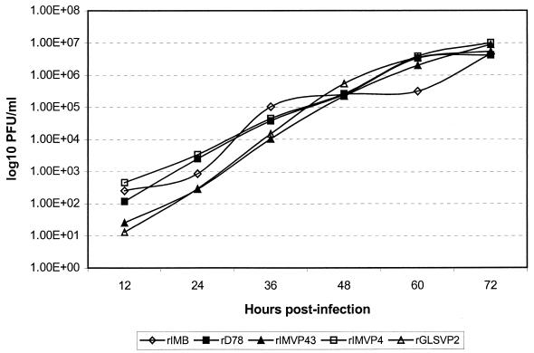
To determine the replication kinetics of rD78, rIMB, rIMVP4, rIMVP43, and rGLSVP2, CEF cells were infected with each virus, and their titers were determined by plaque assay. Figure 2 depicts the growth curve of each virus (expressed as log PFU per milliliter) on different days postinfection. Our results indicate that the final titers of the chimeric viruses were approximately the same as that of the recovered rD78 parent virus (107 PFU/ml). Furthermore, the chimeric viruses were purified by sucrose gradient, and their proteins were analyzed by Western blot analysis using anti-IBDV polyclonal serum. Qualitatively, viral structural proteins (VP2, VP4, and VP3) produced by the chimeric viruses were identical to the proteins synthesized by the recovered rD78 virus (data not shown).
FIG. 2.
Growth curves of rD78, rIMB, rIMVP4, rIMVP43, and rGLSVP2 IBDVs. Monolayers of CEF cells were infected with the indicated viruses at a multiplicity of infection of 0.1 and harvested at the indicated time points, and infectious titers were determined by plaque assay.
In vivo studies and histopathological examination of the bursa.
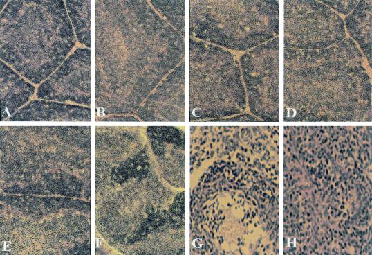
To assess the virulence and pathogenic phenotype of the recovered viruses, groups of 3-week-old chickens were inoculated with 1,000 PFU of rD78, rIMB, rIMVP4, rIMVP43, or rGLSVP2 virus or with 1,000 EID50 of rIMVP2 or IM virus by the ocular route. Three days postinoculation, the bursa of each chicken was excised and analyzed for pathological lesions. Table 3 and Fig. 3 summarize the results of gross pathology and histopathological examination of bursae obtained from different groups of chickens. Chickens inoculated with the rD78, rIMB, rIMVP4, and rIMVP43 viruses showed no gross bursal lesions but had mild-to-moderate (for rIMVP43) microscopic lesions (Table 3 and Fig. 3B to E, respectively). These results suggest the amino acid changes in the VP1, VP4, and VP3 proteins (Table 1) do not contribute to the virulence or pathogenic phenotype of IBDV in our system. However, chickens inoculated with rGLSVP2, rIMVP2, and the parental IM strain exhibited bursal atrophy (rGLSVP2) or gross hemorrhagic lesions with 20 to 25% mortality in the rIMVP2 and IM groups (Table 3). Moreover, the sections derived from these groups showed lymphocytic necrosis and follicular (B-lymphocyte) depletions due to extensive accumulation of mononuclear cells, resulting in the loss of distinction between the cortex and medulla of the infected bursal section (Fig. 3F to H). In contrast, no gross or microscopic lesions were observed in chickens that were inoculated with the control medium (Fig. 3A). These results clearly demonstrate that the virulence and pathogenic-phenotype markers of the U.S. virulent and variant IBDV strains reside in VP2.
TABLE 3.
Gross and microscopic lesions in BFs from chickens infected with recovered IBDVs
| Group | Pathology
|
|
|---|---|---|
| Grossa | Microscopicb | |
| Control | − | −(3/3) |
| rD78 | − | +(4/4) |
| rIMB | − | +(5/5) |
| rIMVP4 | − | +(5/5) |
| rIMVP43 | − | ++(3/5) |
| rGLSVP2 | ++; atrophy | +++(5/5) |
| rIMVP2 | +++; hemorrhagic | ++++(5/5) |
| IM | +++; hemorrhagic | ++++(4/4) |
+, gross bursal lesion; −, no gross lesion. ++ to +++ was graded on the degree of visible, gross bursal lesion formation.
−, no histological lesion; + to ++++ was graded on the degree of extent of multifocal purulent necrotizing bursitis with follicular depletion. The numbers in parentheses represent the number of bursae with lesions/number of bursae examined. The result is representative of two independent experiments.
FIG. 3.
Histopathological appearance of sections (hematoxylin and eosin) of BFs derived from groups of chickens infected with chimeric viruses at day 3 postinfection. (A) Cortical lymphocytes (dark-gray cells adjacent to connective tissue that separates follicles) and medullary lymphocytes (light-gray cells in follicle centers) in portions of follicles from an uninfected chicken are normal. In addition, the interfollicular connective tissues are normal. (B, C, and D) Follicles and interfollicular connective tissues from chickens infected with rD78, rIMB, and rIMVP4 viruses, respectively, appear normal and cannot be differentiated from their control counterparts. (E) Slight damage to the medullary lymphocytes in the bursa from a chicken infected with rIMVP43 virus. (F) There is lymphocytic necrosis and heterophilic inflammation in follicles of the bursa from a chicken infected with rGLSVP2 virus. (G and H) Severe lymphocytic necrosis accompanied by hemorrhagic lesions in bursae from chickens infected with rIMVP2 and IM viruses. Notice the loss of distinction between the cortex and the medulla and the bands of interfollicular connective tissue that are infiltrated by myriad heterophils and macrophages. (Magnifications, ×100).
Detection of viral antigen from the bursa.
To detect the presence of virus in the bursae of infected chickens, bursae from each group were pooled and homogenized in M199 medium, and the filtered homogenate was analyzed by AC-ELISA using a panel of strain-specific IBDV MAbs. Table 4 shows the reactivity patterns of various IBDVs recovered from groups of infected chickens. It should be noted that MAb 21 recognizes all the virulent strains tested so far, and it is evident that this epitope was present in chimeric rIMVP2 virus, which is also virulent. Neutralizing MAb 57 is specific for the GLS variant, and this epitope was also mapped to the VP2 protein.
TABLE 4.
Detection of virus in BFs of chickens infected with various IBDVs by AC-ELISA and RT-PCR
| Group | Results
|
|||||
|---|---|---|---|---|---|---|
| AC-ELISAa
|
RT-PCRb | |||||
| B69 | R63 | 57 | 21 | B29 | ||
| Control | − | − | − | − | − | − |
| rD78 | + | + | − | − | + | + |
| rIMB | + | + | − | − | + | + |
| IM | + | + | − | + | + | + |
| rIMVP2 | + | + | − | + | + | + |
| rIMVP4 | + | + | − | − | + | + |
| rIMVP43 | + | + | − | − | + | + |
| GLS | − | − | + | − | + | + |
| rGLSVP2 | − | − | + | − | + | + |
BFs from each group were pooled, and bursal homogenates were tested for reactivity with a panel of strain-specific MAbs. +, viral antigen detected by AC-ELISA; −, no viral antigen could be detected by AC-ELISA.
+, viral RNA was detected by RT-PCR; −, no viral RNA was detected by RT-PCR.
To determine the genetic stability of chimeric IBDVs in vivo, chickens were inoculated with these viruses, and their bursae were collected 3 days postinfection. Total nucleic acid was extracted from bursal tissue, and the genomic segments A and B were amplified by RT-PCR using a primer pair specific for segment A or B. Sequence analysis of the cloned PCR products confirmed the expected nucleotide substitutions or changes in various genes of the chimeric viruses. These results clearly demonstrate that the chimeric viruses replicated in the bursae of chickens but did not revert to the parental D78 or IM viruses.
Determinants of cell tropism, virulence, and pathogenic phenotype of IBDV.
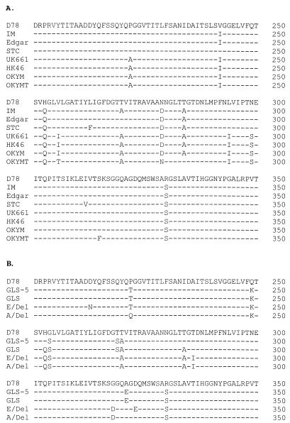
To identify putative amino acids involved in cell tropism, and possibly binding to the cell receptor, we compared the amino acid sequences of the VP2 protein (variable region) of D78, IM, and other selected IBDV strains (Fig. 4A). Comparison of the D78 sequence (tissue culture) with the bursa-derived IM, Edgar, and STC sequences showed only four amino acid differences at positions 253, 279, 284, and 330, which are common to other bursa-derived UK661, HK46, and OKYM sequences, suggesting the involvement of these residues in cell tropism. To further narrow down the amino acids involved in cell tropism, we constructed another clone (pUC19ΔD78VP2), in which amino acids at positions 279, 284, and 330 were replaced with the D78 sequence in plasmid pUC19IMVP2 (Table 1). However, transfection of CEF cells with transcripts derived from clones pUC19ΔD78VP2 and pUCD78B did not yield a viable virus, implying that residue Q at position 253 (Q253) is also involved in cell tropism. This result is in agreement with the results of Mundt, who demonstrated that a Q→H substitution at position 253 is sufficient to allow tissue culture infectivity of the E/Del virus (23). Furthermore, comparison of the bursa-derived GLS sequence of genomic segment A with the tissue culture-adapted GLS-5 sequence (Fig. 4B) reveals only Q→H and A→T substitutions at positions 253 and 284, respectively, suggesting the importance of residues Q253 and A284, but not residue S330, in cell tropism. Taken together, our results indicate that residues Q253, D279, and A284 of VP2 are involved in the cell tropism of virulent IBDV strains.
FIG. 4.
Comparison of the deduced amino acid sequences of selected IBDV strains in the VP2 variable region. (A) Comparison of the deduced amino acid sequence of the D78 strain with those of highly virulent IBDV strains. IM and Edgar, highly virulent strains from the United States; STC, standard challenge strain in the United States; UK661, very virulent strain from the United Kingdom; HK46, very virulent strain from Hong Kong; OKYM, very virulent strain from Japan; OKYMT, attenuated tissue culture virus of virulent OKYM strain. (B) Comparison of the deduced amino acid sequence of the D78 strain with those of the U.S. variant strains GLS-5 (tissue culture), GLS (bursa-derived), E/Del, and A/Del. The dashes indicate amino acid identity.
To identify the putative amino acids involved in virulence and the pathogenic phenotype, we used MAb 21. This MAb recognizes a conformational epitope in VP2 and reacts only with bursa-derived virulent IBDV (22, 41). MAb 21 reacts with rIMVP2 and fails to react with the classic attenuated D78 or other chimeric viruses (rIMVP4 or rIMVP43), which strongly suggests that the determinants for virulence reside in the VP2 region (Table 4). Comparison of the D78 sequence with the bursa-derived IM, Edgar, STC, and UK661 sequences (all of which react with MAb 21) revealed only three amino acid differences at positions 253, 279, and 284, which are common to all other virulent IBDV sequences; this suggests the involvement of these residues in virulence (Fig. 4A). In fact, these are the same residues that are involved in cell tropism. Based on the reactivity with MAb 21, it appears that residues Q253, D279, and A284 are critical in binding to this MAb and imparting the pathogenic phenotype to virulent IBDV.
DISCUSSION
Lack of a reverse-genetics system that can generate virulent IBDV has hampered studies of virulence, cell tropism, and pathogenesis. To overcome this limitation, we modified our transfection procedure and developed an efficient method of transfecting Vero or CEF cells by electroporation, followed by passage in the CAM to recover virus that would not propagate in tissue culture (virulent IBDV). Using this technique, we generated five chimeric viruses, including the virulent rIMVP2, and demonstrated that the virulence, cell tropism, and pathogenic-phenotype markers of IBDV reside in VP2. The fact that we recovered the virulent virus after transfection in CEF cells and propagation in embryonated eggs implies that CEF cells do not possess the receptors for virulent IBDV, which replicates only in B lymphoid cells. In addition, we recovered a chimeric virus containing VP1 of the virulent IM strain in CEF cells but not in Vero cells, suggesting that VP1 contains the determinants for cell-specific replication in Vero cells, which is not receptor mediated.
In recent years, a number of investigators have shown that mutations in the viral genome often lead to changes in the virulence, pathogenesis, and cell tropism of animal viruses (7, 8, 14, 16, 20, 29). For example, a single-amino-acid change in the capsid protein VP1 of coxsackievirus was responsible for the virulence phenotype (7). In swine vesicular disease virus (a picornavirus), the genetic determinants of pathogenicity and plaque phenotype were mapped to a single amino acid residue in VP1 and the 2A proteinase, respectively (14). Similarly, the determinants of virulence and enteric tropism in transmissible gastroenteritis virus were mapped to the spike protein of the virus (29). For rotaviruses of groups A and C, a substitution of two or three amino acid residues in the nonstructural protein gene NSP4 was implicated in reduced virulence (8). In the case of Sindbis virus, the genetic determinants responsible for the pathogenic properties were mapped to the E2 glycoprotein and the 5′ noncoding region (16). In our case, the 5′ noncoding region of the virulent IM strain does not contain the determinants for virulence, since the rIMVP2 virus, lacking the two nucleotide changes in the 5′ noncoding region of wild-type IM, was still virulent and caused hemorrhagic bursal lesions.
To the best of our knowledge, this is the first report demonstrating that the polymerase (VP1) of virulent IBDV carries the determinant for cell-specific viral replication, since we could not recover the virus (rIMB) in Vero cells. However, once we recovered this virus in CEF cells, it could be propagated in Vero cells. Sequence analysis of the VP1 gene of the rIMB virus showed reversion of two amino acid residues at positions 115 and 653 to those of the D78 vaccine strain, which suggests that these residues may be necessary for replication in Vero cell culture (Table 1). This finding is somewhat analogous to the case of lymphocytic choriomeningitis virus, where it was shown that a single-amino-acid substitution in the polymerase gene could enhance viral replication in macrophages (20). Furthermore, our results indicate that the rIMB virus does not induce hemorrhagic lesions in the bursae of infected chickens (Table 3 and Fig. 3C), suggesting that VP1 of the IM strain is not responsible for virulence or the pathogenic phenotype, in agreement with the results of Boot et al. (4). This result is also in accord with that for infectious pancreatic necrosis virus (another birnavirus), where it was shown by generating reassortants between virulent and avirulent strains that the virulence of infectious pancreatic necrosis virus is associated with segment A, which encodes the structural proteins, and not segment B, which encodes VP1 (31).
From earlier studies of IBDV, Yamaguchi and coworkers identified amino acid residues responsible for attenuation of the very virulent OKYM strain by comparing the sequence of the virus, which was obtained after serial passage in non-B lymphoid chicken cells (42). Comparison of the deduced amino acid sequences of the very virulent (OKYM) and attenuated (OKYMT) strains showed specific amino acid substitutions within the hypervariable region of the VP2 protein. Based on these findings, Lim and coworkers mutated residues 279 and 284 of VP2 by site-directed mutagenesis and demonstrated that the very virulent HK46 strain of IBDV could be adapted to CEF cell culture (17). Similarly, Mundt reported that residues 253 and 284 of the VP2 proteins of the variant virus are necessary for tissue culture infectivity (23). However, none of these viruses were tested in chickens to verify the roles of these residues in IBDV virulence and pathogenicity. Our results indicate that VP2 carries the major determinant of cell tropism in IBDV, since we could not recover the rIMVP2 virus in Vero or CEF cells but were able to recover it after passage in the CAM. This was expected, since virulent IBDV does not grow in tissue culture and can only replicate in bursal cells. These results imply that bursal cells possess virus receptors in addition to the one that is present in tissue culture cells which VP2 interacts with. Furthermore, our results clearly demonstrate that VP2 contains the determinants for the virulence and the pathogenic phenotype of IBDV. However, these results are in contrast to those reported by Boot and coworkers, who stated that VP2 is not the sole determinant of the very virulent phenotype due to the fact that their recombinant virus containing VP2 of the very virulent strain did not induce morbidity or mortality (4). In our case, rIMVP2 virus containing the VP2 region of the virulent strain caused hemorrhagic bursal lesions and mortality equivalent to those of the parental IM virus, suggesting that the virulence and pathogenic-phenotype markers of IBDV reside in VP2. This is further supported by the results for chimeric rGLSVP2 virus (containing VP2 of the variant strain), which caused bursal atrophy, a characteristic phenotype of a variant virus (Table 3). Comparison of the GLS-5 sequence with the D78 sequence showed five unique amino acid differences at positions 222, 249, 254, 270, and 330 (Fig. 4B), which are common to other variant GLS, E/Del, and A/Del sequences, suggesting the involvement of these residues in the variant phenotype. In earlier studies, we mapped the classic MAb B69 neutralization epitope (which is absent in the variant viruses) to residues at positions 222, 249, and 254 (38, 41). Therefore, it is apparent that residues T/Q222, K249, and S254 are important in inducing a characteristic phenotype of the variant IBDVs.
In conclusion, we have demonstrated that VP1 carries the determinants for cell-specific replication of IBDV in Vero cells and VP2 carries the determinants for cell tropism. The amino acids involved in binding with the B lymphoid cells are Q253, D279, and A284. Furthermore, the virulence factor of IBDV strain IM resides in VP2 and, based on the reactivity with conformation-dependent MAb 21, residues Q253, D279, and A284 are most likely involved in maintaining this conformation and inducing the pathogenic phenotype (hemorrhagic lesions) of virulent IBDV. However, it is possible that VP4 and VP3 carry additional determinants for virulence, as the rIMVP43 virus also induced slightly more microscopic lesions than the parental D78 virus. In addition, the cooperative effect due to the expression of the 17-kDa NS protein in virulence should not be ruled out, as we have shown that the 17-kDa knockout mutant of strain D78 is nonpathogenic (43). It would be interesting to see whether the NS-deficient mutant of the IM or GLS virus would induce similar pathological lesions and cause immunosuppression.
ACKNOWLEDGMENTS
We thank Donald L. Nuss for reviewing the manuscript, and we thank Gerard H. Edwards, Yi Liu, Subbiah Elankumaran, and Ruby Paramadhas for technical assistance.
This work was supported by a grant from the U.S. Department of Agriculture (NRICGP 97-02492) to V.N.V.
REFERENCES
- 1.Azad A A, Barrett S A, Fahey K J. The characterization and molecular cloning of the double-stranded RNA genome of an Australian strain of infectious bursal disease virus. Virology. 1985;143:35–44. doi: 10.1016/0042-6822(85)90094-7. [DOI] [PubMed] [Google Scholar]
- 2.Becht H. Infectious bursal disease virus. Curr Top Microbiol Immunol. 1980;90:107–121. doi: 10.1007/978-3-642-67717-5_5. [DOI] [PubMed] [Google Scholar]
- 3.Birghan C, Mundt E, Gorbalenya A E. A non-canonical lon proteinase lacking the ATPase domain employs the Ser-Lys catalytic dyad to exercise broad control over the life cycle of a double-stranded RNA virus. EMBO J. 2000;19:114–123. doi: 10.1093/emboj/19.1.114. [DOI] [PMC free article] [PubMed] [Google Scholar]
- 4.Boot H J, ter Huurne A A, Hoekman A J, Peeters B P, Gielkens A L. Rescue of very virulent and mosaic infectious bursal disease virus from cloned cDNA: VP2 is not the sole determinant of the very virulent phenotype. J Virol. 2000;74:6701–6711. doi: 10.1128/jvi.74.15.6701-6711.2000. [DOI] [PMC free article] [PubMed] [Google Scholar]
- 5.Böttcher B, Kiselev N A, Stel'Mashchuk V Y, Perevozchikova N A, Borisov A V, Crowther R A. Three-dimensional structure of infectious bursal disease virus determined by electron cryomicroscopy. J Virol. 1997;71:325–330. doi: 10.1128/jvi.71.1.325-330.1997. [DOI] [PMC free article] [PubMed] [Google Scholar]
- 6.Brown M D, Green P, Skinner M A. VP2 sequences of recent European ‘very virulent’ isolates of infectious bursal disease virus are closely related to each other but are distinct from those of ‘classical’ strains. J Gen Virol. 1994;75:675–680. doi: 10.1099/0022-1317-75-3-675. [DOI] [PubMed] [Google Scholar]
- 7.Caggana M, Chan P, Ramsingh A. Identification of a single amino acid residue in the capsid protein VP1 of coxsackievirus B4 that determines the virulent phenotype. J Virol. 1993;67:4797–4803. doi: 10.1128/jvi.67.8.4797-4803.1993. [DOI] [PMC free article] [PubMed] [Google Scholar]
- 8.Chang K O, Kim Y J, Saif L J. Comparisons of nucleotide and deduced amino acid sequences of NSP4 genes of virulent and attenuated pairs of group A and C rotaviruses. Virus Genes. 1999;18:229–233. doi: 10.1023/a:1008068218966. [DOI] [PubMed] [Google Scholar]
- 9.Dobos P, Berthiaume L, Leong J A, Kibenge F S, Müller H, Nicholoson B L. Family Birnaviridae. In: Murphy F A, Fauquet C M, Bishop D H L, Ghabrial S A, Jarvis A W, Martielli G P, Mayo M O, Summers M D, editors. Virus taxonomy. Vienna, Austria: Springer-Verlag; 1995. pp. 240–244. [Google Scholar]
- 10.Dobos P, Hill B J, Hallett R, Kells D T, Becht H, Teninges D. Biophysical and biochemical characterization of five animal viruses with bisegmented double-stranded RNA genomes. J Virol. 1979;32:593–605. doi: 10.1128/jvi.32.2.593-605.1979. [DOI] [PMC free article] [PubMed] [Google Scholar]
- 11.Fahey K J, Erny K, Crooks J. A conformational immunogen on VP-2 of infectious bursal disease virus that induces virus-neutralizing antibodies that passively protect chickens. J Gen Virol. 1989;70:1473–1481. doi: 10.1099/0022-1317-70-6-1473. [DOI] [PubMed] [Google Scholar]
- 12.Hudson P J, McKern N M, Power B E, Azad A A. Genomic structure of the large RNA segment of infectious bursal disease virus. Nucleic Acids Res. 1986;14:5001–5012. doi: 10.1093/nar/14.12.5001. [DOI] [PMC free article] [PubMed] [Google Scholar]
- 13.Jagadish M N, Staton V J, Hudson P J, Azad A A. Birnavirus precursor polyprotein is processed in Escherichia coli by its own virus-encoded polypeptide. J Virol. 1988;62:1084–1087. doi: 10.1128/jvi.62.3.1084-1087.1988. [DOI] [PMC free article] [PubMed] [Google Scholar]
- 14.Kanno T, Mackay D, Inoue T, Wilsden G, Yamakawa M, Yamazoe R, Yamaguchi S, Shirai J, Kitching P, Murakami Y. Mapping the genetic determinants of pathogenicity and plaque phenotype in swine vesicular disease virus. J Virol. 1999;73:2710–2716. doi: 10.1128/jvi.73.4.2710-2716.1999. [DOI] [PMC free article] [PubMed] [Google Scholar]
- 15.Kibenge F S, Dhillon A S, Russell R G. Biochemistry and immunology of infectious bursal disease virus. J Gen Virol. 1988;69:1757–1775. doi: 10.1099/0022-1317-69-8-1757. [DOI] [PubMed] [Google Scholar]
- 16.Kobiler D, Rice C M, Brodie C, Shahar A, Dubuisson J, Halevy M, Lustig S. A single nucleotide change in the 5′ noncoding region of Sindbis virus confers neurovirulence in rats. J Virol. 1999;73:10440–10446. doi: 10.1128/jvi.73.12.10440-10446.1999. [DOI] [PMC free article] [PubMed] [Google Scholar]
- 17.Lim B L, Cao Y, Yu T, Mo C W. Adaptation of very virulent infectious bursal disease virus to chicken embryonic fibroblasts by site-directed mutagenesis of residues 279 and 284 of viral coat protein VP2. J Virol. 1999;73:2854–2862. doi: 10.1128/jvi.73.4.2854-2862.1999. [DOI] [PMC free article] [PubMed] [Google Scholar]
- 18.Lin Z, Kato A, Otaki Y, Nakamura T, Sasmaz E, Ueda S. Sequence comparisons of a highly virulent infectious bursal disease virus prevalent in Japan. Avian Dis. 1993;37:315–323. [PubMed] [Google Scholar]
- 19.Lombardo E, Maraver A, Castón J R, Rivera J, Fernández-Arías A, Serrano A, Carrascosa J L, Rodriguez J F. VP1, the putative RNA-dependent RNA polymerase of infectious bursal disease virus, forms complexes with the capsid protein VP3, leading to efficient encapsidation into virus-like particles. J Virol. 1999;73:6973–6983. doi: 10.1128/jvi.73.8.6973-6983.1999. [DOI] [PMC free article] [PubMed] [Google Scholar]
- 20.Matloubian M, Kolhekar S R, Somasundaram T, Ahmed R. Molecular determinants of macrophage tropism and viral persistence: importance of single amino acid changes in the polymerase and glycoprotein of lymphocytic choriomeningitis virus. J Virol. 1993;67:7340–7349. doi: 10.1128/jvi.67.12.7340-7349.1993. [DOI] [PMC free article] [PubMed] [Google Scholar]
- 21.McFerran J B, McFerran M S, McKillop E R, Conner T J, McCracken R M, Collins D S, Allan G M. Isolation and serological studies with infectious bursal disease from fowl, turkeys, and ducks: demonstration of a second serotype. Avian Pathol. 1980;9:384–392. doi: 10.1080/03079458008418423. [DOI] [PubMed] [Google Scholar]
- 22.Mengel-Whereat S A. Development of a monoclonal antibody for the detection of highly virulent classic infectious bursal disease virus. M.S. thesis. College Park: University of Maryland; 1995. [Google Scholar]
- 23.Mundt E. Tissue culture infectivity of different strains of infectious bursal disease virus is determined by distinct amino acids in VP2. J Gen Virol. 1999;80:2067–2076. doi: 10.1099/0022-1317-80-8-2067. [DOI] [PubMed] [Google Scholar]
- 24.Mundt E, Beyer J, Müller H. Identification of a novel viral protein in infectious bursal disease virus-infected cells. J Gen Virol. 1995;76:437–443. doi: 10.1099/0022-1317-76-2-437. [DOI] [PubMed] [Google Scholar]
- 25.Mundt E, Vakharia V N. Synthetic transcripts of double-stranded Birnavirus genome are infectious. Proc Natl Acad Sci USA. 1996;93:11131–11136. doi: 10.1073/pnas.93.20.11131. [DOI] [PMC free article] [PubMed] [Google Scholar]
- 26.Nick H, Cursiefen D, Becht H. Structural and growth characteristics of infectious bursal disease virus. J Virol. 1976;18:227–234. doi: 10.1128/jvi.18.1.227-234.1976. [DOI] [PMC free article] [PubMed] [Google Scholar]
- 27.Sambrook J, Fritsch E F, Maniatis T. Molecular cloning: a laboratory manual. 2nd ed. Cold Spring Harbor, N.Y: Cold Spring Harbor Laboratory; 1989. [Google Scholar]
- 28.Sanchez A B, Rodriguez J F. Proteolytic processing in infectious bursal disease virus: identification of the polyprotein cleavage sites by site-directed mutagenesis. Virology. 1999;262:190–199. doi: 10.1006/viro.1999.9910. [DOI] [PubMed] [Google Scholar]
- 29.Sanchez C M, Izeta A, Sanchez-Morgado J M, Alonso S, Sola I, Balasch M, Plana-Duran J, Enjuanes L. Targeted recombination demonstrates that the spike gene of transmissible gastroenteritis coronavirus is a determinant of its enteric tropism and virulence. J Virol. 1999;73:7607–7618. doi: 10.1128/jvi.73.9.7607-7618.1999. [DOI] [PMC free article] [PubMed] [Google Scholar]
- 30.Sanger F, Nicklen S, Coulson A R. DNA sequencing with chain terminating inhibitor. Proc Natl Acad Sci USA. 1977;74:5463–5467. doi: 10.1073/pnas.74.12.5463. [DOI] [PMC free article] [PubMed] [Google Scholar]
- 31.Sano M, Okamoto N, Fukuda H, Saneyoshi M, Sano T. Virulence of infectious pancreatic necrosis virus is associated with the larger RNA segment (RNA segment A) J Fish Dis. 1992;15:283–293. [Google Scholar]
- 32.Snyder D B, Lana D P, Savage P K, Yancey F S, Mengel S A, Marquardt W W. Differentiation of infectious bursal disease viruses directly from infected tissues with neutralizing monoclonal antibodies: evidence of a major antigenic shift in recent field isolates. Avian Dis. 1988;32:535–539. [PubMed] [Google Scholar]
- 33.Snyder D B, Vakharia V N, Savage P K. Naturally occurring neutralizing monoclonal antibody escape variants define the epidemiology of infectious bursal disease viruses in the United States. Arch Virol. 1992;127:89–101. doi: 10.1007/BF01309577. [DOI] [PubMed] [Google Scholar]
- 34.Spies U, Müller H. Demonstration of enzyme activities required for cap structure formation in infectious bursal disease virus, a member of the birnavirus group. J Gen Virol. 1990;71:977–981. doi: 10.1099/0022-1317-71-4-977. [DOI] [PubMed] [Google Scholar]
- 35.Spies U, Müller H, Becht H. Nucleotide sequence of infectious bursal disease virus genome segment A delineates two major open reading frames. Nucleic Acids Res. 1989;17:7982. doi: 10.1093/nar/17.19.7982. [DOI] [PMC free article] [PubMed] [Google Scholar]
- 36.Spies U, Müller H, Becht H. Properties of RNA polymerase activity associated with infectious bursal disease virus and characterization of its reaction products. Virus Res. 1987;8:127–140. doi: 10.1016/0168-1702(87)90024-4. [DOI] [PubMed] [Google Scholar]
- 37.Tsukamoto K, Matsumura T, Mase M, Imai K. A highly sensitive, broad-spectrum infectivity assay for infectious bursal disease virus. Avian Dis. 1995;39:575–586. [PubMed] [Google Scholar]
- 38.Vakharia V N. Development of recombinant vaccines against infectious bursal disease. Biotechnol Annu Rev. 1997;3:151–168. [Google Scholar]
- 39.Vakharia V N, Snyder D B, He J, Edwards G H, Savage P K, Mengel-Whereat S A. Infectious bursal disease virus structural proteins expressed in a baculovirus recombinant confer protection in chickens. J Gen Virol. 1993;74:1201–1206. doi: 10.1099/0022-1317-74-6-1201. [DOI] [PubMed] [Google Scholar]
- 40.Vakharia V N, He J, Ahamed B, Snyder D B. Molecular basis of antigenic variation in infectious bursal disease virus strains. Virus Res. 1994;31:265–273. doi: 10.1016/0168-1702(94)90009-4. [DOI] [PubMed] [Google Scholar]
- 41.Vakharia, V. N., D. B. Snyder, and S. A. Mengel-Whereat. December 2000. Chimeric infectious bursal disease virus cDNA clones, expression products and vaccines based thereon. U.S. patent 6,156,314.
- 42.Yamaguchi T, Ogawa M, Inoshima Y, Miyoshi M, Fukushi H, Hirai K. Identification of sequence changes responsible for the attenuation of highly virulent infectious bursal disease virus. Virology. 1996;223:219–223. doi: 10.1006/viro.1996.0470. [DOI] [PubMed] [Google Scholar]
- 43.Yao K, Goodwin M A, Vakharia V N. Generation of a mutant infectious bursal disease virus that does not cause bursal lesions. J Virol. 1998;72:2647–2654. doi: 10.1128/jvi.72.4.2647-2654.1998. [DOI] [PMC free article] [PubMed] [Google Scholar]


