Abstract
BACKGROUND
Emphysematous pancreatitis (EP) is a rare, severe form of acute necrotizing pancreatitis characterized by gas in pancreatic or peripancreatic tissue, with a high mortality rate.
AIM
To assess the diagnosis, treatment, and outcomes of EP through a series of case studies.
METHODS
This case series was conducted in intensive care units at the Second Affiliated Hospital of Anhui Medical University. Patients were included if they were diagnosed with pancreatic necrosis and gas via computed tomography from June 2018 to June 2024. Patients were categorized into early and late EP groups based on the timing of the appearance of the bubble sign and into extensive and common types based on the distribution range of the bubble sign. The data recorded included sex, age, aetiology, Acute Physiology and Chronic Health Evaluation II score, Sequential Organ Failure Assessment score, Bedside Index for Severity in Acute Pancreatitis score, subtype, gas distribution extent, aetiological diagnostic basis, pathogen categories, intervention measures, and prognosis.
RESULTS
Among the 15 patients, 66.7% had a biliary aetiology and extensive type of EP, 47.1% had early-onset EP, and 73.3% had confirmed aetiological evidence [6 based on bacterial culture, 4 based on both routine culture and next-generation sequencing (NGS), and 1 solely based on NGS]. The common pathogens were Escherichia coli and Klebsiella pneumoniae. Six patients survived. Among the 2 patients who did not undergo percutaneous drainage or surgical treatment, 1 survived. Of the 6 patients who underwent percutaneous drainage, 2 survived, 2 survived after subsequent surgery, and 2 died without surgery. Among the 6 patients who underwent surgery alone, 5 died and 1 survived. Among the early-onset EP patients, 4 survived; among the late-onset EP patients, 2 survived. Among the common EP types, 4 survived; among the extensive EP types, only 1 survived.
CONCLUSION
The mortality rate among patients with EP is considerable, and NGS enhances pathogen identification accuracy. Despite the debate on conservative vs surgical management, the STEP-UP strategy remains viable. Aggressive antimicrobial therapy, early percutaneous catheter drainage, and other minimally invasive interventions, along with delayed surgical intervention, may improve patient prognosis.
Keywords: Emphysematous pancreatitis, Diagnosis, Treatment, Prognosis, Next-generation sequencing
Core Tip: Emphysematous pancreatitis (EP) is a rare and severe condition with high mortality. This study highlights the role of next-generation sequencing in improving pathogen identification and emphasizes early percutaneous drainage, aggressive antimicrobial therapy, and minimally invasive interventions. Delayed surgery, when necessary, may improve outcomes, especially in early-onset and common-type EP patients.
INTRODUCTION
Approximately 20% to 30% of patients with acute pancreatitis (AP) will develop acute necrotizing pancreatitis (ANP)[1,2], and approximately one-third of these patients will develop infected pancreatic necrosis (IPN)[3], making it a potentially fatal disease. If gas is present within the necrotic tissue, it is referred to as EP. EP, a condition that can severely impair one's quality of life, is associated with alarmingly high mortality rates, estimated to be between 32% and 40%[4]. However, there are contradictions in the literature regarding the causes of gas formation in emphysematous pancreatitis, diagnostic criteria, treatment, and prognosis. The most common cause of gas production in or around the pancreas is a bacterial infection, typically by gas-forming bacteria[5]. However, when gas is detected on a computed tomography (CT) scan, there should be a high suspicion of infection. The cause of gas production could also be a gastrointestinal fistula (stomach, colon, or duodenum) or external gas entry caused by percutaneous puncture. There is currently no consensus on the diagnostic criteria for EP. Some scholars classify gas in the pancreas or around it caused by gastrointestinal fistula as EP[6], whereas others exclude it[7]. Although most experts define this entity as IPN[8], some patients show no evidence of infection in clinical practice[9], so one aim of this study was to determine the actual proportion of these patients with infected necrosis and to analyse the reason. Traditionally, EP is considered a critical form of ANP and is characterized by a very poor prognosis and a strong indication for surgical intervention[5]. However, with the tremendous progress made in critical care medicine over the past few decades, the clinical outcomes of some EP patients treated conservatively have significantly improved[10]. Therefore, this study aims to provide a comprehensive description of the clinical characteristics and outcomes of patients with EP in our centre and to explore its clinical diagnosis, treatment, and prognosis by integrating the relevant literature.
MATERIALS AND METHODS
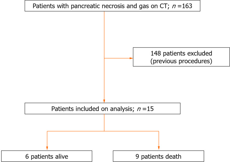
We conducted a retrospective study in which the clinical data of patients diagnosed with EP at the First Department of Critical Medicine, the Second Affiliated Hospital of Anhui Medical University, were recorded from June 2018 to June 2024. The inclusion criteria for patients were as follows: (1) Met the diagnosis of AP; (2) CT scan results revealed the presence of air in the pancreatic parenchyma and/or peripancreatic areas; and (3) Complete clinical and imaging data. The exclusion criteria were as follows: (1) Air accumulation in the pancreas and peripancreatic areas caused by pancreatic surgery or catheterization; (2) A gastrointestinal fistula confirmed by imaging examination; and (3) Missing clinical and imaging data. Figure 1 shows the flowchart of the included patients. EP patients were categorized into early-onset and late-onset subtypes according to a cut-off of 2 weeks from disease onset when air bubble signs were detected on CT. EP can also be categorized based on intra-pancreatic or peri-pancreatic air bubble distribution into extensive EP (when more than 50% of the pancreatic/pancreatic necrosis is involved and connected into sheets) or common EP (when less than 50% is involved). Detailed records of the patients' sex, age, aetiology, Acute Physiology and Chronic Health Evaluation II (APACHE II) score, Sequential Organ Failure Assessment (SOFA) score, Bedside Index for Severity in Acute Pancreatitis (BISAP) score upon admission, subtypes of EP, extent of gas distribution, basis for aetiological diagnosis, categories of pathogens, surgical intervention measures and survival status were obtained. All patients received standardized treatment in accordance with the most recent international guidelines. Upon admission, they were comprehensively evaluated by a multidisciplinary team consisting of pancreatic surgeons, intensivists, gastroenterologists, and radiologists. Timely administration of broad-spectrum or empirical antibiotics was provided to patients with confirmed or suspected infections. For those who do not respond to conservative intensive treatment, an escalated approach, including percutaneous catheter drainage (PCD) followed by minimally invasive or open pancreatic necrosectomy, is the preferred strategy for managing IPN. Once peritoneal infection was considered, retroperitoneal fine-needle aspiration was performed. After performing fine-needle aspiration culture, clinical physicians provided empirical anti-infective treatment, which was then adjusted based on the results of aetiological examinations and clinical manifestations. Depending on the effectiveness of the anti-infective treatment and the CT findings, a multidisciplinary team discussion was held to decide whether to perform PCD and surgical treatment.
Figure 1.
Flowchart of patient inclusion in the study of empysematous pancreatitis. CT: Computed tomography.
RESULTS
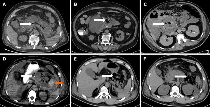
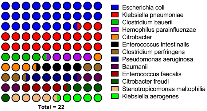
A total of 15 patients were included in the study (Table 1), with a median age of 71.0 (63.0 to 76.0), an APACHE II score of 17.9 ± 5.0, a SOFA score of 6.4 ± 3.7, and a median BISAP score of 3 (3 to 4). There were 10 cases of biliary pancreatitis, 2 cases of hypertriglyceridaemia pancreatitis, 2 cases of pancreatitis for unknown reasons and 1 case of drug-induced pancreatitis. There were 7 cases of early-onset EP and ten common subtypes of EP. CT revealed that emphysema was distributed in different parts of the pancreas, as shown in Figure 2. There was no evidence of pathogen infection in 4 patients. Eleven patients had pathogen examination results, of which 4 patients had both bacterial culture and next-generation sequencing (NGS) results confirming the diagnosis of infected necrosis, 1 had only NGS results, and 6 patients had bacterial culture results only. Among the bacteria isolated, Escherichia coli (E. coli) was the most common, followed by Klebsiella pneumoniae (K. pneumoniae) (Figure 3). Among the 15 patients, 9 died (7 due to septic shock and multiple organ dysfunction, 2 due to abdominal hemorrhage), and 6 survived. Among the 2 patients who did not undergo percutaneous drainage or surgical treatment, 1 survived and 1 died. Six patients underwent PCD, of whom 2 survived, 2 survived after subsequent surgical treatment, and 2 died without additional surgery. Among the 6 patients who underwent surgery alone, 5 died and 1 survived. Four out of 7 patients in the early-onset group survived, 2 out of 8 patients in the late-onset group survived, 4 out of 10 patients in the common group survived, and 2 out of 5 patients in the extensive group survived.
Table 1.
Clinical characteristics and outcomes of patients with oemphysematous pancreatitis
|
No.
|
Age
|
Sex
|
Aetiology
|
Subtype
|
Distribution range
|
APACHE II score
|
SOFA score
|
BISAP score
|
Microbiological basis
|
Percutaneous puncture frequency
|
Surgery frequency
|
Outcome
|
| 1 | 72 | Female | Biliary | Early-onset | Common | 22 | 6 | 3 | Routine culture + NGS | 2 | 0 | Death |
| 2 | 75 | Female | Biliary | Late-onset | Common | 22 | 6 | 5 | Routine culture + NGS | 1 | 0 | Death |
| 3 | 48 | Male | Hyperlipidaemic | Early-onset | Common | 16 | 6 | 3 | Routine culture + NGS | 0 | 1 | Alive |
| 4 | 69 | Male | Biliary | Late-onset | Common | 26 | 15 | 5 | Routine culture + NGS | 0 | 1 | Death |
| 5 | 35 | Male | Unknown | Early-onset | Extensive | 8 | 0 | 0 | NGS | 1 | 0 | Alive |
| 6 | 76 | Male | Unknown | Early-onset | Extensive | 16 | 3 | 4 | Routine culture | 1 | 1 | Alive |
| 7 | 72 | Male | Drug-induced | Early-onset | Extensive | 14 | 5 | 4 | Routine culture | 0 | 2 | Death |
| 8 | 70 | Female | Biliary | Early-onset | Common | 22 | 9 | 4 | None | 0 | 1 | Death |
| 9 | 70 | Male | Biliary | Late-onset | Common | 22 | 9 | 4 | None | 0 | 0 | Death |
| 10 | 63 | Male | Biliary | Late-onset | Extensive | 20 | 9 | 4 | Routine culture | 0 | 2 | Death |
| 11 | 81 | Male | Biliary | Late-onset | Common | 18 | 5 | 4 | None | 0 | 0 | Alive |
| 12 | 39 | Male | Hyperlipidaemic | Late-onset | Common | 10 | 3 | 1 | Routine culture | 1 | 0 | Alive |
| 13 | 82 | Male | Biliary | Late-onset | Common | 20 | 11 | 4 | Routine culture | 0 | 1 | Death |
| 14 | 85 | Male | Biliary | Late-onset | Extensive | 20 | 4 | 4 | None | 0 | 0 | Death |
| 15 | 71 | Male | Biliary | Early-onset | Common | 13 | 5 | 2 | Routine culture | 1 | 1 | Alive |
APACHE II score: Acute Physiology and Chronic Health Evaluation II score; SOFA score: Sequential Organ Failure Assessment score; BISAP score: Bedside Index for Severity in Acute Pancreatitis score; NGS: Next-generation sequencing.
Figure 2.
Computed tomography imaging findings in patients with hyperematous pancreatitis. A: Computed tomography (CT) scan showing gas confined to the pancreatic parenchyma (white arrow) with pancreatic swelling; B: Gas confined to the body of the pancreas (white arrow) with significant swelling of the pancreatic head; C: Gas confined to the pancreatic head (white arrow) with multiple gas cavities and exudate involving the right hepatorenal space; D: Gas confined to the body and tail of the pancreas (orange arrow) with haemorrhage in the pancreatic tail (broad white arrow) and exudate involving the perisplenic and perirenal areas; E: Gas confined to the body and tail of the pancreas with larger gas cavities (white arrow); F: Gas confined to the tail of the pancreas with larger gas cavities (white arrow) and exudate involving the anterior fascia of the left kidney.
Figure 3.
Aetiological distribution of emphysema pancreatitis.
DISCUSSION
In our study, we found that EP is a severe and often fatal condition with a poor prognosis. E. coli and K. pneumoniae are common causative pathogens, and NGS helps to improve the detection rate of pathogens. The precise influence of EP classification on patient prognosis remains a subject of ambiguity. The choice between surgical intervention and conservative treatment hinges on the extent of the patient's condition and their unique health profile, and the STEP-UP protocol represents an endorsed approach for addressing such complexities in patient care.
In patients with AP, IPN is the most lethal complication with an extremely high mortality rate. Due to the rarity of EP as a medical emergency, a special subtype of IPN[5], it is impractical to collect data prospectively. Although the number of relevant literature reports has increased in recent years, there are few reports of large-scale case studies domestically and internationally, and most previous research has been based on retrospective analysis; therefore, there is a lack of relevant data to support the actual incidence of EP[11]. EP is uniquely identified by the presence of gas within the pancreas or peripancreatic region[5,12]. This condition often occurs in patients with diabetes, chronic kidney disease, cardiovascular diseases, or other individuals with compromised immune systems, as well as in long-term alcohol consumers. Patients typically exhibit symptoms such as abdominal pain, nausea, vomiting, and fever[13,14].
The diagnosis of EP is typically confirmed by a CT scan within the proper clinical context[15]. However, there are contradictions in the literature regarding the diagnosis and pathogenesis of EP. Certain studies posit that EP could be a subtype of INP[5] and that the production of gas may be related to infection. The common pathogenic bacteria are E. coli and K. pneumoniae[11], which is consistent with the findings of this study. Some scholars categorize gas produced by gastrointestinal fistulas as EP, whereas others hold the opposite view[7]. The presence of gas in the pancreas and/or peripancreatic tissues on CT imaging is a primary characteristic of EP. However, some scholars believe that the condition where only a small amount of gas is present in the peripancreatic tissues, but no gas is found within the pancreatic tissue itself should not be classified as EP[7]. In our study, we are inclined to exclude the presence of gastrointestinal fistulas at the time of diagnosis and include cases where gas is present in the peripancreatic tissues but not in the pancreatic tissue in the diagnostic category of EP. With respect to aetiological diagnosis, some scholars may question whether EP is the result of secondary infection of necrotic tissue because operating room culture or aspiration culture of the pancreas and peripancreatic tissue is negative in some patients[9]. Indeed, in four patients, no pathological evidence was obtained in our study. This was due to the absence of operations such as fine-needle aspiration in three patients, and in one patient, the operating room culture also failed to yield a positive result. A negative result for the pathogen does not rule out the possibility of secondary infection of necrotic tissue. NGS technology has been used for examination since 2021 to improve the detection rate of pathogens. Due to the advantages of high accuracy, short time consumption, and wide detection range, NGS has extremely important significance for the detection of unknown species or pathogens that are difficult to cultivate[16,17]. Among the five patients, the presence of bacterial infection was confirmed by both routine culture and NGS in four patients; however, in one patient, the pathogen was not detected via routine culture but was via NGS examination, and the same bacteria were also detected via routine blood culture. Therefore, NGS helps to improve the detection rate of pathogens. The failure to detect the pathogen in the literature could be attributed to a variety of factors, such as the inaccuracy of sampling techniques, a low bacterial count, the use of unsuitable culture media, or suboptimal culture conditions. Additionally, other contributing factors may also play a role.
EP can be addressed through two principal methodologies: A conservative approach, encompassing antibiotic therapy, percutaneous drainage, and endoscopic interventions, and a surgical approach, which may be conducted either through open or laparoscopic debridement[18]. Currently, there is no consensus on the treatment of EP. However, in most of the literature, open necrosectomy is considered the standard approach. A majority of experts agree that the presence of gas signs on a CT scan is a clear indication for surgery. The study by Wig et al[5], the largest series published to date, reported on 11 patients with gas-forming pancreatic necrosis, all of whom (100%) underwent surgical treatment. Nonetheless, some studies have noted that certain patients diagnosed with EP can be relieved through conservative management. However, in specific cases, such as those requiring debridement of necrotic tissue for organ failure, conservative treatment may fail. At present, a universal consensus on the treatment strategy for these patients has not yet been reached. Although most experts tend to consider that the presence of gas within the necrotic area is not an absolute indication for surgery, a consensus on this view has not been reached. This study aims to explore the following question: Is the gas in the pancreas found on CT scans indeed a clear indication for surgery? In our study, among the 6 surviving patients, 1 received only conservative management, 2 improved after PCD, and 3 survived after PCD combined with surgery or surgery alone. Among the 8 surgical patients, only 2 survived. This finding supports a view that is not yet widely accepted: in certain patient populations, the gas in pancreatic necrosis may be treatable by conservative treatment and can achieve good therapeutic effects. The current standard approach for infected necrotizing pancreatitis is a minimally invasive step-up approach with catheter drainage as the first step,Compared to PCD, surgery may increase the risk of infection and multiple organ dysfunction[9].
In our institution, the standard treatment for INP basically follows the STEP-UP protocol, and the presence of gas in pancreatic necrosis is not an absolute indication for surgical treatment; however, active antimicrobial therapy, early PCD, other minimally invasive treatments, and delayed surgery are helpful for improving the prognosis[19].
EP is an exceedingly rare medical emergency with a grim prognosis and a high mortality rate of up to 34.5%[20]. As a subtype of IPN, the main causes of death for EP remain septic shock, multiple organ dysfunction, and abdominal hemorrhage. In this study, among the 9 patients who died, 7 succumbed to septic shock and multiple organ dysfunction, while 2 died from abdominal hemorrhage. In our study, the mortality rate of patients with EP was 60%, which was significantly higher than the 23.6% mortality rate of patients with IPN during the same period. The age of the EP patients included in our study was generally greater, which may be the reason why our patient mortality rate was higher than that reported in other studies. In this research, EP was categorized based on the timing of disease onset into the early-onset form (occurring within two weeks of initial onset) and the late-onset form (occurring more than two weeks after initial onset). The early-onset subtype is associated with an extremely poor prognosis and significantly increased mortality[21-23]. However, in our research, among the 7 patients with early-onset EP, 4 survived, indicating that the timing of onset is not the decisive factor for patient prognosis. Some studies have classified EP into common and extensive types based on whether the bubble sign accounts for more than 50% of the pancreatic and peripancreatic necrotic region; patients with extensive bubble signs appearing early have a mortality rate as high as 100%[21]. However, in our study, 2 out of 3 patients with early-onset, extensive EP survived, whereas 4 patients with late-onset, common EP died, suggesting that the timing of onset or the extent of the disease is not the determining factor for mortality. In this study, all 6 surviving patients had APACHE-II scores below the threshold of 18. In contrast, among the 9 patients who unfortunately died, only 1 had an APACHE-II score below this critical value. This observation implies a potential correlation between the APACHE-II score and the eventual prognosis of patients. Patients with EP, as a subtype of INP, benefit from the use of NGS for aetiological diagnosis, which increases the positive detection rate. The most common causative pathogens identified are E. coli and K. pneumoniae. The impact of EP classification on patient prognosis remains unclear. Successful treatment necessitates the early and aggressive use of broad-spectrum antibiotics. The debate over the necessity of surgical intervention vs conservative treatment alone continues to be a subject of discussion. The STEP-UP protocol for the debridement of necrotic tissue remains a recognized and viable option. However, it is important to acknowledge the limitations of this study, which was confined to a single centre and involved a relatively small and older patient sample. The findings, therefore, may not be broadly generalizable. It is anticipated that future multicentre, large-sample studies will yield more comprehensive and reliable clinical insights.
EP is a serious and potentially life-threatening medical condition characterized by a generally unfavourable prognosis. NGS has shown promise in enhancing the likelihood of identifying the underlying cause, with E. coli and K. pneumoniae frequently implicated as causative agents. The impact of the classification of EP on prognosis is still unclear, and more research may be needed to elucidate its specific effects on prognosis. In terms of treatment, the early use of broad-spectrum antibiotics is crucial for controlling infection and improving outcomes, but the decision to undergo surgical treatment or to manage conservatively depends on the severity of the condition and the patient's individual circumstances. The STEP-UP approach currently remains the recommended method[24]. Given its associated high mortality rate, early and prompt recognition and treatment of EP are crucial and require individualized treatment with the involvement of a multidisciplinary team[25]. Since this study is a single-centre, small-sample study with an older patient population, the generalizability of the conclusions is limited. To obtain more reliable and guiding conclusions, multicentre, large-sample studies are needed. These findings could help assess the effectiveness of different treatment methods more accurately and provide stronger evidence for clinical decision-making.
CONCLUSION
The mortality rate among patients with EP is considerable, and NGS enhances pathogen identification accuracy. Despite the debate on conservative vs surgical management, the STEP-UP strategy remains viable. Aggressive antimicrobial therapy, early PCD, and other minimally invasive interventions, along with delayed surgical intervention, may improve patient prognosis.
Footnotes
Institutional review board statement: The study was conducted in accordance with the Declaration of Helsinki and was approved by the Ethics Committee of the Second Affiliated Hospital of Anhui Medical University (Approval No. YX2023-136).
Informed consent statement: Written informed consent was obtained from all participants involved in the study.
Conflict-of-interest statement: All the authors report no relevant conflicts of interest for this article.
Provenance and peer review: Unsolicited article; Externally peer reviewed.
Peer-review model: Single blind
Specialty type: Gastroenterology and hepatology
Country of origin: China
Peer-review report’s classification
Scientific Quality: Grade B, Grade B, Grade C
Novelty: Grade B, Grade B, Grade B
Creativity or Innovation: Grade B, Grade B, Grade B
Scientific Significance: Grade A, Grade A, Grade B
P-Reviewer: Dong W; Zhang R S-Editor: Li L L-Editor: A P-Editor: Cai YX
Contributor Information
Li-Jun Cao, The First Department of Critical Care Medicine, The Second Affiliated Hospital of Anhui Medical University, Hefei 230601, Anhui Province, China.
Zhong-Hua Lu, The First Department of Critical Care Medicine, The Second Affiliated Hospital of Anhui Medical University, Hefei 230601, Anhui Province, China.
Pin-Jie Zhang, The First Department of Critical Care Medicine, The Second Affiliated Hospital of Anhui Medical University, Hefei 230601, Anhui Province, China.
Xiang Yang, The First Department of Critical Care Medicine, The Second Affiliated Hospital of Anhui Medical University, Hefei 230601, Anhui Province, China.
Wei-Li Yu, The First Department of Critical Care Medicine, The Second Affiliated Hospital of Anhui Medical University, Hefei 230601, Anhui Province, China.
Yun Sun, The First Department of Critical Care Medicine, The Second Affiliated Hospital of Anhui Medical University, Hefei 230601, Anhui Province, China. sunyun9653@126.com.
Data sharing statement
Data supporting the results of this study are available from the corresponding author upon reasonable request at sunyun9653@126.com.
References
- 1.Baron TH, DiMaio CJ, Wang AY, Morgan KA. American Gastroenterological Association Clinical Practice Update: Management of Pancreatic Necrosis. Gastroenterology. 2020;158:67–75.e1. doi: 10.1053/j.gastro.2019.07.064. [DOI] [PubMed] [Google Scholar]
- 2.Boxhoorn L, Voermans RP, Bouwense SA, Bruno MJ, Verdonk RC, Boermeester MA, van Santvoort HC, Besselink MG. Acute pancreatitis. Lancet. 2020;396:726–734. doi: 10.1016/S0140-6736(20)31310-6. [DOI] [PubMed] [Google Scholar]
- 3.Forsmark CE, Vege SS, Wilcox CM. Acute Pancreatitis. N Engl J Med. 2016;375:1972–1981. doi: 10.1056/NEJMra1505202. [DOI] [PubMed] [Google Scholar]
- 4.Waller A, Long B, Koyfman A, Gottlieb M. Acute Pancreatitis: Updates for Emergency Clinicians. J Emerg Med. 2018;55:769–779. doi: 10.1016/j.jemermed.2018.08.009. [DOI] [PubMed] [Google Scholar]
- 5.Wig JD, Kochhar R, Bharathy KG, Kudari AK, Doley RP, Yadav TD, Kalra N. Emphysematous pancreatitis. Radiological curiosity or a cause for concern? JOP. 2008;9:160–166. [PubMed] [Google Scholar]
- 6.Komatsu H, Yoshida H, Hayashi H, Sakata N, Morikawa T, Onogawa T, Motoi F, Rikiyama T, Katayose Y, Egawa S, Hirota M, Shimosegawa T, Unno M. Fulminant type of emphysematous pancreatitis has risk of massive hemorrhage. Clin J Gastroenterol. 2011;4:249–254. doi: 10.1007/s12328-011-0229-6. [DOI] [PubMed] [Google Scholar]
- 7.Mei WT, Cao F, Ding YX, Jia YC, Lu JD, Wang S, Jiang Z, Li F. [CT features and diagnosis and treatment of emphysema pancreatitis] Zhonghua Xiaohua Waike Zazhi. 2021;20:701–707. [Google Scholar]
- 8.Kvinlaug K, Kriegler S, Moser M. Emphysematous pancreatitis: a less aggressive form of infected pancreatic necrosis? Pancreas. 2009;38:667–671. doi: 10.1097/MPA.0b013e3181a9f12a. [DOI] [PubMed] [Google Scholar]
- 9.Barreda L, Targarona J, Pando E, Reynel M, Portugal J, Barreda C. Medical versus surgical management for emphysematous pancreatic necrosis: is gas within pancreatic necrosis an absolute indication for surgery? Pancreas. 2015;44:808–811. doi: 10.1097/MPA.0000000000000322. [DOI] [PubMed] [Google Scholar]
- 10.Nadkarni N, D'Cruz S, Kaur R, Sachdev A. Successful outcome with conservative management of emphysematous pancreatitis. Indian J Gastroenterol. 2013;32:242–245. doi: 10.1007/s12664-013-0322-5. [DOI] [PubMed] [Google Scholar]
- 11.Li J, Lin C, Ning C, Wei Q, Chen L, Zhu S, Shen D, Huang G. Early-onset emphysematous pancreatitis indicates poor outcomes in patients with infected pancreatic necrosis. Dig Liver Dis. 2022;54:1527–1532. doi: 10.1016/j.dld.2022.04.001. [DOI] [PubMed] [Google Scholar]
- 12.Chou CY, Su YJ, Yang HW, Chang CW. Risk factors for mortality in emphysematous pancreatitis. J Drug Assess. 2020;9:1–7. doi: 10.1080/21556660.2019.1684927. [DOI] [PMC free article] [PubMed] [Google Scholar]
- 13.Bhattacharjee U, Saroch A, Pannu AK, Wadhera S. Emphysematous pancreatitis. QJM. 2020;113:127–128. doi: 10.1093/qjmed/hcz123. [DOI] [PubMed] [Google Scholar]
- 14.Bul V, Yazici C, Staudacher JJ, Jung B, Boulay BR. Multiorgan Failure Predicts Mortality in Emphysematous Pancreatitis: A Case Report and Systematic Analysis of the Literature. Pancreas. 2017;46:825–830. doi: 10.1097/MPA.0000000000000834. [DOI] [PMC free article] [PubMed] [Google Scholar]
- 15.Sandhu S, Alhankawi D, Chintanaboina J, Prajapati D. Emphysematous Pancreatitis Mimicking Bowel Perforation. ACG Case Rep J. 2021;8:e00641. doi: 10.14309/crj.0000000000000641. [DOI] [PMC free article] [PubMed] [Google Scholar]
- 16.Duan H, Li X, Mei A, Li P, Liu Y, Li X, Li W, Wang C, Xie S. The diagnostic value of metagenomic next⁃generation sequencing in infectious diseases. BMC Infect Dis. 2021;21:62. doi: 10.1186/s12879-020-05746-5. [DOI] [PMC free article] [PubMed] [Google Scholar]
- 17.Yang L, Haidar G, Zia H, Nettles R, Qin S, Wang X, Shah F, Rapport SF, Charalampous T, Methé B, Fitch A, Morris A, McVerry BJ, O'Grady J, Kitsios GD. Metagenomic identification of severe pneumonia pathogens in mechanically-ventilated patients: a feasibility and clinical validity study. Respir Res. 2019;20:265. doi: 10.1186/s12931-019-1218-4. [DOI] [PMC free article] [PubMed] [Google Scholar]
- 18.Martínez D, Belmonte MT, Kośny P, Ghitulescu MA, Florencio I, Aparicio J. Emphysematous Pancreatitis: A Rare Complication. Eur J Case Rep Intern Med. 2018;5:000955. doi: 10.12890/2018_000955. [DOI] [PMC free article] [PubMed] [Google Scholar]
- 19.Cao LJ, Zhang PJ, Hu QY, Chen H, Sun Y. [Emphysematous pancreatitis: a report of three cases and literature review] Zhongguo Putong Waike Zazhi. 2020;29:1098–1104. [Google Scholar]
- 20.Su YJ, Lai YC, Chou CY, Yang HW, Chang CW. Emphysematous Pancreatitis in the Elderly. Am J Med Sci. 2020;359:334–338. doi: 10.1016/j.amjms.2020.03.007. [DOI] [PubMed] [Google Scholar]
- 21.Li J, Zhu S, Cao X, Lin C, Ning C, Huang G. Classification of emphysematous pancreatitis and its relation to prognosis. Zhong Nan Da Xue Xue Bao Yi Xue Ban. 2020;45:1348–1354. doi: 10.11817/j.issn.1672-7347.2020.200678. [DOI] [PubMed] [Google Scholar]
- 22.Darawsha B, Mansour S, Fahoum T, Azzam N, Kluger Y, Assalia A, Khuri S. Fulminant Emphysematous Pancreatitis: Diagnosis Time Counts. Gastroenterology Res. 2024;17:32–36. doi: 10.14740/gr1671. [DOI] [PMC free article] [PubMed] [Google Scholar]
- 23.Xi Terence LY, Jia GY, Madhavan K. Fulminant Emphysematous Pancreatitis. Clin Gastroenterol Hepatol. 2019;17:A32. doi: 10.1016/j.cgh.2018.04.037. [DOI] [PubMed] [Google Scholar]
- 24.Shen D, Ning C, Huang G, Liu Z. Outcomes of infected pancreatic necrosis complicated with duodenal fistula in the era of minimally invasive techniques. Scand J Gastroenterol. 2019;54:766–772. doi: 10.1080/00365521.2019.1619831. [DOI] [PubMed] [Google Scholar]
- 25.Filipović A, Mašulović D, Bulatović D, Zakošek M, Igić A, Filipović T. Emphysematous Pancreatitis as a Life-Threatening Condition: A Case Report and Review of the Literature. Medicina (Kaunas) 2024;60:406. doi: 10.3390/medicina60030406. [DOI] [PMC free article] [PubMed] [Google Scholar]
Associated Data
This section collects any data citations, data availability statements, or supplementary materials included in this article.
Data Availability Statement
Data supporting the results of this study are available from the corresponding author upon reasonable request at sunyun9653@126.com.


