Abstract
Wnt/β-catenin is a signaling pathway associated with embryonic development, organ formation, cancer, and fibrosis. Its activation can repair kidney damage during acute kidney injury (AKI) and accelerate the occurrence of renal fibrosis after chronic kidney disease (CKD). Interestingly, p53 has also been found as a key modulator in AKI and CKD in recent years. Meantime, some studies have found crosstalk between Wnt/β-catenin signaling pathways and p53, but more evidence is required on whether they have synergistic effects in renal disease progression. This article reviews the role and therapeutic targets of Wnt/β-catenin and p53 in AKI and CKD and proposes for the first time that Wnt/β-catenin and p53 have a synergistic effect in the treatment of renal injury.
Keywords: Acute kidney injury, Chronic renal insufficiency, Crosstalk, p53, Wnt/β-catenin
Introduction
According to clinical statistics in recent years, kidney disease has significantly increased the burden on national health systems. Kidney disease can be divided into acute kidney injury (AKI) and chronic kidney disease (CKD) according to the course of disease. Among them, AKI refers to kidney disease with a rapid decline in renal function caused by a variety of reasons such as ischemia and hypoxia, nephrotoxicity, and sepsis [1]. AKI is often accompanied by severe structural renal damage and renal insufficiency, with high mortality and disability rates. It is reported that about 13.3 million patients worldwide develop AKI every year [2]. Recent studies have shown that there is a causal relationship between AKI and CKD, and AKI is an important risk factor for CKD [3,4]. Incomplete repair after AKI can lead to renal fibrosis and eventual progression to CKD. A recent meta-analysis (including 2,017,437 participants, most of which were conducted in Europe) showed that patients with hospitalized AKI were almost three times more at risk of developing new or progressive CKD than those without AKI [5]. A large cross-sectional survey study in 2023 showed that the prevalence of CKD in Chinese adults from 2018 to 2019 was about 8.2% [6]. However, to date, no clinical treatment has been shown to be effective in preventing AKI from developing into CKD. The “progression from AKI to CKD” series of studies highlights the urgent need for new therapeutic strategies in the field of clinical kidney disease treatment.
The Wnt/β-catenin signaling pathway is a complex signaling pathway that plays an important role in embryonic development, cancer, and fibrosis, as well as normal physiological processes. The highly conserved secreted proteins of the Wnt family are key agents of cell-cell signaling events and are regulated by a variety of cytoplasmic and nuclear regulators [7,8]. The mammalian genome contains the same 19 Wnt genes, encoding proteins consisting of 350 to 400 amino acid residues [9]. In damaged kidneys, the silenced Wnt/β-catenin signaling is reactivated, which is subsequently involved in the repair of AKI and fibrosis of CKD.
It is well known that p53 is involved in the regulation of tumorigenesis, cell cycle, and apoptosis. Notably, p53 has a synergistic effect with Wnt/β-catenin signaling. There are evidence suggests that p53 may determine cell fate by interacting with the Wnt signaling pathway [10–12]. In addition, in the field of tumor research, the interaction between the Wnt signaling pathway and p53 can drive the development of tumors [13]. In U251 glioma cells, p53 transcription targets ST7 antisense RNA1 (ST7-AS1) promoter, down-regulates polypyrimidine tract-binding protein 1 (PTBP1) expression and ultimately inhibits glioma progression by inhibiting Wnt/β-catenin pathway [14]. Vice versa, the loss of p53 in cancer cells can activate tumor-associated macrophages by promoting the secretion of Wnt ligand, ultimately inducing systemic inflammation [15]. Besides tumors, the two have synergistic effects in other pathophysiological processes. For instance, the Wnt/β-catenin signaling pathway may mediate bone marrow mesenchymal stem cell aging in patients with systemic lupus erythematosus by regulating p53 expression [16]. In mouse embryonic stem cells, p53 can directly induce the expression of Wnt ligands to achieve anti-differentiation [17]. In the stage of organ development, p53 directly induces the expression of Wnt3 and frizzled protein 1 (FZD1) to affect the activation of key genes in mesoderm differentiation [18]. Conversely, there was a study revealed that p53 negatively regulates the expression of β-catenin in fibroblast cells. Increasing the expression of p53 can significantly reduce the expression level and transcriptional activity of β-catenin, whereas increasing the expression of β-catenin can induce the activation of p53, and then facilitate its inhibitory effect on β-catenin [19].
These studies suggest that p53 and Wnt/β-catenin may play a synergistic role in different cells and tissues. In recent years, many studies have confirmed that p53 and Wnt/β-catenin play important roles in kidney-related diseases. p53 and Wnt/β-catenin cooperate to promote renal fibrosis, but their role in AKI remains unclear. This article reviews the key roles and potential mechanisms of the Wnt/β-catenin signaling pathway and p53 in AKI and CKD, and explores the potential of synergistic inhibition of the two pathways as a treatment strategy for kidney diseases, hoping to provide more inspiration and direction for the mechanism research in the field of AKI and CKD.
Wnt/β-catenin signaling pathway
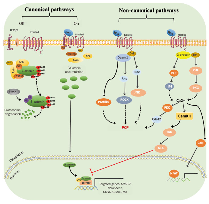
The Wnt signaling pathway plays a crucial role in various biological processes, including early embryonic development, organ formation, cancer, and epithelial-mesenchymal transition (EMT), which is a highly conserved genetic signaling pathway. The first identified Wnt gene, known as Wnt1, was discovered in mouse mammary tumors in 1982 [20]. Wnt signaling includes both classical and non-classical signaling pathways. The classical signaling pathway depends on β-catenin (Fig. 1). Wnt ligands indirectly regulate the intracellular and nuclear expression of β-catenin by affecting the assembly of a destructive complex consisting of axin, glycogen synthase kinase 3 (GSK3), dishevelled (Dvl) protein, adenomatous polyposis coli (APC), and casein kinase 1 (CK1), which finally affects the transcriptional regulation of target genes and the initiation of downstream signaling cascades. Axin is a scaffold protein with multiple interaction sites with other proteins, which can bind APC, CK1, GSK3β, and β-catenin together. The main role of APC is to enhance the affinity of the degradation complex with β-catenin [21]. CK1 and GSK3β function as protein kinases, CK1 phosphorylates β-catenin N-terminal Ser45, whereas GSK3β phosphorylates β-catenin N-terminal Thr41, Ser33, and Ser37 [22]. Phosphorylated Ser33 and Ser37 are ubiquitinated after recognition by β-transducin repeat-containing protein (β-TrCP) and finally degraded by the proteasome [23].
Figure 1. Wnt signaling pathway.
In the absence of Wnt ligands, the classical signaling pathway is off. At that time, β-catenin is phosphorylated at Ser45, Ser33, Ser37, and Thr41 by a destruction complex consisting of axin, GSK3β, Dvl, APC, and CK1. And then phosphorylated β-catenin is ubiquitylated by β-TrCP and degraded by the proteasome. When Wnt ligands are present and activated, they bind to coreceptors consisting of FZD protein and LRP5 or LRP6 on the cell surface, recruiting Dvl which induces dissociation of the destruction complex, and in turn results in intracellular accumulation of β-catenin. The accumulated β-catenin enters the nucleus and binds to TCF/LEF, thereby promoting target gene transcription. In the Wnt/PCP signaling pathway, Wnt ligands bind to frizzled receptors or their coreceptors (such as ROR-frizzled) which then recruit and activate Dvl proteins, which mediate Rho activation through Daam1. Activation of Rho in turn activates ROCK. Dvl also mediates Rac activation and then activates JNK. In addition, Daam1 can also mediate actin polymerization through the actin-binding protein Profilin. In the Wnt/Ca2+ pathway, Wnt ligands mediate G protein activation upon binding to frizzled receptors, which then recruit to activate Dvl, thereby activating the acid diesterase PDE. PKG can block the release of Ca2+, while PDE can increase the release of Ca2+ by inhibiting PKG. In addition, Dvl and G proteins can also activate PLC, which can activate IP3. The activation of IP3 induces the release of Ca2+ from the cell, thereby leading to an increase in Ca2+ levels. Ca2+ flux induced by these two pathways can activate second messengers such as PKC, CamKII, or CaN. Among them, CamKII activates TAK-NLK signaling and then antagonizes classical Wnt/β-catenin signaling through competing TCF. Activated CaN dephosphorylates NFAT, which then enters the nucleus and increases the transcription of its target genes. PKC can also engage in the Wnt/PCP pathway by activating the small GTPase Cdc42.
APC, adenomatous polyposis coli; β-TrCP, β-transducin repeat-containing protein; CamKII, calmodulin-dependent kinase II; CBP, cyclic adenosine monophosphate response-element binding protein; CK1, casein kinase 1; Daam1, dishevelled-associated activator of morphogenesis 1; Dvl, dishevelled; FZD, frizzled protein; GSK3β, glycogen synthase kinase 3β; IP, inositol triphosphate; JNK, c-Jun N-terminal kinase; LEF, lymphoid enhancer binding factor; LRP, lipoprotein receptor protein; NFAT, nuclear factor of activated T-cells; NLK, nemo-like kinase; PCP, planar cell polarity; PDE, phosphodiesterase; PKC, protein kinase C; PKG, cGMP-dependent protein kinase; PLC, phospholipase C; Rac, Ras-related C3 botulinum toxin substrate; ROCK, Rho-associated protein kinase; TAK, TGFβ-activated kinase; TCF, T-cell factor.
Non-canonical pathways mainly include Wnt/planner cell polarity (PCP) and Wnt/Ca2+ signaling pathways. The Wnt/PCP pathway primarily regulates cytoskeletal dynamics and directs the asymmetric distribution and migration of cellular components [24,25]. In Wnt/PCP signaling, activated Wnt ligands regulate cell polarity by inducing Rho-associated protein kinase (ROCK), profilin, and c-Jun N-terminal kinase (JNK) activation by recruiting Dvl and Damm1 proteins after binding to frizzled receptors. Notably, the Wnt/PCP signaling and the canonical Wnt/β-catenin signaling are mutually antagonistic, with the activation of one often accompanied by inhibition of the other.
The Wnt/Ca2+ pathway plays a key role mainly in cell adhesion, gastrulation, and heart development [24,25]. In the Wnt/Ca2+ signaling pathway, activated Wnt ligands bind to frizzled receptors by recruiting G proteins, causing intracellular calcium release and eventually triggering a downstream signaling cascade.
The role of Wnt/β-catenin signaling in acute kidney injury and chronic kidney disease
Wnt signaling is involved in a variety of pathological processes of kidney injury. It is now generally accepted that transient activation of Wnt signaling contributes to AKI recovery. Mild ischemic AKI induces a transient increase in the expression of most Wnt ligands and β-catenin. For example, in the AKI model of rats, ischemia-reperfusion (I/R) induces Wnt4 expression and activates β-catenin, which in turn promotes cell cycle progression by increasing the transcription of the target gene CCND1, thus repairing the damaged kidney [26]. In addition, some studies have found that macrophage-derived Wnt7b also has a protective effect on renal I/R injury [27]. In vitro, constitutively expressed active β-catenin can inhibit the activation and translocation of B-cell lymphoma 2-associated X protein (Bax) through the phosphatidylinositol-3 kinase/protein kinase B (AKT) signaling pathway, thereby inhibiting apoptosis and improving cell survival in renal tubular epithelial cells under metabolic stress [28]. In AKI induced by I/R or folic acid, the loss of tubular β-catenin significantly aggravates renal functional and structural damage, increases the expression of p53 and Bax, and induces cell apoptosis. Consistent with this, exogenous Wnt1 can protect renal tubular epithelial cells from apoptotic damage by activating β-catenin or stabilizing β-catenin to inhibit p53 and Bax expression [29]. In conclusion, moderate activation of Wnt/β-catenin signaling plays a renoprotective role in ischemic or nephrotoxin-induced AKI.
However, when severe ischemic AKI progresses to CKD, the Wnt/β-catenin signaling pathway is continuously activated. However, its continuous activation accelerates the progression from AKI to CKD. Sustained high expression of Wnt1 can significantly promote renal function decline and fibrosis after severe AKI, while inhibition of the Wnt/β-catenin signaling pathway can effectively alleviate the progression of AKI to CKD [29].
Severe or recurrent AKI can lead to incomplete renal recovery and eventually progress to CKD characterized by renal fibrosis. The current mainstream theory suggests that excessive and persistent activation of Wnt/β-catenin signaling can activate the EMT system, leading to AKI-CKD transition. It has been found that mild ischemic reperfusion injury (IRI; 20-minute ischemia) leads to transient activation of Wnt/β-catenin and acute renal injury. Severe IRI (40-minute ischemia) leads to continuous over-activation of Wnt/β-catenin, accompanied by renal fibrosis. In addition, Wnt1-induced β-catenin activation can accelerate the progression of AKI to CKD. In contrast, indirect catenin inhibitor-gamma-001 (ICG-001), a Wnt signaling inhibitor, can delay the transition from AKI to CKD by blocking Wnt/β-catenin [29]. However, in contrast to the results of most studies, Nlandu-Khodo et al. [30] found that the continuous and stable expression of active β-catenin in the proximal renal tubules has therapeutic potential in the progression of ischemic AKI to CKD. This study is the first to propose that proximal tubular β-catenin may be forkhead box protein O3 (FoxO3)-dependent to exert renoprotective effects during the transformation from AKI to CKD, suggesting that β-catenin in different cells may play completely opposite roles in AKI and CKD.
In the CKD model, the Wnt signaling pathway remains activated, and inhibition of Wnt signaling contributes to the repair of damaged kidneys. Activation of Wnt/β-catenin signaling occurs in almost all forms of CKD, including adriamycin nephropathy, polycystic kidney disease, 5/6 post-nephrectomy CKD, and diabetic nephropathy [31–34]. Wnt1 and β-catenin are upregulated in clinical diabetic nephropathy and focal segmental glomerulosclerosis specimens [35]. This was also confirmed in another study in which Wnt/β-catenin signaling was activated in glomeruli and podocytes of patients with diabetic nephropathy [36]. Active components of the Wnt/β-catenin signaling pathway have also been detected in patients with lupus nephritis [37]. Inhibition of β-catenin activation after unilateral ureteral obstruction (UUO) has been reported to attenuate renal fibrosis [38]. In vitro, stable expression of β-catenin in renal tubular cells suppressed cadherin expression and induced the expression of fibrosis-related genes. In vivo, ICG-001, a Wnt signaling inhibitor, ameliorated renal interstitial fibrosis by suppressing β-catenin driven gene transcription and fibrosis-related gene expression in a dose-dependent manner [39].
In conclusion, activation of Wnt signaling after AKI and CKD may be essential for kidney repair and regeneration but may also contribute to the progression of human kidney disease and fibrosis.
Therapeutic strategies targeting Wnt/β-catenin signaling
In AKI induced by I/R or folic acid, specific ablation of β-catenin in renal tubular epithelial cells aggravated renal lesions and increased mortality in mice [40]. In addition, macrophage-derived Wnt7b may stimulate injured kidney regeneration by regulating epithelial cell cycle progression and basement membrane repair in the early stage of renal injury induced by I/R [27]. In contrast, inhibition of Wnt/β-catenin signaling had renoprotective effects in chronic kidney injury models. Specific deletion of β-catenin in renal macrophages reduced M2 macrophage infiltration and alleviated fibrosis in UUO injury model mice [41]. The above studies suggest that the use of activators or inhibitors of Wnt signaling may play a renoprotective role during AKI or CKD.
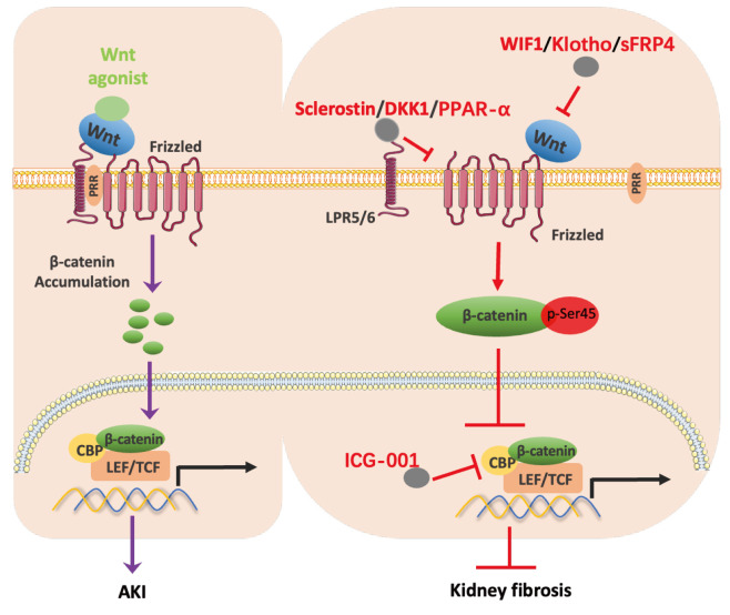
Several antagonists of the Wnt/β-catenin signaling pathway have been identified, among which Dickkopf-related protein 1 (DKK1), sclerostin and Wnt inhibitor 1 (WIF1) are recognized upstream antagonists of Wnt/β-catenin signaling (Fig. 2). DKK1 and sclerostin could competitively bind lipoprotein receptor protein (LRP) 5 and LRP6, blocking their interaction with FZD and ultimately inhibiting the downstream signal transmission [42]. In contrast, DKK2 binds to LRP5/LRP6 on the cell surface and enhances Wnt signaling. Another DKK family protein, DKK3, is released in response to stimulation by renal tubular epithelial cells. Recent studies have found that DKK3 drives renal fibrosis and is associated with the short-term risk of CKD progression and AKI, suggesting that DKK3 may be another potential target for the treatment of kidney-related diseases [43]. WIF1 binds directly to Wnt ligands, hindering its interaction with its receptors LRP5/LRP6, and ultimately keeps the entire signaling pathway in an inactive state [44]. Consistent with previous studies, transient activation of Wnt/β-catenin signaling plays a protective role in the AKI model. DKK2 recombinant protein as an agonist can promote renal cortical regeneration in the early stage of I/R and play a renoprotective role [27]. A study using Wnt agonists of synthetic pyrimidines showed that administration of Wnt agonists 1 hour before ischemia in rats alleviated ischemia-induced renal structural damage and decreased renal function [45].
Figure 2. Therapeutic strategies targeting Wnt/β-catenin signaling.
In the AKI model, β-catenin can be activated and accumulated in cells by Wnt agonist and then transferred to the nucleus. With the involvement of CBP and LEF/TCF, β-catenin increases the transcription of target genes and finally alleviates the early renal function decline and renal structural damage caused by AKI. In the CKD-induced fibrosis model, sclerostin, DKK1 or PPAR-α can competitively bind to LRP5/6, thereby inhibiting β-catenin and its downstream signaling pathways which finally alleviate renal fibrosis. In addition, WIF, Klotho, or sFRP4 can competitively bind to Wnt ligands and frizzled receptors, thereby blocking the binding of Wnt ligands and frizzled receptors and ultimately inhibiting β-catenin and its downstream signaling pathways. Furthermore, ICG-001 competitively binds to CBP, which then inhibits β-catenin mediated gene expression.
AKI, acute kidney injury; CBP, cyclic adenosine monophosphate response-element binding protein; CKD, chronic kidney disease; DKK-1, Dickkopf-1; ICG-001, indirect catenin inhibitor-gamma-001; LEF, lymphoid enhancer binding factor; LRP, lipoprotein receptor protein; PPAR-α, peroxisome proliferator-activated receptor-α; sFRP4, secreted frizzled-related protein 4; TCF, T-cell factor; WIF, Wnt inhibitory factor.
In a 2009 study using intravenous delivery of the Wnt antagonist DKK1 to mice, exogenous DKK1 expression in vivo significantly reduced UUO induced β-catenin overexpression, and ultimately inhibited renal interstitial extracellular matrix deposition and renal fibrosis [46]. Another study in 2013 also confirmed this conclusion. In the angiotensin (Ang) II-induced podocyte injury experiment in mice, exogenous DKK1 could alleviate Ang II-induced kidney injury by inhibiting β-catenin activation [47]. In addition to DKK1, many non-Wnt signaling molecules have been found to play antagonistic roles, such as Klotho, peroxisome proliferator-activated receptor-α (PPAR-α), ICG-001, and secreted frizzled-related protein 4 (sFRP4). Among them, Klotho inhibits myofibroblast activation and reduces renal tubulointerstitial fibrosis by inhibiting the Wnt/β-catenin signaling pathway [48,49]. PPAR-α interacts with LRP6 to inhibit Wnt/β-catenin signaling, leading to reduced oxidative stress [50]. ICG-001, a peptide-mimetic small molecule, can prevent the complex formation consisting of the β-catenin and cyclic adenosine monophosphate response-element binding protein (CBP) through competitively binding to CBP, and then inhibit β-catenin mediated gene expression. Studies have found that ICG-001 inhibits EMT of tubular cells induced by stable expression of β-catenin in vitro, and also inhibits the expression of fibrosis-related molecules and alleviates the degree of renal interstitial fibrosis in an in vivo model of obstructive renal fibrosis [39]. The sFRP can bind to Wnt proteins and frizzled and may inhibit the function of Wnt proteins by interfering with their interaction. Five sFRPs have been identified, among which sFRP4 can alleviate renal fibrosis in UUO model mice by inhibiting Wnt/β-catenin downstream signaling [38].
Collectively, targeting Wnt/β-catenin signaling agonists or upstream antagonists is a potential therapeutic strategy for AKI and CKD.
p53 signal
The p53 gene family in human encompasses tumor protein 53 (TP53), TP63, and TP73, which share similar DNA-binding domains and are able to bind similar DNA-specific sequences to regulate gene transcription. p53 is the encoded product of TP53 gene, which has many biological activities such as DNA damage repair, inducing apoptosis and inhibiting tumorigenesis [51,52]. In addition, p53 is involved in physiological and pathophysiological processes such as anti-infection, immune response, aging, and tissue I/R.
The protein level and activity of p53 gene are strictly regulated, and there is less p53 protein in cells under non-stress conditions. p53 is activated when intracellular triphosphate ribonucleotides levels or ribosomes are too small to maintain external stimuli such as cell cycle, shock, hypoxia, nutrient depletion, apoptosis, aging, and oncogene expression [51,53,54]. Tissue inflammation and its associated nitric oxide signals can also activate p53, which binds to its response elements to transcriptionally regulate the expression of target genes and initiate many cellular responses [55]. Different types of stress signals also lead to different modes of p53 protein modification, such as phosphorylation, acetylation, methylation, and ubiquitination [55,56].
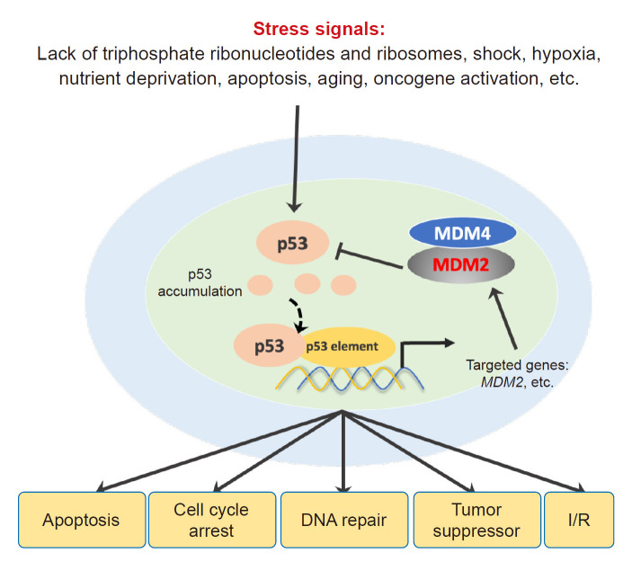
p53 tightly controls cell survival in mammals by regulating DNA damage processes (Fig. 3). Excessive expression or inappropriate activation of p53 can lead to cell apoptosis, while too little expression or continuous inhibition of p53 will lead to tumorigenesis. The E3 ubiquitin ligases mouse double minute 2 (MDM2) and MDM4 are negative regulators of p53 signaling. Among them, MDM2 can stabilize p53 content in cells by directly binding to p53 to inhibit its transcription factor activity and increase the degradation of p53 by promoting its ubiquitination. However, MDM4 indirectly maintains the stability of p53 content in cells by forming a complex with MDM2 and then inhibiting the degradation of MDM2 protein. Moreover, MDM2 is also a target gene of p53, which can form a negative feedback loop of MDM2-p53 [57].
Figure 3. p53 signaling.
p53 is activated when cells are subjected to stimuli such as deficiency of triphosphate ribonucleotides and ribosomes, shock, hypoxia, nutrient deprivation, apoptosis, aging, and oncogene expression. Accumulated p53 recognizes and binds to p53 response elements in the nucleus and participates in apoptosis, cell cycle, DNA damage, tumorigenesis, and tissue I/R by regulating the expression of its target genes. MDM2 and MDM4 are negative regulatory proteins of p53. Intracellular p53 content is tightly regulated by MDM2 and MDM4. MDM2 maintains the content of p53 in cells by ubiquitination and degradation of p53, while MDM4 inhibits p53 by stabilizing the content of MDM2 in cells. In addition, MDM2 is also a target gene of p53.
I/R, ischemia-reperfusion; MDM, mouse double minute.
Role of p53 in acute kidney injury and chronic kidney disease
Proximal tubule cell p53 is activated by DNA damage, reactive oxygen species, and hypoxia during AKI development and renal repair. In 2003, Kelly et al. [58] first proposed that p53 was involved in renal I/R injury. Their study showed that after renal I/R, the expression of p53 in the renal medulla increased sharply 24 hours later, and inhibition of p53 could alleviate the apoptosis of renal tubule cells and the decline of renal function caused by I/R [58]. Ying and Kim [59] confirmed that specific deletion of p53 in the proximal renal tubules had a protective effect on renal IRI. I/R-induced AKI facilitates the opening and subsequent cell death of mitochondrial permeability transition pores (mPTP), in which cyclophilin D (CypD) is an important component. Recent studies have shown that the p53-CypD complex mediates I/R-AKI tubular cell apoptosis through mPTP opening [60]. Nephrotoxicity is another major cause of AKI. Cisplatin can inhibit the proliferation of tumor cells and is widely used in the clinical treatment of cancer. However, the toxic side effects of cisplatin, such as inducing AKI, have gradually attracted attention. N-acetylcysteine, an antioxidant, can delay the progression of cisplatin-induced AKI to CKD by inducing p53 deacetylation, suggesting that p53 deacetylation plays an important role in AKI damage repair and subsequent CKD [61]. Furthermore, it has been shown that cisplatin induced p53 expression and phosphorylation in proximal tubular epithelial cells, while p53 inhibitor pifithrin-α or p53 mutation significantly attenuated cisplatin-induced apoptosis [62,63]. Consistent with this, another study found that inhibition of p53 could also attenuate cisplatin-induced AKI by upregulating miR-142-5p [64]. Zhang et al. [65] constructed renal I/R injury models and cisplatin-induced AKI models in p53-specific knockout mice in the proximal renal tubule, distal renal tubule, Henle ring, and medullary collecting duct, respectively. Their results showed that only p53-specific knockout in the proximal renal tubule could effectively resist renal injury induced by I/R and cisplatin, which revealed the important function of p53 in proximal tubular cells in renal AKI [65]. It is worth noting that p53 is also associated with folic acid, endotoxin, aristolochic acids, glycerol injection, and polymyxin B-induced AKI [66–70].
p53 is associated with poor repair of damaged kidneys, which is the main cause of AKI progression to CKD. There are studies suggesting that pharmacological or genetic blocking of p53 can effectively inhibit the inflammatory response, apoptosis, and renal fibrosis caused by UUO [71]. Moreover, specific deletion of p53 in proximal tubule cells alleviates renal interstitial fibrosis induced by I/R [59]. Interestingly, conflicting views have emerged in recent years. In a long-term observational study in rats after ischemic AKI, Dagher et al. [72] found that although p53 inhibitor pifithrin-α had an acute protective effect in the ischemic AKI model, it accelerated the progression of renal fibrosis after 8 weeks of I/R. Of note, a 2013 study pointed out that p53 inhibition and deletion play a role in aggravating damage in both ischemic AKI and CKD stages in mice [73]. The authors noted that in the rat model, inhibition of p53 exerted a renoprotective effect by reducing renal tubular cell apoptosis, but in mice, leukocyte p53 exerted a renoprotective effect by reducing the degree and duration of inflammation and by promoting an anti-inflammatory M2 macrophage phenotype. The above studies suggest that p53 may play opposite functions in different species and different cells. The role of p53 in ischemic AKI and subsequent CKD is extremely complex, and more in-depth studies are needed to clarify the mechanism of p53.
Therapeutic strategies targeting p53 signals
Targeting p53-regulated pathways related to apoptosis, cell cycle arrest, autophagy, and cell senescence may be a potential strategy for the treatment of AKI and AKI-progressive CKD. Molitoris et al. [74] found that p53 short interfering RNA intravenous injection at 4 hours after renal ischemia could attenuate apoptosis and renal function damage caused by I/R. The E3 ubiquitin ligase MDM2 is a p53-specific cellular antagonist, which has been shown that reducing p53 levels by targeting MDM2 can inhibit the transcription of apoptotic cells, thereby partially protecting hypoxic AKI cells from apoptosis [75]. In addition, direct inhibition of p53 activation can also alleviate renal function decline, renal structural damage, and renal tubular cell apoptosis during renal I/R in mice [76]. It has been reported that p53 inhibitor pifithrin-α can reduce renal tubular cell apoptosis and renal function decline induced by I/R [58]. Moreover, mesenchymal stem cell-derived exosomes may reduce the apoptosis of renal tubular epithelial cells after AKI by inhibiting p53, thereby reducing DNA damage and apoptosis pathway activity [77]. In line with this, in vitro studies have shown that inhibition of p53 can reduce hypoxia-reoxygenation induced apoptosis in human proximal tubule cells [78]. The main mechanism is that p53 forms a complex with CypD to mediate the opening of mPTP and promote the apoptosis of renal tubular cells after AKI [60]. Taken together, direct inhibition of p53 activation or increased p53 degradation are potential therapeutic strategy to mitigate apoptotic injury in renal tubular cells during AKI.
Cell cycle arrest plays a critical role in the repair of damaged kidneys after AKI, while p53 is a major mediator regulating the cell cycle. It has been shown that G1 arrest in renal tubular cells is associated with p53 induction in a rat model of septic AKI induced by cecal ligation and puncture [79]. Targeted knockdown of p53 in proximal tubular cells can alleviate G2/M phase arrest and attenuate the decline in renal function, renal inflammation, apoptosis, and the occurrence of regeneration-phase renal fibrosis after AKI [59]. Bypassing G2/M phase arrest with the p53 inhibitor pifithrin-α also alleviated fibrosis in the unilateral ischemia-injured kidney [80]. In ischemic AKI, exosomes derived from mesenchymal stem cells highly enriched miR-125b-5p in proximal renal tubular cells, inhibited the expression of p53 protein, resulting in the upregulation of cyclin-dependent kinase 1 and cyclin B1, thereby rescuing G2/M arrest, improving ischemic AKI, and promoting renal tubular repair [81]. In conclusion, p53 induces renal fibrosis by participating in cell cycle arrest after AKI, and targeted inhibition of p53 may alleviate renal fibrosis in CKD by rescuing cell cycle arrest after AKI.
A large amount of evidence has confirmed that autophagy plays a renoprotective role in AKI. Jiang et al. [82] further demonstrated that blocking autophagy aggravated AKI by using autophagy inhibitors and autophagy-deficient mice. It is well known that p53 has a dual role in regulating autophagy, mainly depending on its subcellular localization. In 2008, Periyasamy-Thandavan et al. [83] first found that cisplatin-induced autophagy of renal tubular cells was inhibited by pifithrin-α which is a chemical inhibitor of p53, suggesting that p53 was involved in the occurrence of autophagy in AKI. In sepsis-induced AKI, inducing p53 deacetylation can alleviate sepsis-induced AKI by promoting autophagy in renal tubular epithelial cells [84]. This suggests that we can induce renal repair after AKI by promoting p53 deacetylation and thus increasing autophagy. Contrary to AKI, the activation of autophagy in proximal tubular cells in CKD leads to tubular atrophy, inflammation, and renal fibrosis [85,86]. However, some studies have confirmed that autophagy plays an anti-fibrotic role in CKD [87,88]. Therefore, the roles of autophagy and p53 in CKD need to be further investigated.
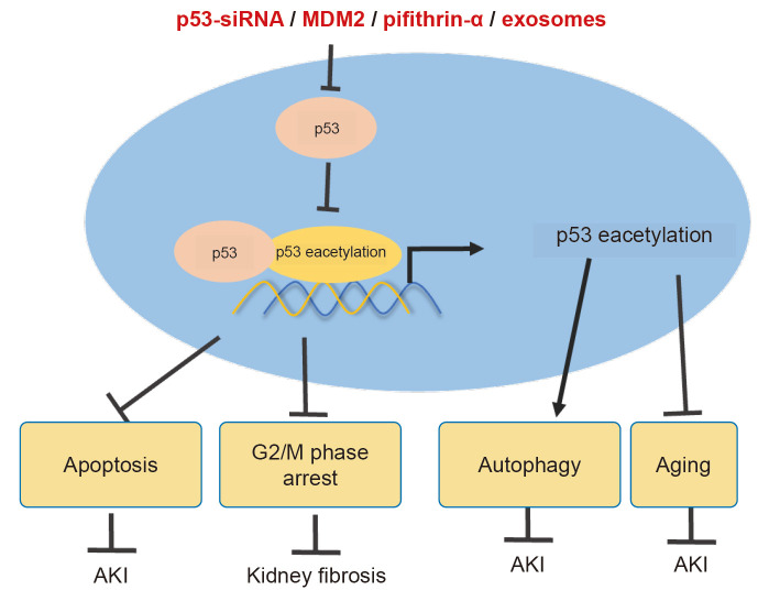
Renal tubular cell senescence is one of the main reasons for the progression from AKI to CKD, and p53 is one of the senescence marker proteins in the damaged kidney and renal tubules of AKI mice [89]. Alpha 2 adrenergic agonists dexmedetomidine pretreatment reduce age-related biomarkers of p53, p21, and p16 protein expression, reduce aging tubule cells, improve renal fibrosis, and slow AKI to CKD [34]. Xenon released into the injured kidney can reduce renal fibrosis and renal function decline induced by I/R, and the mechanism is related to the decreased protein expression of cell senescence markers p53 and p16 [90]. The above studies confirm that p53 is involved in the cellular senescence of damaged renal tubules. Premature aging plays an important role in sepsis AKI, inhibit the premature aging of the p53/p21 pathway may contribute to the recovery of renal function after sepsis AKI [91]. In addition, p53 deacetylation may be a potential target for delaying premature tubular cell senescence and CKD progression after AKI [61]. These results suggest that targeted inhibition of p53 expression or promotion of p53 deacetylation may be an effective strategy for the treatment of renal tubular cell senescence after AKI and CKD (Fig. 4).
Figure 4. Therapeutic strategies targeting p53 signals.
Targeted inhibition or knockdown of p53 or induction of p53 deacetylation may alleviate renal injury or fibrosis caused by AKI by inhibiting apoptosis, rescuing cell cycle arrest, inducing autophagy, or inhibiting cell senescence.
AKI, acute kidney injury; MDM2, mouse double minute 2; siRNA, short interfering RNA.
Interaction of Wnt/β-catenin signaling pathway with p53
There may be a tight interaction between the Wnt/β-catenin signaling pathway and p53. Observations in a variety of tumor models have shown that the Wnt signaling pathway and p53 cooperate to drive the occurrence and development of tumors. In 1999, Damalas et al. [92] found that overexpression of β-catenin inhibited MDM2-mediated degradation of p53 protein, resulting in accumulation of transcriptionally active p53. Webster et al. [93] found that high levels of Wnt5a in melanoma can stabilize wild-type p53 and increase its half-life. Vice versa, p53 activates β-catenin by increasing the expression of Wnt7b through transcription, thereby affecting tumor proliferation and metastasis of hepatocellular carcinoma [94]. Additionally, in colorectal cancer patient-derived tumor organoids and tumor cells, wild-type p53 can induce Wnt/β-catenin pathway activation by promoting Wnt3 transcription [13]. In addition to tumors, recent studies have found a close relationship between them in the kidney. In I/R or folate-induced AKI, the loss of renal tubule β-catenin aggravates renal injury, while the expression of p53 and Bax increases. On the contrary, activating or stabilizing β-catenin can protect tubule epithelial cells from apoptosis and inhibit the expression of p53 and Bax [40]. In the lipopolysaccharide and sterile AKI model of bilateral I/R, β-catenin alleviates renal function decline, renal structural damage, apoptosis, and necrosis after AKI by activating AKT and inhibiting p53 signaling [95]. These results suggest that Wnt/β-catenin may act as an upstream signal of p53 and play a renoprotective role by inhibiting p53 expression in AKI.
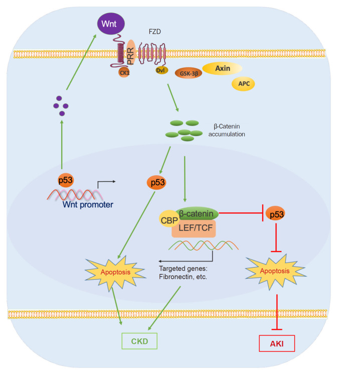
In the kidneys of diabetic rats, GSK3β is activated and p-LRP6 is down-regulated, resulting in increased interaction between GSK3β and p53 and promoting podocyte apoptosis [96]. In high-glucose treated podocytes, Wnt/β-catenin signals and p53 were increased simultaneously after β-arrestin 1/2 treatment, promoting podocyte apoptosis [97]. In addition, our study showed that in UUO and adenine-induced renal fibrosis models, p53 may regulate Wnt7a expression through transcription, activate Wnt/β-catenin signal, and participate in the process of renal fibrosis [98]. In human proximal tubule epithelial cell lines and primary renal tubule cells, Wnt9a significantly upregulated the levels of aging-related p16, p19, p53, and p21, promoting renal fibrosis [99]. These results revealed that the crosstalk between the Wnt/β-catenin signaling pathway and p53 may play diametrically opposite roles in AKI and CKD. In AKI, the Wnt/β-catenin signaling pathway may play a renoprotective role by inhibiting the expression of p53. In CKD, p53 targets to upregulate the transcription level of the Wnt/β-catenin signaling pathway, and the increased Wnt/β-catenin signaling in turn promotes the expression of p53. Finally, the two synergistically promote the occurrence of renal fibrosis (Fig. 5).
Figure 5. Interaction of Wnt/β-catenin signaling pathway with p53 in AKI and CKD.
In the AKI model, the Wnt/β-catenin signaling pathway is transiently activated, and activated β-catenin can protect the kidney from apoptosis by inhibiting p53 expression or inhibiting p53 activity. In the CKD model, p53 can increase the expression of Wnt ligands through transcriptional regulation, thereby inducing the continuous activation of the Wnt/β-catenin signaling pathway, which leads to the dissociation of the destruction complex composed of axin, CK1, Dvl, APC, and GSK3β, and finally leads to the accumulation of β-catenin in the cytoplasm. The accumulated β-catenin enters the nucleus and promotes renal fibrosis by regulating the transcription of target genes. In addition, the accumulation of β-catenin may also increase the expression of p53, thereby inducing apoptosis and aggravating kidney damage in the process of CKD.
AKI, acute kidney injury; APC, adenomatous polyposis coli; CBP, cyclic adenosine monophosphate response-element binding protein; CKD, chronic kidney disease; CK1, casein kinase 1; Dvl, dishevelled; FZD, frizzled protein; GSK3β, glycogen synthase kinase 3β; LEF, lymphoid enhancer binding factor; PRR, pattern recognition receptor; TCF, T-cell factor.
Conclusions
Increasing evidence shows that the Wnt/β-catenin signaling pathway and p53 play an important role in AKI and subsequent renal fibrosis. A deeper understanding of their mechanisms will provide new targets for the treatment of kidney diseases. Although Wnt/β-catenin signaling and p53 play synergistic roles in tumor and embryonic development, whether they play a synergistic role in kidney disease needs to be further elucidated. In this review, we found that the β-catenin signaling pathway may play a renoprotective role in AKI by inhibiting the expression of p53, while in CKD, the β-catenin signaling pathway and p53 may synergistically promote renal fibrosis. Based on the characteristics of the biological effects of β-catenin and p53, finding ways to regulate the activity of β-catenin and p53 is expected to be transformed into an effective strategy for the prevention and treatment of kidney diseases, and will also bring unprecedented opportunities for the prevention and treatment of kidney diseases.
Footnotes
Conflicts of interest
All authors have no conflicts of interest to declare.
Funding
This work was supported by the Natural Science Foundation of Liaoning Province, China 2022-MS-326 (to ZLL). We are also grateful for the support from the Liaoning BaiQianWan Talents Program.
Data sharing statement
The data presented in this study are available from the corresponding author upon reasonable request.
Authors’ contributions
Conceptualization: ZLL
Data curation: LW, RFQ, YZ, BWS, YNB, PG, ZLL
Funding acquisition: ZLL
Investigation: All authors
Writing–original draft: WHM, LW, WJH
Writing–review & editing: WHM, ZLL
All authors read and approved the final manuscript.
References
- 1.Menon S, Symons JM, Selewski DT. Acute kidney injury. Pediatr Rev. 2023;44:265–279. doi: 10.1542/pir.2021-005438. [DOI] [PubMed] [Google Scholar]
- 2.Mehta RL, Cerdá J, Burdmann EA, et al. International Society of Nephrology\'s 0by25 initiative for acute kidney injury (zero preventable deaths by 2025): a human rights case for nephrology. Lancet. 2015;385:2616–2643. doi: 10.1016/S0140-6736(15)60126-X. [DOI] [PubMed] [Google Scholar]
- 3.Jiang M, Bai M, Lei J, et al. Mitochondrial dysfunction and the AKI-to-CKD transition. Am J Physiol Renal Physiol. 2020;319:F1105–F1116. doi: 10.1152/ajprenal.00285.2020. [DOI] [PubMed] [Google Scholar]
- 4.Kurzhagen JT, Dellepiane S, Cantaluppi V, Rabb H. AKI: an increasingly recognized risk factor for CKD development and progression. J Nephrol. 2020;33:1171–1187. doi: 10.1007/s40620-020-00793-2. [DOI] [PubMed] [Google Scholar]
- 5.See EJ, Jayasinghe K, Glassford N, et al. Long-term risk of adverse outcomes after acute kidney injury: a systematic review and meta-analysis of cohort studies using consensus definitions of exposure. Kidney Int. 2019;95:160–172. doi: 10.1016/j.kint.2018.08.036. [DOI] [PubMed] [Google Scholar]
- 6.Wang L, Xu X, Zhang M, et al. Prevalence of chronic kidney disease in China: results from the sixth China Chronic Disease and Risk Factor Surveillance. JAMA Intern Med. 2023;183:298–310. doi: 10.1001/jamainternmed.2022.6817. [DOI] [PMC free article] [PubMed] [Google Scholar]
- 7.Nusse R, Clevers H. Wnt/β-catenin signaling, disease, and emerging therapeutic modalities. Cell. 2017;169:985–999. doi: 10.1016/j.cell.2017.05.016. [DOI] [PubMed] [Google Scholar]
- 8.Liu J, Xiao Q, Xiao J, et al. Wnt/β-catenin signalling: function, biological mechanisms, and therapeutic opportunities. Signal Transduct Target Ther. 2022;7:3. doi: 10.1038/s41392-021-00762-6. [DOI] [PMC free article] [PubMed] [Google Scholar]
- 9.MacDonald BT, Tamai K, He X. Wnt/beta-catenin signaling: components, mechanisms, and diseases. Dev Cell. 2009;17:9–26. doi: 10.1016/j.devcel.2009.06.016. [DOI] [PMC free article] [PubMed] [Google Scholar]
- 10.Kim NH, Kim HS, Kim NG, et al. p53 and microRNA-34 are suppressors of canonical Wnt signaling. Sci Signal. 2011;4:ra71. doi: 10.1126/scisignal.2001744. [DOI] [PMC free article] [PubMed] [Google Scholar]
- 11.Cha YH, Kim NH, Park C, Lee I, Kim HS, Yook JI. MiRNA-34 intrinsically links p53 tumor suppressor and Wnt signaling. Cell Cycle. 2012;11:1273–1281. doi: 10.4161/cc.19618. [DOI] [PubMed] [Google Scholar]
- 12.Han D, Xu Y, Peng WP, et al. Citrus alkaline extracts inhibit senescence of A549 cells to alleviate pulmonary fibrosis via the β-catenin/P53 pathway. Med Sci Monit. 2021;27:e928547. doi: 10.12659/MSM.928547. [DOI] [PMC free article] [PubMed] [Google Scholar]
- 13.Cho YH, Ro EJ, Yoon JS, et al. 5-FU promotes stemness of colorectal cancer via p53-mediated WNT/β-catenin pathway activation. Nat Commun. 2020;11:5321. doi: 10.1038/s41467-020-19173-2. [DOI] [PMC free article] [PubMed] [Google Scholar]
- 14.Sheng J, He X, Yu W, et al. p53-targeted lncRNA ST7-AS1 acts as a tumour suppressor by interacting with PTBP1 to suppress the Wnt/β-catenin signalling pathway in glioma. Cancer Lett. 2021;503:54–68. doi: 10.1016/j.canlet.2020.12.039. [DOI] [PubMed] [Google Scholar]
- 15.Wellenstein MD, Coffelt SB, Duits DE, et al. Loss of p53 triggers WNT-dependent systemic inflammation to drive breast cancer metastasis. Nature. 2019;572:538–542. doi: 10.1038/s41586-019-1450-6. [DOI] [PMC free article] [PubMed] [Google Scholar]
- 16.Gu Z, Tan W, Feng G, et al. Wnt/β-catenin signaling mediates the senescence of bone marrow-mesenchymal stem cells from systemic lupus erythematosus patients through the p53/p21 pathway. Mol Cell Biochem. 2014;387:27–37. doi: 10.1007/s11010-013-1866-5. [DOI] [PubMed] [Google Scholar]
- 17.Lee KH, Li M, Michalowski AM, et al. A genomewide study identifies the Wnt signaling pathway as a major target of p53 in murine embryonic stem cells. Proc Natl Acad Sci U S A. 2010;107:69–74. doi: 10.1073/pnas.0909734107. [DOI] [PMC free article] [PubMed] [Google Scholar]
- 18.Wang Q, Zou Y, Nowotschin S, et al. The p53 family coordinates Wnt and nodal inputs in mesendodermal differentiation of embryonic stem cells. Cell Stem Cell. 2017;20:70–86. doi: 10.1016/j.stem.2016.10.002. [DOI] [PMC free article] [PubMed] [Google Scholar]
- 19.Sadot E, Geiger B, Oren M, Ben-Ze’ev A. Down-regulation of beta-catenin by activated p53. Mol Cell Biol. 2001;21:6768–6781. doi: 10.1128/MCB.21.20.6768-6781.2001. [DOI] [PMC free article] [PubMed] [Google Scholar]
- 20.Nusse R, Varmus HE. Many tumors induced by the mouse mammary tumor virus contain a provirus integrated in the same region of the host genome. Cell. 1982;31:99–109. doi: 10.1016/0092-8674(82)90409-3. [DOI] [PubMed] [Google Scholar]
- 21.Tanneberger K, Pfister AS, Kriz V, Bryja V, Schambony A, Behrens J. Structural and functional characterization of the Wnt inhibitor APC membrane recruitment 1 (Amer1) J Biol Chem. 2011;286:19204–19214. doi: 10.1074/jbc.M111.224881. [DOI] [PMC free article] [PubMed] [Google Scholar]
- 22.Liu C, Li Y, Semenov M, et al. Control of beta-catenin phosphorylation/degradation by a dual-kinase mechanism. Cell. 2002;108:837–847. doi: 10.1016/s0092-8674(02)00685-2. [DOI] [PubMed] [Google Scholar]
- 23.van Noort M, van de Wetering M, Clevers H. Identification of two novel regulated serines in the N terminus of beta-catenin. Exp Cell Res. 2002;276:264–272. doi: 10.1006/excr.2002.5520. [DOI] [PubMed] [Google Scholar]
- 24.Wang Y, Zhou CJ, Liu Y. Wnt signaling in kidney development and disease. Prog Mol Biol Transl Sci. 2018;153:181–207. doi: 10.1016/bs.pmbts.2017.11.019. [DOI] [PMC free article] [PubMed] [Google Scholar]
- 25.Ma L, Wang HY. Mitogen-activated protein kinase p38 regulates the Wnt/cyclic GMP/Ca2+ non-canonical pathway. J Biol Chem. 2007;282:28980–28990. doi: 10.1074/jbc.M702840200. [DOI] [PubMed] [Google Scholar]
- 26.Terada Y, Tanaka H, Okado T, et al. Expression and function of the developmental gene Wnt-4 during experimental acute renal failure in rats. J Am Soc Nephrol. 2003;14:1223–1233. doi: 10.1097/01.asn.0000060577.94532.06. [DOI] [PubMed] [Google Scholar]
- 27.Lin SL, Li B, Rao S, et al. Macrophage Wnt7b is critical for kidney repair and regeneration. Proc Natl Acad Sci U S A. 2010;107:4194–4199. doi: 10.1073/pnas.0912228107. [DOI] [PMC free article] [PubMed] [Google Scholar]
- 28.Wang Z, Havasi A, Gall JM, Mao H, Schwartz JH, Borkan SC. Beta-catenin promotes survival of renal epithelial cells by inhibiting Bax. J Am Soc Nephrol. 2009;20:1919–1928. doi: 10.1681/ASN.2009030253. [DOI] [PMC free article] [PubMed] [Google Scholar]
- 29.Xiao L, Zhou D, Tan RJ, et al. Sustained activation of Wnt/β-catenin signaling drives AKI to CKD progression. J Am Soc Nephrol. 2016;27:1727–1740. doi: 10.1681/ASN.2015040449. [DOI] [PMC free article] [PubMed] [Google Scholar]
- 30.Nlandu-Khodo S, Osaki Y, Scarfe L, et al. Tubular β-catenin and FoxO3 interactions protect in chronic kidney disease. JCI Insight. 2020;5:e135454. doi: 10.1172/jci.insight.135454. [DOI] [PMC free article] [PubMed] [Google Scholar]
- 31.Zhou L, Mo H, Miao J, et al. Klotho ameliorates kidney injury and fibrosis and normalizes blood pressure by targeting the renin-angiotensin system. Am J Pathol. 2015;185:3211–3223. doi: 10.1016/j.ajpath.2015.08.004. [DOI] [PMC free article] [PubMed] [Google Scholar]
- 32.Zhou T, He X, Cheng R, et al. Implication of dysregulation of the canonical wingless-type MMTV integration site (WNT) pathway in diabetic nephropathy. Diabetologia. 2012;55:255–266. doi: 10.1007/s00125-011-2314-2. [DOI] [PubMed] [Google Scholar]
- 33.Xiao L, Wang M, Yang S, Liu F, Sun L. A glimpse of the pathogenetic mechanisms of Wnt/β-catenin signaling in diabetic nephropathy. Biomed Res Int. 2013;2013:987064. doi: 10.1155/2013/987064. [DOI] [PMC free article] [PubMed] [Google Scholar]
- 34.Li Q, Chen C, Chen X, Han M, Li J. Dexmedetomidine attenuates renal fibrosis via α2-adrenergic receptor-dependent inhibition of cellular senescence after renal ischemia/reperfusion. Life Sci. 2018;207:1–8. doi: 10.1016/j.lfs.2018.05.003. [DOI] [PubMed] [Google Scholar]
- 35.Dai C, Stolz DB, Kiss LP, Monga SP, Holzman LB, Liu Y. Wnt/beta-catenin signaling promotes podocyte dysfunction and albuminuria. J Am Soc Nephrol. 2009;20:1997–2008. doi: 10.1681/ASN.2009010019. [DOI] [PMC free article] [PubMed] [Google Scholar]
- 36.Kato H, Gruenwald A, Suh JH, et al. Wnt/β-catenin pathway in podocytes integrates cell adhesion, differentiation, and survival. J Biol Chem. 2011;286:26003–26015. doi: 10.1074/jbc.M111.223164. [DOI] [PMC free article] [PubMed] [Google Scholar]
- 37.Wang XD, Huang XF, Yan QR, Bao CD. Aberrant activation of the WNT/β-catenin signaling pathway in lupus nephritis. PLoS One. 2014;9:e84852. doi: 10.1371/journal.pone.0084852. [DOI] [PMC free article] [PubMed] [Google Scholar]
- 38.Surendran K, Schiavi S, Hruska KA. Wnt-dependent beta-catenin signaling is activated after unilateral ureteral obstruction, and recombinant secreted frizzled-related protein 4 alters the progression of renal fibrosis. J Am Soc Nephrol. 2005;16:2373–2384. doi: 10.1681/ASN.2004110949. [DOI] [PubMed] [Google Scholar]
- 39.Hao S, He W, Li Y, et al. Targeted inhibition of β-catenin/CBP signaling ameliorates renal interstitial fibrosis. J Am Soc Nephrol. 2011;22:1642–1653. doi: 10.1681/ASN.2010101079. [DOI] [PMC free article] [PubMed] [Google Scholar]
- 40.Zhou D, Li Y, Lin L, Zhou L, Igarashi P, Liu Y. Tubule-specific ablation of endogenous β-catenin aggravates acute kidney injury in mice. Kidney Int. 2012;82:537–547. doi: 10.1038/ki.2012.173. [DOI] [PMC free article] [PubMed] [Google Scholar]
- 41.Feng Y, Ren J, Gui Y, et al. Wnt/β-catenin-promoted macrophage alternative activation contributes to kidney fibrosis. J Am Soc Nephrol. 2018;29:182–193. doi: 10.1681/ASN.2017040391. [DOI] [PMC free article] [PubMed] [Google Scholar]
- 42.Joiner DM, Ke J, Zhong Z, Xu HE, Williams BO. LRP5 and LRP6 in development and disease. Trends Endocrinol Metab. 2013;24:31–39. doi: 10.1016/j.tem.2012.10.003. [DOI] [PMC free article] [PubMed] [Google Scholar]
- 43.Schunk SJ, Floege J, Fliser D, Speer T. WNT-β-catenin signalling: a versatile player in kidney injury and repair. Nat Rev Nephrol. 2021;17:172–184. doi: 10.1038/s41581-020-00343-w. [DOI] [PubMed] [Google Scholar]
- 44.Malinauskas T, Aricescu AR, Lu W, Siebold C, Jones EY. Modular mechanism of Wnt signaling inhibition by Wnt inhibitory factor 1. Nat Struct Mol Biol. 2011;18:886–893. doi: 10.1038/nsmb.2081. [DOI] [PMC free article] [PubMed] [Google Scholar]
- 45.Kuncewitch M, Yang WL, Corbo L, et al. WNT agonist decreases tissue damage and improves renal function after ischemia-reperfusion. Shock. 2015;43:268–275. doi: 10.1097/SHK.0000000000000293. [DOI] [PMC free article] [PubMed] [Google Scholar]
- 46.He W, Dai C, Li Y, Zeng G, Monga SP, Liu Y. Wnt/beta-catenin signaling promotes renal interstitial fibrosis. J Am Soc Nephrol. 2009;20:765–776. doi: 10.1681/ASN.2008060566. [DOI] [PMC free article] [PubMed] [Google Scholar]
- 47.Jiang L, Xu L, Song Y, et al. Calmodulin-dependent protein kinase II/cAMP response element-binding protein/Wnt/β-catenin signaling cascade regulates angiotensin II-induced podocyte injury and albuminuria. J Biol Chem. 2013;288:23368–23379. doi: 10.1074/jbc.M113.460394. [DOI] [PMC free article] [PubMed] [Google Scholar]
- 48.Satoh M, Nagasu H, Morita Y, Yamaguchi TP, Kanwar YS, Kashihara N. Klotho protects against mouse renal fibrosis by inhibiting Wnt signaling. Am J Physiol Renal Physiol. 2012;303:F1641–F1651. doi: 10.1152/ajprenal.00460.2012. [DOI] [PMC free article] [PubMed] [Google Scholar]
- 49.Zhou L, Li Y, Zhou D, Tan RJ, Liu Y. Loss of Klotho contributes to kidney injury by derepression of Wnt/β-catenin signaling. J Am Soc Nephrol. 2013;24:771–785. doi: 10.1681/ASN.2012080865. [DOI] [PMC free article] [PubMed] [Google Scholar]
- 50.Cheng R, Ding L, He X, Takahashi Y, Ma JX. Interaction of PPARα with the canonic Wnt pathway in the regulation of renal fibrosis. Diabetes. 2016;65:3730–3743. doi: 10.2337/db16-0426. [DOI] [PMC free article] [PubMed] [Google Scholar]
- 51.Liu J, Zhang C, Hu W, Feng Z. Tumor suppressor p53 and metabolism. J Mol Cell Biol. 2019;11:284–292. doi: 10.1093/jmcb/mjy070. [DOI] [PMC free article] [PubMed] [Google Scholar]
- 52.Zhang C, Liu J, Xu D, Zhang T, Hu W, Feng Z. Gain-of-function mutant p53 in cancer progression and therapy. J Mol Cell Biol. 2020;12:674–687. doi: 10.1093/jmcb/mjaa040. [DOI] [PMC free article] [PubMed] [Google Scholar]
- 53.Levine AJ. The many faces of p53: something for everyone. J Mol Cell Biol. 2019;11:524–530. doi: 10.1093/jmcb/mjz026. [DOI] [PMC free article] [PubMed] [Google Scholar]
- 54.Hu W. The role of p53 gene family in reproduction. Cold Spring Harb Perspect Biol. 2009;1:a001073. doi: 10.1101/cshperspect.a001073. [DOI] [PMC free article] [PubMed] [Google Scholar]
- 55.Harris SL, Levine AJ. The p53 pathway: positive and negative feedback loops. Oncogene. 2005;24:2899–2908. doi: 10.1038/sj.onc.1208615. [DOI] [PubMed] [Google Scholar]
- 56.Vogelstein B, Lane D, Levine AJ. Surfing the p53 network. Nature. 2000;408:307–310. doi: 10.1038/35042675. [DOI] [PubMed] [Google Scholar]
- 57.Karni-Schmidt O, Lokshin M, Prives C. The roles of MDM2 and MDMX in cancer. Annu Rev Pathol. 2016;11:617–644. doi: 10.1146/annurev-pathol-012414-040349. [DOI] [PMC free article] [PubMed] [Google Scholar]
- 58.Kelly KJ, Plotkin Z, Vulgamott SL, Dagher PC. P53 mediates the apoptotic response to GTP depletion after renal ischemia-reperfusion: protective role of a p53 inhibitor. J Am Soc Nephrol. 2003;14:128–138. doi: 10.1097/01.asn.0000040596.23073.01. [DOI] [PubMed] [Google Scholar]
- 59.Ying Y, Kim J, Westphal SN, Long KE, Padanilam BJ. Targeted deletion of p53 in the proximal tubule prevents ischemic renal injury. J Am Soc Nephrol. 2014;25:2707–2716. doi: 10.1681/ASN.2013121270. [DOI] [PMC free article] [PubMed] [Google Scholar]
- 60.Yang H, Li R, Zhang L, et al. p53-cyclophilin D mediates renal tubular cell apoptosis in ischemia-reperfusion-induced acute kidney injury. Am J Physiol Renal Physiol. 2019;317:F1311–F1317. doi: 10.1152/ajprenal.00072.2019. [DOI] [PubMed] [Google Scholar]
- 61.Li C, Xie N, Li Y, Liu C, Hou FF, Wang J. N-acetylcysteine ameliorates cisplatin-induced renal senescence and renal interstitial fibrosis through sirtuin1 activation and p53 deacetylation. Free Radic Biol Med. 2019;130:512–527. doi: 10.1016/j.freeradbiomed.2018.11.006. [DOI] [PubMed] [Google Scholar]
- 62.Jiang M, Yi X, Hsu S, Wang CY, Dong Z. Role of p53 in cisplatin-induced tubular cell apoptosis: dependence on p53 transcriptional activity. Am J Physiol Renal Physiol. 2004;287:F1140–F1147. doi: 10.1152/ajprenal.00262.2004. [DOI] [PubMed] [Google Scholar]
- 63.Wei Q, Dong G, Yang T, Megyesi J, Price PM, Dong Z. Activation and involvement of p53 in cisplatin-induced nephrotoxicity. Am J Physiol Renal Physiol. 2007;293:F1282–F1291. doi: 10.1152/ajprenal.00230.2007. [DOI] [PMC free article] [PubMed] [Google Scholar]
- 64.Chen G, Xue H, Zhang X, Ding D, Zhang S. p53 inhibition attenuates cisplatin-induced acute kidney injury through microRNA-142-5p regulating SIRT7/NF-κB. Ren Fail. 2022;44:368–380. doi: 10.1080/0886022X.2022.2039195. [DOI] [PMC free article] [PubMed] [Google Scholar]
- 65.Zhang D, Liu Y, Wei Q, et al. Tubular p53 regulates multiple genes to mediate AKI. J Am Soc Nephrol. 2014;25:2278–2289. doi: 10.1681/ASN.2013080902. [DOI] [PMC free article] [PubMed] [Google Scholar]
- 66.Li X, Zou Y, Fu YY, et al. A-lipoic acid alleviates folic acid-induced renal damage through inhibition of ferroptosis. Front Physiol. 2021;12:680544. doi: 10.3389/fphys.2021.680544. [DOI] [PMC free article] [PubMed] [Google Scholar]
- 67.Lv W, Xue L, Liang L, et al. Endotoxin induced acute kidney injury modulates expression of AQP1, P53 and P21 in rat kidney, heart, lung and small intestine. PLoS One. 2023;18:e0288507. doi: 10.1371/journal.pone.0288507. [DOI] [PMC free article] [PubMed] [Google Scholar]
- 68.Zhou L, Fu P, Huang XR, Liu F, Lai KN, Lan HY. Activation of p53 promotes renal injury in acute aristolochic acid nephropathy. J Am Soc Nephrol. 2010;21:31–41. doi: 10.1681/ASN.2008111133. [DOI] [PMC free article] [PubMed] [Google Scholar]
- 69.Yuqiang C, Lisha Z, Jiejun W, Qin X, Niansong W. Pifithrin-α ameliorates glycerol induced rhabdomyolysis and acute kidney injury by reducing p53 activation. Ren Fail. 2022;44:473–481. doi: 10.1080/0886022X.2022.2048857. [DOI] [PMC free article] [PubMed] [Google Scholar]
- 70.Li H, Wang B, Wu S, et al. Ferroptosis is involved in polymyxin B-induced acute kidney injury via activation of p53. Chem Biol Interact. 2023;378:110479. doi: 10.1016/j.cbi.2023.110479. [DOI] [PubMed] [Google Scholar]
- 71.Yang R, Xu X, Li H, et al. p53 induces miR199a-3p to suppress SOCS7 for STAT3 activation and renal fibrosis in UUO. Sci Rep. 2017;7:43409. doi: 10.1038/srep43409. [DOI] [PMC free article] [PubMed] [Google Scholar]
- 72.Dagher PC, Mai EM, Hato T, et al. The p53 inhibitor pifithrin-α can stimulate fibrosis in a rat model of ischemic acute kidney injury. Am J Physiol Renal Physiol. 2012;302:F284–F291. doi: 10.1152/ajprenal.00317.2011. [DOI] [PMC free article] [PubMed] [Google Scholar]
- 73.Sutton TA, Hato T, Mai E, et al. p53 is renoprotective after ischemic kidney injury by reducing inflammation. J Am Soc Nephrol. 2013;24:113–124. doi: 10.1681/ASN.2012050469. [DOI] [PMC free article] [PubMed] [Google Scholar]
- 74.Molitoris BA, Dagher PC, Sandoval RM, et al. siRNA targeted to p53 attenuates ischemic and cisplatin-induced acute kidney injury. J Am Soc Nephrol. 2009;20:1754–1764. doi: 10.1681/ASN.2008111204. [DOI] [PMC free article] [PubMed] [Google Scholar]
- 75.Guan C, Li C, Shen X, et al. Hexarelin alleviates apoptosis on ischemic acute kidney injury via MDM2/p53 pathway. Eur J Med Res. 2023;28:344. doi: 10.1186/s40001-023-01318-w. [DOI] [PMC free article] [PubMed] [Google Scholar]
- 76.Liu Y, Fu Y, Liu Z, et al. Irisin is induced in renal ischemia-reperfusion to protect against tubular cell injury via suppressing p53. Biochim Biophys Acta Mol Basis Dis. 2020;1866:165792. doi: 10.1016/j.bbadis.2020.165792. [DOI] [PubMed] [Google Scholar]
- 77.Wang SY, Xu Y, Hong Q, Chen XM, Cai GY. Mesenchymal stem cells ameliorate cisplatin-induced acute kidney injury via let-7b-5p. Cell Tissue Res. 2023;392:517–533. doi: 10.1007/s00441-022-03729-3. [DOI] [PubMed] [Google Scholar]
- 78.Che H, He W, Feng J, et al. WWP2 ameliorates acute kidney injury by mediating p53 ubiquitylation and degradation. Cell Biochem Funct. 2020;38:695–701. doi: 10.1002/cbf.3533. [DOI] [PubMed] [Google Scholar]
- 79.Yang QH, Liu DW, Long Y, Liu HZ, Chai WZ, Wang XT. Acute renal failure during sepsis: potential role of cell cycle regulation. J Infect. 2009;58:459–464. doi: 10.1016/j.jinf.2009.04.003. [DOI] [PubMed] [Google Scholar]
- 80.Yang L, Besschetnova TY, Brooks CR, Shah JV, Bonventre JV. Epithelial cell cycle arrest in G2/M mediates kidney fibrosis after injury. Nat Med. 2010;16:535–43. doi: 10.1038/nm.2144. [DOI] [PMC free article] [PubMed] [Google Scholar]
- 81.Cao JY, Wang B, Tang TT, et al. Exosomal miR-125b-5p deriving from mesenchymal stem cells promotes tubular repair by suppression of p53 in ischemic acute kidney injury. Theranostics. 2021;11:5248–5266. doi: 10.7150/thno.54550. [DOI] [PMC free article] [PubMed] [Google Scholar]
- 82.Jiang M, Wei Q, Dong G, Komatsu M, Su Y, Dong Z. Autophagy in proximal tubules protects against acute kidney injury. Kidney Int. 2012;82:1271–1283. doi: 10.1038/ki.2012.261. [DOI] [PMC free article] [PubMed] [Google Scholar]
- 83.Periyasamy-Thandavan S, Jiang M, Wei Q, Smith R, Yin XM, Dong Z. Autophagy is cytoprotective during cisplatin injury of renal proximal tubular cells. Kidney Int. 2008;74:631–640. doi: 10.1038/ki.2008.214. [DOI] [PubMed] [Google Scholar]
- 84.Sun M, Li J, Mao L, et al. p53 deacetylation alleviates sepsis-induced acute kidney injury by promoting autophagy. Front Immunol. 2021;12:685523. doi: 10.3389/fimmu.2021.685523. [DOI] [PMC free article] [PubMed] [Google Scholar]
- 85.Livingston MJ, Ding HF, Huang S, Hill JA, Yin XM, Dong Z. Persistent activation of autophagy in kidney tubular cells promotes renal interstitial fibrosis during unilateral ureteral obstruction. Autophagy. 2016;12:976–998. doi: 10.1080/15548627.2016.1166317. [DOI] [PMC free article] [PubMed] [Google Scholar]
- 86.Yan Q, Song Y, Zhang L, et al. Autophagy activation contributes to lipid accumulation in tubular epithelial cells during kidney fibrosis. Cell Death Discov. 2018;4:2. doi: 10.1038/s41420-018-0065-2. [DOI] [PMC free article] [PubMed] [Google Scholar]
- 87.Ding Y, Kim SL, Lee SY, Koo JK, Wang Z, Choi ME. Autophagy regulates TGF-β expression and suppresses kidney fibrosis induced by unilateral ureteral obstruction. J Am Soc Nephrol. 2014;25:2835–2846. doi: 10.1681/ASN.2013101068. [DOI] [PMC free article] [PubMed] [Google Scholar]
- 88.Shi M, Flores B, Gillings N, et al. αKlotho mitigates progression of AKI to CKD through activation of autophagy. J Am Soc Nephrol. 2016;27:2331–2345. doi: 10.1681/ASN.2015060613. [DOI] [PMC free article] [PubMed] [Google Scholar]
- 89.Yang L, Wang B, Guo F, et al. FFAR4 improves the senescence of tubular epithelial cells by AMPK/SirT3 signaling in acute kidney injury. Signal Transduct Target Ther. 2022;7:384. doi: 10.1038/s41392-022-01254-x. [DOI] [PMC free article] [PubMed] [Google Scholar]
- 90.Yang J, Chen C, Miao X, et al. Injury site specific xenon delivered by platelet membrane-mimicking hybrid microbubbles to protect against acute kidney injury via inhibition of cellular senescence. Adv Healthc Mater. 2023;12:e2203359. doi: 10.1002/adhm.202203359. [DOI] [PubMed] [Google Scholar]
- 91.Chen C, Qiu R, Yang J, et al. Lipoxin A4 restores septic renal function via blocking crosstalk between inflammation and premature senescence. Front Immunol. 2021;12:637753. doi: 10.3389/fimmu.2021.637753. [DOI] [PMC free article] [PubMed] [Google Scholar]
- 92.Damalas A, Ben-Ze’ev A, Simcha I, et al. Excess beta-catenin promotes accumulation of transcriptionally active p53. EMBO J. 1999;18:3054–3063. doi: 10.1093/emboj/18.11.3054. [DOI] [PMC free article] [PubMed] [Google Scholar]
- 93.Webster MR, Fane ME, Alicea GM, et al. Paradoxical role for wild-type p53 in driving therapy resistance in melanoma. Mol Cell. 2020;77:633–644. doi: 10.1016/j.molcel.2019.11.009. [DOI] [PMC free article] [PubMed] [Google Scholar]
- 94.Tang N, Cai X, Peng L, Liu H, Chen Y. TCP1 regulates Wnt7b/β-catenin pathway through P53 to influence the proliferation and migration of hepatocellular carcinoma cells. Signal Transduct Target Ther. 2020;5:169. doi: 10.1038/s41392-020-00278-5. [DOI] [PMC free article] [PubMed] [Google Scholar]
- 95.Li H, Leung JC, Yiu WH, et al. Tubular β-catenin alleviates mitochondrial dysfunction and cell death in acute kidney injury. Cell Death Dis. 2022;13:1061. doi: 10.1038/s41419-022-05395-3. [DOI] [PMC free article] [PubMed] [Google Scholar]
- 96.Peixoto EB, Papadimitriou A, Teixeira DA, et al. Reduced LRP6 expression and increase in the interaction of GSK3β with p53 contribute to podocyte apoptosis in diabetes mellitus and are prevented by green tea. J Nutr Biochem. 2015;26:416–430. doi: 10.1016/j.jnutbio.2014.11.012. [DOI] [PubMed] [Google Scholar]
- 97.Wang Y, Li H, Song SP. β-arrestin 1/2 aggravates podocyte apoptosis of diabetic nephropathy via Wnt/β-catenin pathway. Med Sci Monit. 2018;24:1724–1732. doi: 10.12659/MSM.905642. [DOI] [PMC free article] [PubMed] [Google Scholar]
- 98.Ming WH, Luan ZL, Yao Y, et al. Pregnane X receptor activation alleviates renal fibrosis in mice via interacting with p53 and inhibiting the Wnt7a/β-catenin signaling. Acta Pharmacol Sin. 2023;44:2075–2090. doi: 10.1038/s41401-023-01113-7. [DOI] [PMC free article] [PubMed] [Google Scholar]
- 99.Luo C, Zhou S, Zhou Z, et al. Wnt9a promotes renal fibrosis by accelerating cellular senescence in tubular epithelial cells. J Am Soc Nephrol. 2018;29:1238–1256. doi: 10.1681/ASN.2017050574. [DOI] [PMC free article] [PubMed] [Google Scholar]


