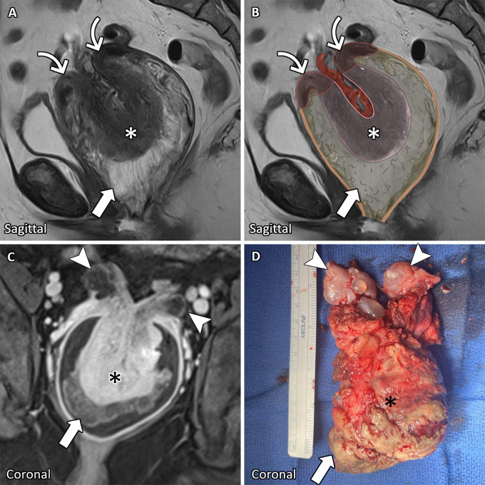
A 57-year-old postmenopausal woman presented with acute-on-chronic vaginal bleeding. Pelvic examination revealed a vaginal mass extending from the cervix to the introitus. Pelvic MRI revealed uterine inversion secondary to an endometrial mass (Figure). Results from radical laparoscopic hysterectomy, bilateral salpingo-oophorectomy, and bilateral lymph node dissection confirmed the MRI findings. Histopathologic findings demonstrated high-grade uterine adenosarcoma.
A 57-year-old postmenopausal woman with uterine inversion from adenosarcoma. (A) Sagittal T2-weighted MR image with (B) illustrated overlay, (C) coronal T1-weighted postcontrast MR image, and (D) photograph of corresponding gross pathologic findings show uterine inversion with transcervical (curved arrows) myometrial (asterisks) protrusion into the vagina. The inverted endometrium and associated frondlike enhancing mass (arrows) are located between the myometrium (asterisks) and vaginal wall (orange line in B). Uterine inversion tethers the ovaries (arrowheads) at the level of the cervix.
Uterine inversion, a rare entity typically associated with childbirth, is characterized by uterine fundal inversion and descent through the cervix. Nonpuerperal uterine inversion is often associated with benign leiomyomas, but uterine malignancies occur in one-third of patients (1,2). The inverted uterine fundus within the vagina may cause clinical and radiologic diagnostic confusion and may be mistaken for a cervical or vaginal mass (2).
Surgical management is the primary treatment. Preoperative imaging guides the surgical type and approach (2). US and CT imaging may aid the diagnosis by showing a uterine U-shaped configuration (2). MRI is the modality of choice for accurately delineating the inverted uterine anatomy and characterizing the underlying cause (2). A U-shaped uterine cavity with a reversed intravaginal uterine fundus can be observed on sagittal and coronal planes, while axial images may show a bull’s-eye appearance demonstrating telescoping of the uterus into the vagina (2,3). Additionally, MRI may help identify the underlying mass and reveal associated pelvic lymphadenopathy. Radiologist awareness and recognition of uterine inversion is critical for correct diagnosis, which guides successful surgical management.
Footnotes
Authors declared no funding for this work.
Disclosures of conflicts of interest: B.V. No relevant relationships. A.B. No relevant relationships. A.M. Grants or contracts, NCI, DOD; consulting fees, Axena Healthcare. T.P. Payment through institution, General Electric, American Roentgen Ray Society, Society of Abdominal Radiology, National Institutes of Health, Food and Drug Administration, Massachusetts Institute of Technology Lincoln Laboratory, Massachusetts General Hospital; royalties, edited article on contrast agent reactions, Wolters Kluwer; consultant, AutonomUS Medical Technologies; honorarium for invited talk, Zhejiang Medical Association; patents, Massachusetts General Hospital, Systems and Methods for Portable Ultrasound Guided Cannulation (pending), Systems and Methods for Guided Intervention (pending), Systems and Methods for Supervised Remote Ultrasound-Guided Intervention (submitted), Automated Conditionally Increased Acoustic Output Algorithm (planned); stock or stock options, AutonomUS Medical Technologies.
Keywords: MR Imaging, Pelvis, Uterus, Uterine Inversion
References
- 1. Witt C , Vranes C , Clark LH . Uterine adenosarcoma presenting as uterine inversion: A case study . Gynecol Oncol Rep 2024. ; 53 : 101398 . [DOI] [PMC free article] [PubMed] [Google Scholar]
- 2. Herath RP , Patabendige M , Rashid M , Wijesinghe PS . Nonpuerperal Uterine Inversion: What the Gynaecologists Need to Know? Obstet Gynecol Int 2020. ; 2020 : 8625186 . [DOI] [PMC free article] [PubMed] [Google Scholar]
- 3. Leconte I , Thierry C , Bongiorno A , Luyckx M , Fellah L . Non-Puerperal Uterine Inversion . J Belg Soc Radiol 2016. ; 100 ( 1 ): 47 . [DOI] [PMC free article] [PubMed] [Google Scholar]


