Abstract
Cervical intradural meningioma are rare central nervous system neoplasms. Surgical resection is the primary treatment due to the tumor’s benign nature and clear demarcation from the spinal cord, although the posterior surgical approach can result in complications such as neurological deficits and cerebrospinal fluid leaks. We present a case of a 78-year-old woman with progressive clumsiness, gait disturbance, and weakness. She was diagnosed with an intradural extramedullary meningioma at the C2 to C3 level through magnetic resonance imaging. The tumor was excised using a cervical biportal endoscopic spine surgery approach, a minimally invasive technique that utilizes 2 small portals for endoscope and instrument access. The procedure, performed under general anesthesia, involved a hemilaminectomy and partial laminectomy to access and remove the tumor. Postoperative assessments indicated significant neurological recovery, with the patient regaining independent mobility and fine motor skills. Follow-up magnetic resonance images at 18 months confirmed the absence of tumor recurrence. This case demonstrates the efficacy of cervical biportal endoscopic spine surgery in managing high cervical intradural tumors, highlighting its potential for reducing surgical complications and promoting rapid patient recovery.
Keywords: spinal meningioma, minimally invasive, endoscopic surgery, intradural tumor, cervical spine
Introduction
Spinal and spinal cord neoplasms are classified into extradural and intradural categories.1 Intradural tumors are further delineated into intramedullary and extramedullary types. Among intradural extramedullary (IDEM) neoplasms, the most prevalent are schwannomas, neurofibromas, and meningiomas.
IDEM tumors are rare central nervous system neoplasms, occurring in only 0.3 per 100,000 individuals annually.2 More than half of these tumors are situated in the thoracic spine, with comparable incidence rates observed in the cervical and lumbosacral spine regions at 22% and 18%, respectively. Histopathological analyses reveal that schwannomas constitute 23% to 48%, meningiomas 9.6% to 35%, neurofibromas 4% to 23%, and metastatic tumors 6.4% to 25% of all cases.3
The majority of IDEM tumors are benign and are typically managed through aggressive surgical resection due to their clear demarcation from the spinal cord.4 Nevertheless, complications frequently linked with the posterior approach, a commonly chosen method for spinal cord tumor removal, encompass neurological deficits (0.18%–2.6%),5 malpositioning of fixation screws (4%–7%),6 vertebral artery injury (1.3%–4%),7 and cerebrospinal fluid leaks (1%).8
Cervical cord meningiomas are benign tumors arising from the protective membranes of the spinal cord in the neck region. They account for about 14% to 27% of all spinal meningiomas, which themselves represent 25% to 46% of primary spinal cord tumors.9 These tumors are more common in adults, particularly in their 50s and 60s, and show a strong female predominance with a female-to-male ratio of 3:1 to 4:1.10 While relatively rare compared with intracranial meningiomas, cervical meningiomas can cause significant problems due to spinal cord compression despite their typically slow growth.11 The exact cause is unknown, but factors, such as genetics, hormones, and prior radiation exposure may increase risk.12
Cervical biportal endoscopic spine surgery (BESS) is a minimally invasive surgical approach employed for treating diverse cervical spinal conditions. This technique involves utilizing 2 small portals or incisions to access the posterior cervical spine—1 for the endoscope and the other for surgical instruments. Here, we present a case detailing the application of cervical BESS for the resection of an IDEM tumor located at a high cervical level.
Case Presentation
A 78-year-old woman presented to our hospital with a 3-week history of progressive clumsiness, tingling in both hands, gait disturbance, and weakness in both lower extremities. Physical examination indicated normal motor strength in all limbs, with a Medical Research Council grade of good, but hyperactive deep tendon reflexes were noted in both knees and ankles. A radiographic evaluation of the cervical spine revealed signs of degenerative spondylosis. Magnetic resonance images (MRI) disclosed an 8.7 × 15.7 × 7.5 mm oval mass, determined to be an IDEM tumor, at the C2 to C3 level. It was characterized by isointensity on T2-weighted imaging (Figure 1A) and displayed heterogeneous gadolinium enhancement on T1-weighted images (Figure 1C). The tumor was situated anterolaterally within the dura mater, and the adjacent spinal cord was compressed and deviated on the contrast-enhanced T2-weighted image (Figure 1B) and T1-weighted image (Figure 1D).
Figure 1.
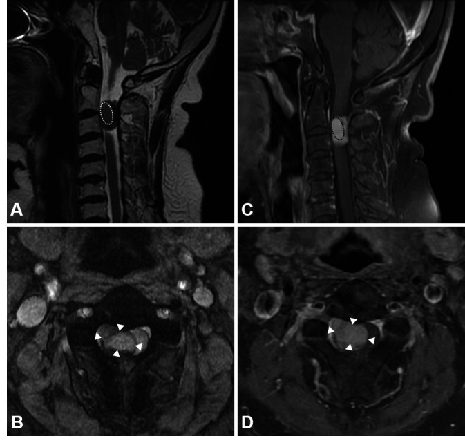
The intradural extramedullary mass at the level of C2–C3 vertebrae. An oval, well-demarcated, isointensity mass (dotted line) was positioned anteriorly in the central canal on the sagittal T2-weighted image (A). The mass had a heterogeneous gadolinium enhancement on the sagittal T1-weighted image (C). The contrast-enhanced mass (white arrowhead) was positioned in the dura mater and deviated from the adjacent spinal cord on the axial images (T2-weighted image on B and T1-weighted images on D).
The IDEM tumor at the C2 to C3 level was excised using a biportal endoscopic approach. Under general anesthesia, the patient was positioned prone. The head was supported with a contoured foam pillow designed to maintain the chin and forehead while allowing access to the eyes, nose, and airways. A transverse skin incision approximately 1 to 1.5 cm long was made on the right side of the midline of the C2 spinous process. A second incision, positioned 2 to 3 cm inferior to the first, was also created. These served as the proximal portal for visualization and the distal portal for manipulation. Access to the C2 to C3 interlaminar space was achieved by carefully using a burr to perform a right-sided C2 hemilaminectomy. Subsequently, a Kerrison punch was employed to partially remove the upper portion of the right C3 lamina. Throughout the procedure, instruments were directed toward the midline and kept within the endoscopic field of view. Removal of the ligamentum flavum exposed the underlying dura mater. A third portal was established mediocaudally to the distal portal to adjust the endoscopic view at this stage (Figure 2). A longitudinal dural incision was meticulously performed using a sharp probe while maintaining the irrigation system pressure at approximately 30 mmHg. Following the excision of the intradural mass, the dura mater was carefully sutured with 7–0 nylon sutures (Figure 3). The irrigation pump was then deactivated, and a thorough inspection for hemostasis was conducted prior to withdrawing the instruments from the 2 portals. Once hemostasis was confirmed, the subcutaneous fascia and skin were closed with sutures. Surgical instruments and any residual irrigation saline were subsequently removed. The histopathological examination confirmed the tumor to be a meningioma (Figure 4).
Figure 2.
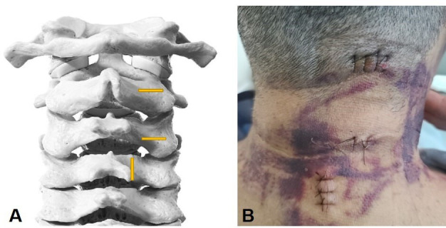
(A) Schematic illustration. A cranial portal was made on the right side of the midline of the C2 spinous process. A caudal portal, positioned 2–3 cm inferior to the first, was also created. A third portal was established mediocaudally to the caudal portal. (B) Sutured state of the portals after surgery.
Figure 3.
(A) Endoscopic view of performing a longitudinal incision (dotted line) following dura mater exposure (sharp probe, arrow). (B) A substantial grayish-white mass (white arrowhead) was being excised through the incised dural margins, with the microforceps positioned inferiorly. (C) Gross appearance of the excised tissues.
Figure 4.
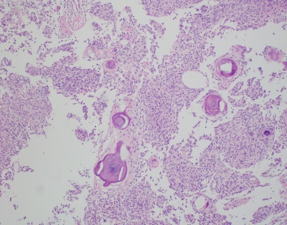
Photomicrograph of the resected tumor. The tumor cells exhibited lobules and whorl formation of spindle-shaped cells. Psammoma bodies were also visible (hematoxylin and eosin staining, ×100 magnification).
A series of postoperative neurological assessments were conducted, focusing on early detection of complications. At 12 hours postsurgery, the patient was allowed to sit up while wearing a soft cervical collar, showing no balance issues. By 24 hours postsurgery, the patient began walking with minimal assistance using a walker. Two days after the procedure, a follow-up MRI and computed tomographic images confirmed substantial removal of the mass (Figure 5). The patient was discharged upon confirming mobility and the absence of surgical complications 7 days postsurgery. Two months after discharge, the patient achieved independent walking, normal fine motor skills in the hands, and self-sufficiency in daily activities. At 18-month follow-up, MRI revealed a clear view of the spinal cord without residual mass, absence of tumor recurrence, and sufficient canal space (Figure 6).
Figure 5.
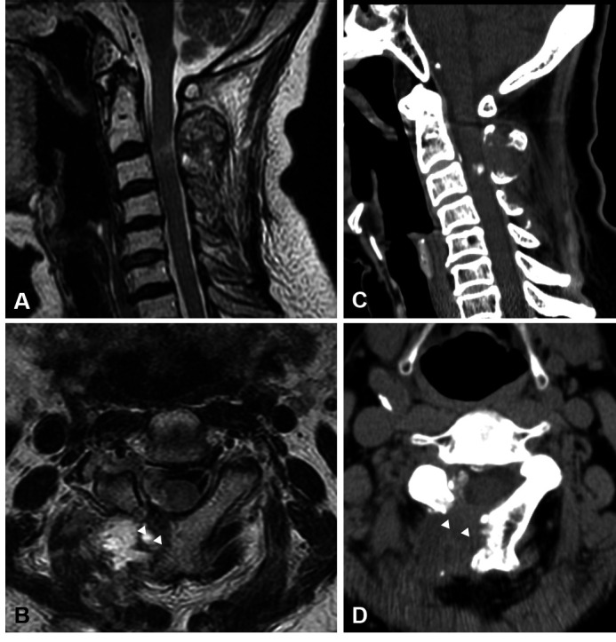
Postoperative magnetic resonance imaging (sagittal T1-weighted image on A and axial T2-weighted image on B) and computed tomographic scans (sagittal view on C and axial view on D) confirmed that the mass was nearly completely removed, and the right-sided laminectomy (white arrowhead) was clearly visible.
Figure 6.
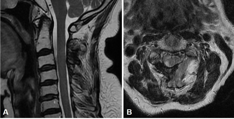
Magnetic resonance imaging 18 months after surgery (sagittal view on A and axial view on B) demonstrated a clear visualization of the spinal cord without evidence of residual mass, absence of tumor recurrence, and adequate central canal space.
Discussion
IDEM tumors encompass a diverse range of neoplastic lesions, varying from benign to malignant entities, but they are relatively uncommon.13 MRI serves as the optimal imaging modality for their evaluation, enabling precise localization that aids in narrowing down diagnostic considerations to specific entities. Among these tumors, meningiomas, arising from arachnoidal cap cells, predominantly occur intracranially, with approximately 10% adherent to the spinal dura, and have an age-adjusted incidence of 0.33 per 100,000, most commonly affecting women in their seventh to eighth decades.14 Around 80% of spinal meningiomas originate in the thoracic region, while the cervical region accounts for the second most common site at approximately 15%.15 Spinal meningiomas are mostly found intradurally and extramedullary on MRI, appearing well-defined with a broad dural attachment and often showing a dural tail sign. They typically appear isointense on T1 and T2 images and enhance uniformly with contrast. Histologically, spinal meningiomas exhibit a lobulated structure with whorls and psammoma bodies, and they commonly stain positive for vimentin.1 Most are classified as World Health Organization grade 1 tumors, with the meningothelial subtype being the most frequently encountered in the spine. World Health Organization grade II tumors, particularly of the clear cell subtype, tend to originate from the denticulate ligaments and are more prevalent in spinal locations.16
The preferred treatment for spinal meningiomas is surgical removal, with adjuvant radiotherapy considered for higher-grade or recurrent tumors. In a retrospective analysis of 22 consecutive patients diagnosed with cervical spinal meningioma,17 the mean duration of preoperative symptoms was 11.2 months, and the most common axial location was ventrolateral (54.5%) followed by dorsolateral (18.2%). In this study, the functional outcome improvement rate after surgery was 86.4%, with significant enhancements observed in Japanese Orthopaedic Association score and the Neck Disability Index.17 The overall recurrence rate in this series was 9.1%. Other studies have also reported that 71.3% to 92% of patients showed improvement in functional status following surgery.11,18–21
The application of cervical BESS for treating cervical spine disorders has been a recent area of study.22,23 This technique offers several advantages, including minimal tissue damage during the approach to pathological sites, enhanced visualization of the microenvironment, and improved maneuverability of surgical instruments.22 The prognosis associated with combining BESS and percutaneous fixation for removing an extradural aneurysmal bone cyst at the L1 level has been notably favorable.24 There are documented cases where BESS facilitated successful resection of spinal canal tumors. For instance, BESS was successfully employed in a 60-year-old woman to remove a T10 IDEM tumor, resulting in significant symptomatic relief, spinal cord decompression, and confirmed pathological diagnosis.25 Additional cases have reported successful removal of an extradural hemangioma at the upper thoracic level and an epidural cyst at the lower thoracic level using BESS, without complications.26
There have been occasional reports of incorporating a third portal to optimize the portal design in cervical BESS. One documented case involved planning a multilevel approach to remove a diffuse hematoma in the lumbar epidural space, employing multiple portals longitudinally and achieving successful removal.27 Another reported instance detailed the establishment of a third portal opposite existing portals to access and decompress severe central stenosis.28 In this scenario, the surgeon (S.B.J.) strategically positioned a third portal caudally and medially to the original distal portal, using it as a working portal for complete resection while minimizing the laminectomy required for instrument access. We recommend careful preoperative planning to assess the necessity of constructing a third portal and conducting a comprehensive cost-benefit analysis to optimize surgical outcomes.
BESS presents numerous advantages for the resection of spinal cord tumors, characterized by reduced iatrogenic trauma to adjacent tissues, enhanced visualization through high-definition magnification, and minimal skin incisions. These factors collectively contribute to diminished postoperative pain, abbreviated hospital stays, and expedited patient recovery. Additionally, BESS is associated with a reduced risk of perioperative complications compared with conventional open surgery. This technique facilitates superior access to intricate spinal anatomy and preserves structural integrity. The minimally invasive nature of BESS also yields favorable cosmetic outcomes, with minimal scarring and prompt patient mobilization, establishing it as an efficacious modality for spinal cord tumor resection.
It is crucial to acknowledge that performing cervical BESS for IDEM tumors in the upper cervical spine requires a substantial learning curve. Surgeons need proficiency in endoscopic posterior approaches specific to the cervical spine. While BESS has shown promising outcomes and favorable attributes, particularly in cases such as schwannomas within our series, its current application should be limited to selected cases. Additionally, further research is needed to determine the optimal location and extent of dural excision for the removal of intradural tumors. Further accumulation of case reports is essential to establish its broader applicability.
References
- 1. Kumar N, Tan WLB, Wei W, Vellayappan BA. An overview of the tumors affecting the spine-inside to out. Neurooncol Pract. 2020;7(Suppl 1):i10–i17. 10.1093/nop/npaa049 [DOI] [PMC free article] [PubMed] [Google Scholar]
- 2. Nittner K, Vinken P, Bruyn G. Spinal meningiomas, neuromas and neurofibromas and hourglass tumors. Handbook of Clinical Neurology. North Holland/America, New York: Elsevier; 1976. [Google Scholar]
- 3. Cheng MK. Spinal cord tumors in the People’s Republic of China: a statistical review. Neurosurg. 1982;10(1):22–24. 10.1227/00006123-198201000-00004 [DOI] [PubMed] [Google Scholar]
- 4. Cohen AR, Wisoff JH, Allen JC, Epstein F. Malignant astrocytomas of the spinal cord. J Neurosurg. 1989;70(1):50–54. 10.3171/jns.1989.70.1.0050 [DOI] [PubMed] [Google Scholar]
- 5. Hojo Y, Ito M, Abumi K, et al. A late neurological complication following posterior correction surgery of severe cervical kyphosis. Eur Spine J. 2011;20(6):890–898. 10.1007/s00586-010-1590-8 [DOI] [PMC free article] [PubMed] [Google Scholar]
- 6. Geck MJ, Macagno A, Ponte A, Shufflebarger HL. The ponte procedure: posterior only treatment of Scheuermann’s kyphosis using segmental posterior shortening and pedicle screw instrumentation. J Spinal Disord Tech. 2007;20(8):586–593. 10.1097/BSD.0b013e31803d3b16 [DOI] [PubMed] [Google Scholar]
- 7. Neo M, Fujibayashi S, Miyata M, Takemoto M, Nakamura T. Vertebral artery injury during cervical spine surgery: a survey of more than 5600 operations. Spine. 2008;33(7):779–785. 10.1097/BRS.0b013e31816957a7 [DOI] [PubMed] [Google Scholar]
- 8. Hannallah D, Lee J, Khan M, Donaldson WF, Kang JD. Cerebrospinal fluid leaks following cervical spine surgery. J Bone Joint Surg Am. 2008;90(5):1101–1105. 10.2106/JBJS.F.01114 [DOI] [PubMed] [Google Scholar]
- 9. Peker S, Cerçi A, Ozgen S, Isik N, Kalelioglu M, Pamir MN. Spinal meningiomas: evaluation of 41 patients. J Neurosurg Sci. 2005;49(1):7–11. [PubMed] [Google Scholar]
- 10. Westwick HJ, Shamji MF. Effects of sex on the incidence and prognosis of spinal meningiomas: a surveillance, epidemiology, and end results study. J Neurosurg Spine. 2015;23(3):368–373. 10.3171/2014.12.SPINE14974 [DOI] [PubMed] [Google Scholar]
- 11. Setzer M, Vatter H, Marquardt G, Seifert V, Vrionis FD. Management of spinal meningiomas: surgical results and a review of the literature. Neurosurg Focus. 2007;23(4). 10.3171/FOC-07/10/E14 [DOI] [PubMed] [Google Scholar]
- 12. Wiemels J, Wrensch M, Claus EB. Epidemiology and etiology of meningioma. J Neurooncol. 2010;99(3):307–314. 10.1007/s11060-010-0386-3 [DOI] [PMC free article] [PubMed] [Google Scholar]
- 13. Beall DP, Googe DJ, Emery RL, et al. Extramedullary intradural spinal tumors: a pictorial review. Curr Probl Diagn Radiol. 2007;36(5):185–198. 10.1067/j.cpradiol.2006.12.002 [DOI] [PubMed] [Google Scholar]
- 14. Kshettry VR, Hsieh JK, Ostrom QT, Kruchko C, Benzel EC, Barnholtz-Sloan JS. Descriptive epidemiology of spinal meningiomas in the United States. Spine. 2015;40(15):E886–E889. 10.1097/BRS.0000000000000974 [DOI] [PubMed] [Google Scholar]
- 15. Osborn AG. Diagnostic neuroradiology. Elsevier Health; 1994. [Google Scholar]
- 16. Abul-Kasim K, Thurnher MM, McKeever P, Sundgren PC. Intradural spinal tumors: current classification and MRI features. Neuroradiology. 2008;50(4):301–314. 10.1007/s00234-007-0345-7 [DOI] [PubMed] [Google Scholar]
- 17. Lee Y-J, Jung J, Hyun S-J, Kim K-J, Jahng T-A, Kim H-J. The clinical and radiological outcome of cervical spinal meningioma. Nerve. 2018;4(2):37–41. 10.21129/nerve.2018.4.2.37 [DOI] [Google Scholar]
- 18. Gezen F, Kahraman S, Canakci Z, Bedük A. Review of 36 cases of spinal cord meningioma. Spine. 2000;25(6):727–731. 10.1097/00007632-200003150-00013 [DOI] [PubMed] [Google Scholar]
- 19. Gottfried ON, Gluf W, Quinones-Hinojosa A, Kan P, Schmidt MH. Spinal meningiomas: surgical management and outcome. Neurosurg Focus. 2003;14(6). 10.3171/foc.2003.14.6.2 [DOI] [PubMed] [Google Scholar]
- 20. Maiti TK, Bir SC, Patra DP, Kalakoti P, Guthikonda B, Nanda A. Spinal meningiomas: clinicoradiological factors predicting recurrence and functional outcome. Neurosurg Focus. 2016;41(2). 10.3171/2016.5.FOCUS16163 [DOI] [PubMed] [Google Scholar]
- 21. Yoon SH, Chung CK, Jahng TA. Surgical outcome of spinal canal meningiomas. J Korean Neurosurg Soc. 2007;42(4):300–304. 10.3340/jkns.2007.42.4.300 [DOI] [PMC free article] [PubMed] [Google Scholar]
- 22. Jung SB, Kim N. Biportal endoscopic spine surgery for cervical disk herniation: a technical notes and preliminary report. Medicine. 2022;101(27). 10.1097/MD.0000000000029751 [DOI] [PMC free article] [PubMed] [Google Scholar]
- 23. Kim J, Heo DH, Lee DC, Chung HT. Biportal endoscopic unilateral laminotomy with bilateral decompression for the treatment of cervical spondylotic myelopathy. Acta Neurochir. 2021;163(9):2537–2543. 10.1007/s00701-021-04921-0 [DOI] [PubMed] [Google Scholar]
- 24. Kim SK, Bendardaf R, Ali M, Kim HA, Heo EJ, Lee SC. Unilateral biportal endoscopic tumor removal and percutaneous stabilization for extradural tumors: technical case report and literature review. Front Surg. 2022;9. 10.3389/fsurg.2022.863931 [DOI] [PMC free article] [PubMed] [Google Scholar]
- 25. Peng W, Zhuang Y, Cui W, et al. Unilateral biportal endoscopy for the resection of thoracic intradural extramedullary tumors: technique case report and literature review. Int Med Case Rep J. 2024;17:301–309. 10.2147/IMCRJ.S444226 [DOI] [PMC free article] [PubMed] [Google Scholar]
- 26. Wang T, Yu H, Zhao S-B, et al. Complete removal of intraspinal extradural mass with unilateral biportal endoscopy. Front Surg. 2022;9. 10.3389/fsurg.2022.1033856 [DOI] [PMC free article] [PubMed] [Google Scholar]
- 27. Kim N, Jung SB. Biportal endoscopic spine surgery in the treatment of multi-level spontaneous lumbar epidural hematoma: case report. J Orthop Sci. 2022;27(1):288–291. 10.1016/j.jos.2019.03.010 [DOI] [PubMed] [Google Scholar]
- 28. Zhu C, Cheng W, Wang D, Pan H, Zhang W. A helpful third portal for unilateral biportal endoscopic decompression in patients with cervical spondylotic myelopathy: a technical note. World Neurosurg. 2022;161:75–81. 10.1016/j.wneu.2022.02.021 [DOI] [PubMed] [Google Scholar]


