Case Presentation
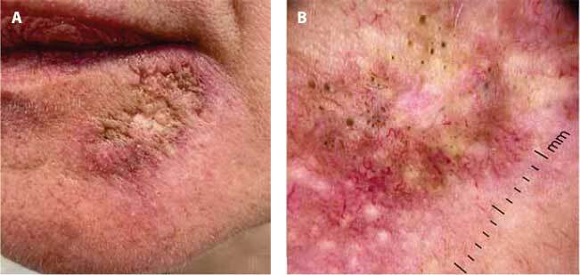
A 52-year-old woman presented with a 3-year history of a scarring erythematous lesion on her left chin. Initially diagnosed with folliculitis, she underwent various topical treatments and systemic isotretinoin, without improvement. On reevaluation, a hypotrophic scarring plaque was noted (Figure 1A). Dermoscopy revealed multiple comedones with diffuse arborizing telangiectasia, partially scarred areas, and white dots (Figure 1B). Histopathologic examination demonstrated a lichenoid inflammation and interface dermatitis with mucin and CD-123 positive cells, leading to a diagnosis of comedogenic lupus erythematosus, a variant of chronic cutaneous lupus erythematosus.
Figure 1.

(A) Hypotrophic scarring plaque on the left chin. (B) Dermoscopic features, including comedones with diffuse arborizing telangiectasia, partially scarred areas, and white dots.
Teaching Point
Comedogenic lupus erythematosus (CLE) is a rare variant of chronic cutaneous lupus erythematosus (CCLE) often initially misdiagnosed as other folliculocentric dermatoses. This initial misdiagnosis can lead to delayed intervention, resulting in disease progression and disfigurement. Dermoscopic examination plays an important role in distinguishing CLE from other conditions, revealing characteristic findings such as comedones, telangiectasia, and scarring. Histologically, CLE is characterized by the identification of lichenoid inflammation with mucin deposition and CD-123 positive cells, aiding in the accurate diagnosis of CLE.
Timely recognition of CLE is paramount to prevent long-term sequelae. Effective management of CLE remains challenging. Photoprotection is extremely important, as in all other variants. Consideration could be given to initiating treatment with topical retinoids or steroids; however, in many cases, systemic treatment with hydroxychloroquine is often required and considered the first line of treatment [1, 2]. Furthermore, understanding the clinical, dermoscopic, and histological features of CLE is essential for dermatologists to provide optimal care.
Footnotes
Funding: None.
Competing Interests: None.
Authorship: All authors have contributed significantly to this publication.
References
- 1.Haroon TS, Fleming KA. An unusual presentation of discoid lupus erythematosus. Br J Dermatol. 1972;87(6):642–645. doi: 10.1111/j.1365-2133.1972.tb07456.x. [DOI] [PubMed] [Google Scholar]
- 2.Hemmati I, Otberg N, Martinka M, Alzolibani A, Restrepo I, Shapiro J. Discoid lupus erythematosus presenting with cysts, comedones, and cicatricial alopecia on the scalp. J Am Acad Dermatol. 2009;60(6):1070–1072. doi: 10.1016/j.jaad.2008.11.882. [DOI] [PubMed] [Google Scholar]


