Abstract
The canal of Nuck is an embryological remnant of the processus vaginalis found in females, and is a potential site for endometriosis seeding. Endometriosis in the canal of Nuck is an exceedingly rare condition. Patients with this condition present with groin swelling or suprapubic pain. We describe a case of a 44-year-old female presenting for left inguinal pain and a lump. Physical examination revealed a 3 cm reducible mass; magnetic resonance imaging (MRI) revealed a 6.2 × 3.0 × 1.7 cm multiloculated cystic structure, extending from the left pelvic region into the left inguinal region near the round ligament. The patient underwent surgical excision of the mass. Pathology revealed a multi-cystic lesion lined with mesothelium cells and a fibrous wall with multiple foci of misplaced endometrial glands and stroma, corroborating a diagnosis of endometriosis in the canal of Nuck.
LEARNING POINTS
Endometriosis of the canal of Nuck is an exceedingly rare entity with an uncertain pathophysiology due to paucity of data available in the medical literature.
Physicians should keep Nuck’s canal endometriosis in their differential diagnosis when approaching a patient with chronic pelvic pain.
Keywords: Endometriosis, canal of Nuck, inguinal pain
INTRODUCTION
The canal of Nuck is the result of a patent processus vaginalis in the inguinal canal of females. The canal of Nuck was first described by Anton Nuck in 1961[1]. A myriad of structures can be found in the canal of Nuck such as cysts, hydroceles and hernias. Endometriosis is the presence of endometrial glands and stroma outside the uterus. Endometriosis can be found in the vulva, vagina, cervix, inguinal canal, perineum, urinary system, gastrointestinal tract, pulmonary system, skin and central nervous system[2]. Endometriosis of the canal of Nuck is exceedingly rare and its pathophysiology remains not fully understood, due to paucity of data in the medical literature. Patients usually present with groin swelling and suprapubic or pelvic pain. Here, we describe a case of a 44-year-old female seeking medical care for left inguinal pain and lump, who was found to have endometriosis of the canal of Nuck. Although rare, physicians should be aware of endometriosis of the canal of Nuck when approaching a patient with groin swelling and suprapubic pain.
CASE DESCRIPTION
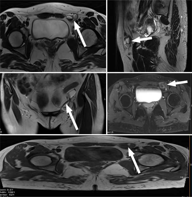
A 44-year-old female, previously healthy, sought medical care for left-sided inguinal pain and a palpable lump of a one-year duration. Her first pain episode was felt while she was exercising at the gym, and it was accompanied by a lump at the groin area. The patient denies any history of lower urinary tract infection. The pain was aggravated by menstruation; her bowel movement was normal. The pain worsened gradually over the year as her lump was gradually increasing in size. Physical examination revealed a 3 cm reducible mass in the left inguinal area with tenderness upon palpation. MRI was performed for an accurate characterisation of the mass, which revealed a 6.2 × 3.0 × 1.7 cm multiloculated cystic structure (Fig. 1). This extended from the left pelvic region into the left inguinal that was medial to the left common femoral vein abutting the round ligament, corroborating a diagnosis of Nuck’s canal endometriosis. Surgery was chosen to excise the mass. Intraoperatively, the mass was soft-to-rubbery and exhibiting a brownish colour. Pathology revealed a multi-cystic lesion lined with mesothelial cells and a fibrous wall with multiple foci of misplaced endometrial glands and stroma. A diagnosis of endometriosis of the canal of Nuck was made. The patient’s post-operative course was uneventful, and she was discharged home after full recovery was achieved; her symptoms had abated.
Figure 1.
Multiplanar and multisequential MRI of the pelvis with IV gadolinium showing a 6.2 × 3.0 × 1.7 cm multiloculated cystic structure (white arrow) extending from the left pelvic region into the left inguinal and exhibiting fluid-fluid levels in its inguinal region representing haemorrhagic content. Findings corroborated a diagnosis of endometriosis of the canal of Nuck.
DISCUSSION
Embryologically, the inguinal canal is comprised of the gubernaculum and the processus vaginalis. The gubernaculum ultimately becomes the round ligament connecting the uterus and labia majora by passing through the inguinal canal. Conversely, the processus vaginalis obliterates before birth. If it remains, the processus vaginalis becomes a patent pouch of peritoneum lining the inguinal canal. The canal of Nuck is located anterior to the round ligament and may extend into the labia majora.
The pathophysiology of extra-pelvic endometriosis is not fully understood. Several theories have been posited to explain the mechanism of extra-pelvic endometriosis: retrograde menstruation, vascular or lymphatic spread and coelomic metaplasia[1]. When endometrial cells adhere to the round ligament they form an inguinal endometriosis, which is an exceedingly rare entity.
Chronic pelvic pain has many aetiologies, such as hernias and hydroceles. The canal of Nuck is rarely considered the culprit behind chronic pelvic pain. Our patient presented with a one-year history of left inguinal pain with a groin bulge that was gradually increasing in size. Physical examination revealed a 3 cm mass that was reducible and tender to palpation. MRI, which has a sensitivity of 90% and a specificity of 98% for pelvic endometriosis[3] was employed for a detailed characterisation of the mass. Although pelvic endometriosis was entertained among our differentials, a soft tissue sarcoma could not be excluded. Thus, surgery was performed as it is both diagnostic and therapeutic.
In summary, this article highlights a case of endometriosis with an unusual location and undiagnostic imaging modalities. Thus, the diagnosis of endometriosis of the canal of Nuck poses a great challenge for physicians when confronted by a case of chronic pelvic pain of unexplained aetiology. This article also underscores that endometriosis of the canal of Nuck should be included in the differential diagnosis when approaching a patient with a palpable mass in the subcutaneous tissue of the pelvis.
CONCLUSION
In conclusion, endometriosis of the canal of Nuck is an exceedingly rare entity with an uncertain pathophysiology, due to paucity of data available in the medical literature. Physicians should keep Nuck’s canal endometriosis in their differential diagnosis when approaching a patient with chronic pelvic pain. With a meticulous history-taking, adequate physical examination, and proper imaging and histologic findings, a diagnosis of endometriosis of the canal of Nuck would not go undiagnosed.
Footnotes
Conflicts of Interests: The Authors declare that there are no competing interests.
Patient Consent: Written signed informed consent was obtained from the patient prior to writing the manuscript.
REFERENCES
- 1.Swatesutipun V, Srikuea K, Wakhanrittee J, Thamwongskul C. Endometriosis in the canal of Nuck presenting with suprapubic pain: a case report and literature review. Urol Case Rep. 2020;34:101497. doi: 10.1016/j.eucr.2020.101497. [DOI] [PMC free article] [PubMed] [Google Scholar]
- 2.Cervini P, Wu L, Shenker R, O’Blenes C, Mahoney J. Endometriosis in the canal of Nuck: atypical manifestations in an unusual location. Can J Plast Surg. 2004;12:73–75. doi: 10.1177/229255030401200202. [DOI] [PMC free article] [PubMed] [Google Scholar]
- 3.Prodromidou A, Pandraklakis A, Rodolakis A, Thomakos N. Endometriosis of the canal of Nuck: a systematic review of the literature. Diagnostics (Basel) 2020;11:3. doi: 10.3390/diagnostics11010003. [DOI] [PMC free article] [PubMed] [Google Scholar]


