Abstract
Objective This study aims to compare the proximal femoral bone density changes in follow-up X-ray imaging and the proximal filling ratios of stems between anatomical and double-tapered wedge stem designs.
Methods Patients aged between 18 and 80 years who received primary total hip arthroplasty using both types of stems between 2017 and 2019 and had follow-up tests for up to a year were included in the study. Canal filling ratios at 3 levels (lesser trochanter [LT], 2 cm above LT, and 7 cm below LT) using the optimal densitometry method. Femoral bone density changes were measured using the Gruen zoning method.
Results A total of 92 patients (76% female and 24% male) met the inclusion criteria for this study. The mean age was 53.86 ± 13.00 years. The canal filling ratio in the double-tapered wedge group (Accolade II) was significantly higher than that in the anatomical stem group (ABGII) ( p < 0.001, p < 0.001, and p = 0.013) for all levels of measurement. No significant difference was observed between both types of stems in femoral bone density changes in zones 1 and 4. However, there were significant differences in femoral bone change, with bone loss being higher in the anatomical stem group in zone 7 (−25% versus −17%; p = 0.010).
Conclusion Double-tapered wedge stem had a significantly higher canal filling ratio than the anatomical stem at all levels but had less femoral bone density loss in the follow-up postoperative imaging in zone 7. Furthermore, in zones 1 and 4, there was no significant difference in femoral bone density loss.
Keywords: bone remodeling, femur, hip prosthesis, prosthesis design
Introduction
Total hip arthroplasty is one of the most common orthopedic surgical procedures. As the number of surgeries increased, the stem design became one of the most important factors affecting overall prosthesis longevity and patient satisfaction. One of the stems that provide good results and long-term outcomes is the cementless one, invented in 1950. 1 Several studies have reported early loosening and instability associated with the initial design, which may be caused by proximal femoral osteopenia resulting from the stress shielding effect. 2 Many modern stems have been developed by promoting proximal engagement, using hydroxyapatite porous coating, which is more compatible with the patient proximal femoral dense bone. By utilizing taper and anatomical designs, they can decrease the distal stem engagement and employ shorter stems, which can reduce proximal bone loss by up to 14%. 3 Moreover, many studies have shown that the stem design revolution reduced stem subsidence, thigh pain, and loosening. 4 5 Nevertheless, no previous research has compared the progression of bone integration and proximal bone loss between a double-tapered wedge stems (Accolade II, Stryker, Portage, MI, USA) and anatomical stems (ABGII, Stryker). Therefore, this study compares proximal femoral filling differences between such stem designs, using immediate postoperative imaging and proximal bone loss utilizing follow-up X-ray. The results will provide a better choice of stem, decrease early complications, and increase satisfaction with the total hip replacement operation.
Materials and Methods
Study Design
This study is a retrospective descriptive-cohort study of immediate and postoperative follow-up imaging from total hip arthroplasty surgery performed from 2017 to 2019. The hospital's Ethics Committee (ID: 62133) approved the research protocol and waived the requirement for informed consent for this study. All patients' collected data and identifiers were made fully anonymous.
Samples
Inclusion Criteria
With permission from the radiology department of Rajavithi, patients aged between 18 and 80 years who received primary total hip arthroplasty using both types of stems between 2017 and 2019 and had follow-up imaging for up to a year were included in this study.
Exclusion Criteria
Patients who were under 18 years of age, received revision hip arthroplasty, had prior hip dysplasia, had any postoperative complication, and with follow-up imaging of less than 1 year were excluded.
Data Collection and Measurement
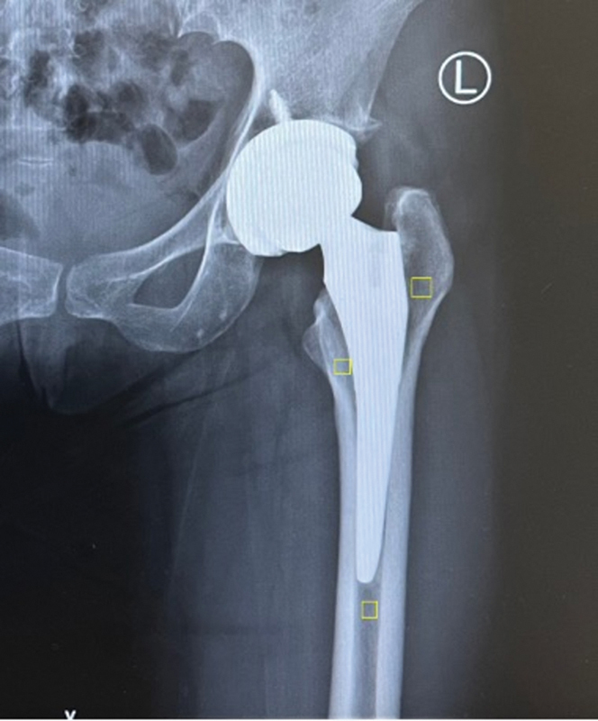
We obtained immediate and postoperative follow-up imaging from the radiology department for patients who underwent total hip arthroplasty, covering a period of up to 1 year. Data analysis was based on the femoral canal filling ratio method, used by the orthopedic surgeon responsible for adult hip and knee reconstruction. 6 The data included measurements of the proximal femoral and stem diameter in the anteroposterior view at three levels: lesser trochanter (LT), 2 cm proximal to the LT, and 6 cm distal to the LT ( Fig. 1 ).
Fig. 1.

Measurement of canal filling ratios at the lesser trochanter (LT), 2 cm proximal to LT, and 6 cm distal to LT.
The follow-up imaging was analyzed for proximal femoral bone density changes using the optimal densitometry method, 7 employing Image J (public domain), a digital optical image analysis software for windows, which measured bone changes in zones 1, 4, and 7 according to the Gruen fixation zone 8 ( Fig. 2 ).
Fig. 2.

Measurement of the proximal femoral bone density using optimal densitometry method.
Statical Analysis
Descriptive statistics (number, percentage, mean, median, standard deviation, as well as minimum and maximum values) were used to describe the characteristics of the samples. The Chi-squared test was employed to compare categorical data. The paired t test was utilized to compare independent data such as stem and femoral types. Furthermore, the t test was employed to compare dependent data such as postoperative imaging. The level of significance was defined as a p -value < 0.05. All statistical analyses were performed using the IBM SPSS Statistics for Windows (IBM Corp., Armonk, NY, USA) software, version 20.0.
Results
Demographics Data
A total sample of 92 patients (22 males and 70 females) was included in this study. The mean age was of 53.86 ± 13.00 years. There were 34 patients in the anatomical stem group (ABGII) and 58 patients in the double-tapered wedge stem group (Accolade II).
When comparing canal filling ratios between both stems, the canal filling ratio in the double-tapered wedge stem was significantly higher than that in the anatomical stem group at all 3 measurement levels ( p < 0.001, p < 0.001, and p = 0.013), as shown in Table 1 .
Table 1. Comparison of canal filling ratios between two types of stems.
| Level | Anatomical stem (n = 34) | Double-tapered wedge stem (n = 58) | Difference (95% confidence interval) | p -value |
|---|---|---|---|---|
| Lesser trochanter | 81.56 | 88.13 | −6.57 (−9.74 to −3.39) | < 0.001* |
| 2 cm above the lesser trochanter | 85.98 | 93.49 | −7.51 (−10.07 to −4.95) | < 0.001* |
| 6 cm below the lesser trochanter | 78.58 | 85.64 | −7.06 (−12.56 to −1.56) | 0.013* |
Note: *Statistical significant ( p -value <0.05).
Table 2 presents the femoral bone density changes in each stem type in Gruen zones 1, 4, and 7. Both stems showed a femoral proximal bone loss from the baseline to every time point.
Table 2. Femoral bone density changes in each stem type in Gruen zones 1, 4, and 7.
| Femur | Anatomical stem (n = 34) | Double-tapered wedge stem (n = 58) | ||||||
|---|---|---|---|---|---|---|---|---|
| Postoperative (baseline) | 6 months | 1 year | 2 years | Postoperative (baseline) | 6 months | 1 year | 2 years | |
|
Zone 1
FBD (%; mean ± SD) Change (%) |
(135.59 ± 12.20) |
(131.15 ± 10.53) −4.44 |
(117.35 ± 14.19) −13.79 |
(84.74 ± 7.29) −34.48 |
(130.33 ± 13.30) |
(122.05 ± 15.14) −8.28 |
(109.57 ± 12.09) −12.48 |
(91.63 ± 9.06) −22.37 |
| p -value | 0.024* | < 0.001* | < 0.001* | < 0.001* | < 0.001* | < 0.001* | ||
|
Zone 4
FBD (%; mean ± SD) Change (%) |
(164.03 ± 16.41) |
(158.29 ± 17.11) −5.74 |
(145.74 ± 16.92) −12.56 |
(135.39 ± 12.74) −13.65 |
(152.86 ± 29.44) |
(148.14 ± 24.19) −4.72 |
(138.64 ± 18.49) −9.50 |
(129.77 ± 11.34) −16.23 |
| p -value | 0.001* | < 0.001* | 0.004* | 0.098 | < 0.001* | < 0.001* | ||
|
Zone 7
FBD (%; mean ± SD) Change (%) |
(157.85 ± 13.84) |
(152.71 ± 11.31) −5.15 |
(132.06 ± 12.33) −20.65 |
(107.83 ± 19.79) −29.57 |
(152.29 ± 13.11) |
(144.29 ± 14.85) −8.00 |
(135.12 ± 14.89) −9.17 |
(126.87 ± 10.59) −11.13 |
| p -value | 0.006 | < 0.001* | < 0.001* | < 0.001* | < 0.001* | < 0.001* | ||
Abbreviations: FBD, femoral bone density; SD, standard deviation.
Note: *Statistical significant ( p -value <0.05).
-
Proximal bone density changes in anatomical stem
At 6-months postoperatively, there were significant differences in femoral bone loss in zones 1, 4, and 7 ( p = 0.024, p < 0.001, and p = 0.006, respectively); the highest femoral bone loss was observed in zone 4 (5.74%). At 1-year postoperatively, there was a significant difference in femoral bone loss in zones 1, 4, and 7 ( p < 0.001); the highest femoral bone loss was found in zone 7 (20.65%). At 2 years postoperatively, significant differences in femoral bone loss were observed in zones 1 ( p < 0.001), 4 ( p = 0.004), and 7 ( p < 0.001); the highest femoral bone loss was seen in zone 1 (34.48%).
-
Proximal bone density changes in double-tapered wedge stem
At 6-months postoperatively, there was a significant difference in femoral bone loss in zones 1, 4, and 7 ( p < 0.001); the highest femoral bone loss was observed in zone 1 (8.28%). At 1-year postoperatively, there was a significant difference in femoral bone loss in zones 1, 4, and 7 ( p < 0.001); the highest femoral bone loss was seen in zone 1 (12.48%). At 2-years postoperatively, a significant difference in the femoral bone loss was found in zones 1, 4, and 7 ( p < 0.001); the highest femoral bone loss was seen in zone 1 (22.37%).
Comparing proximal femoral bone loss between both designs, the double-tapered wedge stem demonstrated significantly less proximal femoral bone loss in the Gruen zone 7 ( Fig. 3 ). However, there was no significant difference in proximal femoral bone loss in zones 1 and 4, as shown in ( Figs. 4 5 ) and Table 3 .
Fig. 3.

Comparison of the proximal femoral bone density changes in the Gruen zone 7 of both stems (A, anatomical stem; B, double-tapered wedge stem).
Fig. 4.

Comparison of the proximal femoral bone density changes in the Gruen zone 1 of both stems (A, anatomical stem; B, double-tapered wedge stem).
Fig. 5.

Comparison of the proximal femoral bone density changes in the Gruen zone 4 of both stems (A, anatomical stem; B, double-tapered wedge stem).
Table 3. Comparison of femoral bone density changes in each zone in both types of stems.
| Femoral bone density | Stem type | p -value | |
|---|---|---|---|
| Anatomical stem (n = 34) | Double-tapered wedge stem (n = 58) | ||
| Zone 1 | |||
| Postoperative (baseline) | 135.59 ± 12.20 | 130.33 ± 13.30 | 0.062 |
| 6 months | 131.15 ± 10.53 | 122.05 ± 15.14 | 0.003* |
| 1 year | 117.35 ± 14.19 | 109.57 ± 12.09 | 0.006* |
| 2 years | 84.74 ± 7.29 | 91.63 ± 9.06 | 0.004* |
| Change (1-year postoperatively) | −18.24 ± 20.48 | −20.76 ± 9.36 | 0.501 |
| Zone 4 | |||
| Postoperative (baseline) | 164.03 ± 16.41 | 152.86 ± 29.44 | 0.022* |
| 6 months | 158.29 ± 17.11 | 148.14 ± 24.19 | 0.021* |
| 1 year | 145.74 ± 16.92 | 138.64 ± 18.49 | 0.070 |
| 2 years | 135.39 ± 12.74 | 129.77 ± 11.34 | 0.096 |
| Change (1-year postoperatively) | −18.29 ± 14.79 | −14.22 ± 24.22 | 0.320 |
| Zone 7 | |||
| Postoperative (baseline) | 157.85 ± 13.84 | 152.29 ± 13.11 | 0.058 |
| 6 months | 152.71 ± 11.31 | 144.29 ± 14.85 | 0.005* |
| 1 year | 132.06 ± 12.33 | 135.12 ± 14.89 | 0.314 |
| 2 years | 107.83 ± 19.79 | 126.87 ± 10.59 | < 0.001* |
| Change (1-year postoperatively) | −25.79 ± 15.85 | −17.17 ± 13.23 | 0.010* |
Note: *Statistical significant ( p -value <0.05).
Table 3 showed that only zone 7 had significant differences in femoral bone density changes between both stems ( p = 0.01).
Discussion
Cementless total hip arthroplasty is a popular procedure, particularly for younger patients, 9 with a good long-term outcome. However, it was reported to have proximal femoral osteopenia and early aseptic loosening in early designs 4 due to the stress shielding effect and proximal micromotion of the stem. Later, the stem was refined by improvement in surface and coating material, decrease in the material's stiffness, and variants of the femoral stem length all greatly improve the survival and lessens chance of complications in the procedure. 10
For canal filling of femoral canal, our study found the canal filling ratio in the double-tapered wedge stem was significantly higher than that in the anatomical stem group at all levels (LT, 2 cm above LT, and 6 cm below LT). It is worth noting that the higher femoral canal filling and canal filling ratio observed in our study may increase the risk of failure of osteointegration, as suggested by the study by Cooper et al., 11 who noted that an increase in the mid and distal filling, as well as and canal-flare index, are the most important risk factors for osteointegration failure.
According to our study on changes in periprosthetic bone density, the immediate postoperative bone mineral density on the operated side should be used as the baseline value to exclude bone loss due to the operation procedure. 12 Despite this method, our study found bone density loss in both stems from the baseline, which we attribute to stress-shielding in the area. This finding is consistent with the Venesmaa et al. 13 study, which reported a general decrease in all regions of interest until 6 months, particularly in the Calcar region, and only minor changes after this time period. However, our study observed a decrease in bone density up to 1 year postoperatively. We found that in zone 7, the anatomical stem had a significantly higher femoral bone density loss than that the double-tapered wedge stem (−25% versus −17%, p = 0.010). Nevertheless, no significant femoral bone density loss was observed in zones 1 and 4.
The main strength of this study is that it was conducted by a single surgeon in a single center, which minimized confounding factors from surgical technique and postoperative patient care. However, this study has limitations. First, its retrospective nature, which limited data collection on all factors that may have influenced bone loss in our patient population. Additionally, the sample size was relatively small, which prevented us from conducting a meaningful subgroup analysis to investigate the impact of various factors on bone loss. Second, the follow-up period was short (12 months), although we believe it was adequate, as periprosthetic bone density loss was most pronounced in the first postoperative year, with minimal changes thereafter. This finding is consistent with previous studies that highlighted the initial periprosthetic bone remodeling process occurring within the first 12 postoperative months. 14
Conclusion
Double-tapered wedge stem design had a significantly higher canal filling ratio than the anatomical stem at all levels, with a lower femoral bone density loss identified in the follow-up postoperative imaging at zone 7. However, in zones 1 and 4, no significant difference in femoral bone density loss was observed.
Funding Statement
Suporte Financeiro Os autores declaram que não receberam suporte financeiro de agências dos setores público, privado ou sem fins lucrativos para a realização deste estudo.
Financial Support The authors declare that they have received no financial support from agencies in the public, private, or nonprofit sectors to conduct the present study.
Conflito de Interesses Os autores não têm conflito de interesses a declarar.
Contribuições dos Autores
TK: Conceituação, metodologia, validação, coleta de dados, experimento-piloto, redação do manuscrito original e redação – revisão e edição. PP: análise formal e redação – revisão e edição. Todos os autores leram e concordaram com a versão publicada do manuscrito.
Trabalho desenvolvido no Departamento de Ortopedia, Faculdade de Medicina, Rajavithi Hospital, Bangcoc, Tailândia.
Author Contributions
TK: Conceptualization, methodology, validation, data collection, pilot experiment, writing of the original draft, and writing—review and editing. PP: formal analysis and writing—review and editing. All authors have read and agreed to the published version of the manuscript.
Work carried out at the Department of Orthopedics, Faculty of Medicine, Rajavithi Hospital, Bangkok, Thailand.
Supplementary Material
Referências
- 1.van der Wal B C, de Kramer B J, Grimm B, Vencken W, Heyligers I C, Tonino A J. Femoral fit in ABG-II hip stems, influence on clinical outcome and bone remodeling: a radiographic study. Arch Orthop Trauma Surg. 2008;128(10):1065–1072. doi: 10.1007/s00402-007-0537-y. [DOI] [PubMed] [Google Scholar]
- 2.Herrera A, Canales V, Anderson J, García-Araujo C, Murcia-Mazón A, Tonino A J. Seven to 10 years followup of an anatomic hip prosthesis: an international study. Clin Orthop Relat Res. 2004;(423):129–137. doi: 10.1097/01.blo.0000128973.73132.0b. [DOI] [PubMed] [Google Scholar]
- 3.Panisello J J, Canales V, Herrero L, Herrera A, Mateo J, Caballero M J. Changes in periprosthetic bone remodelling after redesigning an anatomic cementless stem. Int Orthop. 2009;33(02):373–379. doi: 10.1007/s00264-007-0501-z. [DOI] [PMC free article] [PubMed] [Google Scholar]
- 4.de Boer F A, Sariali E. Comparison of anatomic vs. straight femoral stem design in total hip replacement - femoral canal fill in vivo. Hip Int. 2017;27(03):241–244. doi: 10.5301/hipint.5000439. [DOI] [PubMed] [Google Scholar]
- 5.Laine H J, Puolakka T J, Moilanen T, Pajamäki K J, Wirta J, Lehto M U. The effects of cementless femoral stem shape and proximal surface texture on ‘fit-and-fill’ characteristics and on bone remodeling. Int Orthop. 2000;24(04):184–190. doi: 10.1007/s002640000150. [DOI] [PMC free article] [PubMed] [Google Scholar]
- 6.Umer M, Sepah Y J, Khan A, Wazir A, Ahmed M, Jawad M U. Morphology of the proximal femur in a Pakistani population. J Orthop Surg (Hong Kong) 2010;18(03):279–281. doi: 10.1177/230949901001800304. [DOI] [PubMed] [Google Scholar]
- 7.Hernandez-Vaquero D, Garcia-Sandoval M A, Fernandez-Carreira J M, Suarez-Vázquez A, Perez-Hernández D. Measurement of bone mineral density is possible with standard radiographs: a study involving total knee replacement. Acta Orthop. 2005;76(06):791–795. doi: 10.1080/17453670510045381. [DOI] [PubMed] [Google Scholar]
- 8.Gruen T A, McNeice G M, Amstutz H C. “Modes of failure” of cemented stem-type femoral components: a radiographic analysis of loosening. Clin Orthop Relat Res. 1979;(141):17–27. [PubMed] [Google Scholar]
- 9.Stea S, Comfort T, Sedrakyan Aet al. Multinational comprehensive evaluation of the fixation method used in hip replacement: interaction with age in context J Bone Joint Surg Am 201496(Suppl 1, Suppl 1)42–51. [DOI] [PMC free article] [PubMed] [Google Scholar]
- 10.Khanuja H S, Vakil J J, Goddard M S, Mont M A. Cementless femoral fixation in total hip arthroplasty. J Bone Joint Surg Am. 2011;93(05):500–509. doi: 10.2106/JBJS.J.00774. [DOI] [PubMed] [Google Scholar]
- 11.Cooper H J, Jacob A P, Rodriguez J A.Distal fixation of proximally coated tapered stems may predispose to a failure of osteointegration J Arthroplasty 201126(6, Suppl)78–83. [DOI] [PubMed] [Google Scholar]
- 12.Inaba Y, Kobayashi N, Oba M, Ike H, Kubota S, Saito T. Difference in postoperative Periprosthetic bone mineral density changes between 3 major designs of Uncemented stems: a 3-year follow-up study. J Arthroplasty. 2016;31(08):1836–1841. doi: 10.1016/j.arth.2016.02.009. [DOI] [PubMed] [Google Scholar]
- 13.Venesmaa P K, Kröger H P, Miettinen H J, Jurvelin J S, Suomalainen O T, Alhava E M. Monitoring of periprosthetic BMD after uncemented total hip arthroplasty with dual-energy X-ray absorptiometry–a 3-year follow-up study. J Bone Miner Res. 2001;16(06):1056–1061. doi: 10.1359/jbmr.2001.16.6.1056. [DOI] [PubMed] [Google Scholar]
- 14.Christiansen J D, Laursen M B, Ejaz A, Nielsen P T. Bone remodelling of the proximal femur after total hip arthroplasty with 2 different hip implant designs: 15 years follow-up of the thrust plate prosthesis and the Bi-Metric stem. Hip Int. 2018;28(06):606–612. doi: 10.1177/1120700018755371. [DOI] [PubMed] [Google Scholar]