Abstract
Background
Streptococcus mutans and Lactobacilli play an important role in the etiopathogenesis and progression of dental caries (DC). Their quantification and identification may be helpful for epidemiological and early intervention measures.
Objectives
We conducted the study to evaluate the colony counts of S. mutans and Lactobacillus with the location of DC and correlate their prevalence with the age of the patient.
Materials and methods
The study population comprised 60 patients with DC. They were divided into two groups according to age, and each group was further divided into three subgroups based on involvement of enamel, dentin, and pulp by DC. The swab samples were collected, and organisms were isolated using Mitis Salivarius Bacitracin (MSB) Agar and Lactobacillus MRS Agar. Manual counting of colonies on plates illuminated by transmitted light was done. Results were summarized and analyzed statistically.
Results
The caries prevalence was found to be higher in children, with females being more affected. In both groups, posterior teeth were more affected, and occlusal/incisal surface caries were more common. The mean colony count of S. mutans (61.3%) and Lactobacillus (63.4%) was significantly higher in group I compared to group II. In both groups, the mean colony counts of S. mutans were higher in enamel, followed by dentin and pulp. In contrast, in both groups, the mean colony counts of Lactobacillus were higher in pulp, followed by dentin and enamel.
Conclusion
Bacterial colony counts may help in taking specific measures against specific organisms and thereby prevent the development of new carious lesions.
How to cite this article
Tandon A, Srivastava A, Singh P, et al. Beyond Decay: Exploring the Age-associated Variations in Streptococcus mutans and Lactobacillus in Dental Caries. Int J Clin Pediatr Dent 2024;17(9):993–998.
Keywords: Colony count, Dental caries, Lactobacillus, Streptococcus mutans
Introduction
Dental caries (DC) is a chronic infection caused by normal oral microbial flora.1 Imbalance between pathological factors leading to the demineralization of tooth tissue and protective factors causing remineralization leads to DC.2Streptococcus mutans, the major cariogenic organism,3 along with Lactobacilli, is implicated as important contributory bacteria in tooth decay and progression of the disease.4
Quantification and identification of S. mutans and Lactobacillus may be helpful for epidemiological and early intervention measures. We conducted the study to evaluate the colony count of S. mutans and Lactobacillus with the location of DC and to correlate their prevalence with the age of the patient.
Materials and Methods
The study population comprised 60 patients divided into two groups. Group I comprised 30 patients aged between 5 and 15, and Group II comprised 30 patients aged 16 years and above. Further, the 30 patients were divided into three subgroups in both Group I and Group II, and the salivary samples were taken accordingly.
Group I (a) and Group II (a) comprised patients affected with caries involving only the enamel.
Group I (b) and Group II (b) comprised patients affected with caries extending to the dentin.
Group I (c) and Group II (c) comprised patients affected with caries extending to the pulp.
The informed consents were obtained after the patients and their parents were informed of the study and the related procedures. Approval from the Institutional Ethics Committee had been obtained prior to the study.
The diagnosis of caries was done clinically by means of visual and tactile methods and by use of intraoral periapical radiographs. Patients having signs and symptoms of DC were included in the study, and those with evidence of periapical infections, periodontal infections, systemic diseases, and those undergoing fluoride treatment were excluded from the study.
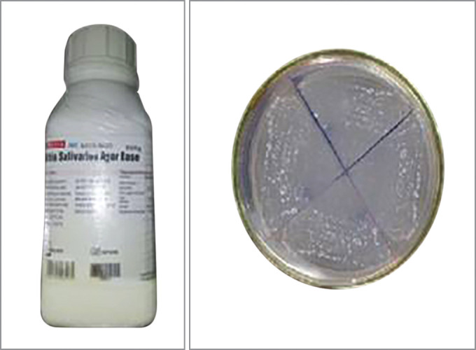
Mitis Salivarius Bacitracin (MSB) Agar Base (HiMedia Laboratories, Mumbai) was used for S. mutans. Mitis salivarius agar is used for the isolation of streptococci (Fig. 1), especially Streptococcus mitis, Streptococcus salivarius, and Enterococcus faecalis from grossly contaminated specimens. It was modified by adding 0.2 units/mL bacitracin to make it a selective medium for S. mutans. Around 90.07 gm of agar powder was suspended in 1000 mL of distilled water. This mixture was heated until boiling to dissolve the medium completely. Autoclaving at 15 lbs pressure (121°C) for 15 minutes was done for sterilization. Then the solution was cooled to 50–55°C, and 1 mL of sterile 1% potassium tellurite solution was added. The mixture was mixed well and poured into sterile Petri plates for bacterial culture.
Fig. 1.

Mitis salivarius agar and colonies of S. mutans on agar plate
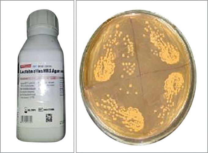
Lactobacillus MRS Agar (HiMedia Laboratories, Mumbai) was used for the cultivation of all Lactobacillus species (Fig. 2). Around 67.15 gm of agar powder was suspended in 1000 mL of distilled water. The mixture was heated until boiling to dissolve the medium completely. Sterilization was done by autoclaving at 15 lbs pressure (121°C) for 15 minutes. Finally, the contents were mixed well and poured into sterile Petri plates for bacterial culture.
Fig. 2.

Lactobacillus MRS Agar and colonies of Lactobacillus on agar plate
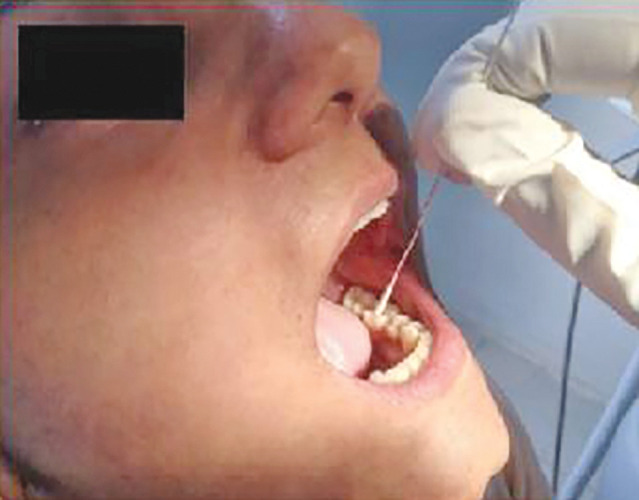
Salivary samples, along with caries debris, were collected under sterile conditions from patients having signs and symptoms of DC using sterile cotton swabs (Fig. 3). The swabs were inserted at carious sites, kept for 1–2 minutes, taken out, and placed in a sterile vial containing 1 mL saline for transportation. A loopful (10 µL) of the saliva along with caries debris was streaked on different isolation media. The inoculated plates were incubated at their respective temperatures, that is, S. mutans for 48 hours at 37°C and Lactobacillus for 24 hours at 37°C. After bacterial cultivation, the bacterial count of S. mutans and Lactobacillus was done in colony forming units (CFUs) and recorded as CFU/mL × 103 for each sample. Manual counting of colonies on plates illuminated by transmitted light was done.
Fig. 3.

Collection of saliva along with caries debris through sterilized aluminum swab stick
The results were tabulated and subjected to appropriate statistical analysis. Data were summarized as Mean ± standard error of the mean (SE). Groups were compared by independent Student's t-test. Groups were also compared by two-way analysis of variance (ANOVA), and the significance of mean difference within and between the groups was determined by Tukey's post hoc test after ascertaining normality by the Shapiro–Wilk test and homogeneity of variances by Levene's test. Categorical groups were compared by Chi-squared (χ2) test. A two-tailed (α = 0.05) p-value < 0.05 was considered statistically significant.
Results
The demographic characteristics of the study population are summarized in Table 1.
Table 1.
Demographic characteristics of two groups
| Demographic characteristics | Group I (n = 30) (%) | Group II (n = 30) (%) |
|---|---|---|
| Age (years) | ||
| Mean ± standard error of the mean (SE) | 10.10 ± 0.45 | 30.30 ± 1.78 |
| Sex | ||
| Male | 13 (43%) | 11 (36%) |
| Female | 17 (57%) | 19 (64%) |
| Teeth involved | ||
| Posterior (%) | 18 (60%) | 20 (67%) |
| Anterior (%) | 12 (40%) | 10 (33%) |
| Surface | ||
| Occlusal/incisal | 18 (60%) | 16 (53%) |
| Smooth surface | 12 (40%) | 14 (47%) |
Bacterial Colony Count
The bacterial colony counts of S. mutans, Lactobacillus, and total (S. mutans + Lactobacillus) according to groups (group I and group II) are summarized in Table 2.
Table 2.
Bacterial colony count (mean ± SE) of two groups
| Bacterial colony count (CFU) | Group I (n = 30) | Group II (n = 30) | t-value | p-value |
|---|---|---|---|---|
| S. mutans | 32180 ± 7309 | 12453 ± 4920 | 2.24 | 0.029 |
| Lactobacillus | 22517 ± 6062 | 8240 ± 3458 | 2.05 | 0.045 |
| Total | 54697 ± 6731 | 20693 ± 5498 | 3.91 | <0.001 |
The bacterial colony counts of S. mutans, Lactobacillus, and total (S. mutans + Lactobacillus) according to groups (group I and group II) and tooth involved (posterior and anterior) are summarized in Table 3. DC incidence was found to be higher in posterior teeth in both group I and group II. The mean colony counts of S. mutans were higher in posterior than anterior teeth and higher in group I than group II, while those of Lactobacillus were higher in anterior than posterior teeth and higher in group I than group II.
Table 3.
Bacterial colony counts according to groups and tooth involved
| Group I | Group II | |||||
|---|---|---|---|---|---|---|
| Bacterial colony count (CFU) | Tooth involved | N | Mean ± SE | N | Mean ± SE | p-value |
| S. mutans | Posterior | 18 | 32856 ± 9771 | 20 | 17375 ± 7167 | 0.999 |
| Anterior | 12 | 31167 ± 11431 | 10 | 2610 ± 986 | 0.685 | |
| p-value | – | 0.513 | – | 0.223 | – | |
| Lactobacillus | Posterior | 18 | 20433 ± 8012 | 20 | 5330 ± 2011 | 0.956 |
| Anterior | 12 | 25642 ± 9589 | 10 | 14060 ± 9636 | 0.842 | |
| p-value | – | 0.331 | – | 0.755 | – | |
| Total | Posterior | 18 | 53289 ± 9538 | 20 | 22705 ± 6879 | 0.993 |
| Anterior | 12 | 56808 ± 9329 | 10 | 16670 ± 9473 | 0.968 | |
| p-value | – | 0.039 | – | 0.040 | ||
The bacterial colony counts of S. mutans, Lactobacillus, and total (S. mutans + Lactobacillus) according to groups (group I and group II) and tooth surface (occlusal/incisal and smooth surface) are summarized in Table 4.
Table 4.
Bacterial colony counts according to groups and tooth surface
| Group I | Group II | |||||
|---|---|---|---|---|---|---|
| Bacterial colony count (CFU) | Tooth surface | N | Mean ± SE | N | Mean ± SE | p-value |
| S. mutans | Occlusal/incisal | 18 | 31394 ± 9194 | 16 | 10381 ± 6066 | 0.999 |
| Smooth surface | 12 | 33358 ± 12489 | 14 | 14821 ± 8148 | 0.985 | |
| p-value | – | 0.302 | – | 0.530 | – | |
| Lactobacillus | Occlusal/incisal | 18 | 19533 ± 6839 | 16 | 11637± 6386 | 0.883 |
| Smooth surface | 12 | 26992 ± 11435 | 14 | 4357 ± 1096 | 0.885 | |
| p-value | – | 0.834 | – | 0.162 | – | |
| Total | Occlusal/incisal | 18 | 50928 ± 8273 | 16 | 22019 ± 7984 | 0.880 |
| Smooth surface | 12 | 60350 ± 11625 | 14 | 19179 ± 7755 | 0.996 | |
| p-value | – | 0.076 | – | 0.017 | ||
Dental caries was found to be more on the occlusal/incisal surface in both group I and group II. The mean colony counts of S. mutans were higher on the smooth surface than on the occlusal/incisal surface and higher in group I than group II. In contrast, the mean colony counts of Lactobacillus were higher on the smooth surface than on the occlusal/incisal surface in group I. However, in group II, the mean colony counts of Lactobacillus were higher on the occlusal/incisal surface than on the smooth surface.
The bacterial colony counts of S. mutans, Lactobacillus, and total (S. mutans + Lactobacillus) according to groups (group I and group II) and tooth site (enamel, dentin, and pulp) are summarized in Table 5.
Table 5.
Bacterial colony counts according to groups and tooth site
| Group I | Group II | |||||
|---|---|---|---|---|---|---|
| Bacterial colony count (CFU) | Tooth site | N | Mean ± SE | N | Mean ± SE | p-value |
| S. mutans | Enamel | 10 | 79500 ± 9788 | 10 | 32500 ± 12829 | <0.001 |
| Dentin | 10 | 15600 ± 6002 | 10 | 4100 ± 1345 | 0.857 | |
| Pulp | 10 | 1440 ± 521 | 10 | 760 ± 88 | 1.000 | |
| Lactobacillus | Enamel | 10 | 1140 ± 442 | 10 | 2000 ± 996 | 1.000 |
| Dentin | 10 | 3410 ± 1234 | 10 | 4020 ± 1382 | 1.000 | |
| Pulp | 10 | 63000 ± 8950 | 10 | 18700 ± 9707 | <0.001 | |
| Total | Enamel | 10 | 80640 ± 9654 | 10 | 34500 ± 12528 | 0.006 |
| Dentin | 10 | 19010 ± 6118 | 10 | 8120 ± 1778 | 0.951 | |
| Pulp | 10 | 64440 ± 8973 | 10 | 19460 ± 9656 | 0.008 | |
For each group, the comparison (p-value) of the mean difference in bacterial colony count between the sites is shown in Table 6.
Table 6.
For each group, comparison (p-value) of mean difference in bacterial colony count between the sites by Tukey's test
| Comparisons | S. mutans | Lactobacillus | Total number of colonies | |||
|---|---|---|---|---|---|---|
| Group I | Group II | Group I | Group II | Group I | Group II | |
| Enamel vs dentin | <0.001 | 0.065 | 1.000 | 1.000 | <0.001 | 0.293 |
| Enamel vs pulp | <0.001 | 0.028 | <0.001 | 0.272 | 0.783 | 0.831 |
| Dentin vs pulp | 0.715 | 0.999 | <0.001 | 0.413 | 0.008 | 0.942 |
Discussion
The oral cavity harbors one of the most complex microbiomes in the body, and oral bacteria are important contributors to the occurrence and progression of DC. DC is a prevalent chronic disease.5 In the oral cavity, there is a biofilm (dental plaque) that comprises >800 species of microorganisms living in a complex community. It changes over time, and the microorganism population can shift between a healthy and pathological environment when factors such as sugar are enhanced.6 The first microorganisms to colonize are termed pioneer species, and collectively they make up the pioneer microbial community. In the mouth, the predominant pioneer organisms are streptococci, in particular S. mutans, S. salivarius, S. mitis, and S. oralis.7
S. mutans is able to metabolize glucose, fructose, sucrose, lactose, galactose, mannose, cellobiose, glucosides, trehalose, maltose, and a previously unrecognized group of sugar-alcohols. In the presence of extracellular glucose and sucrose, S. mutans synthesizes intracellular glycogen-like polysaccharides (IPSs). S. mutans also produces mutacins (bacteriocins), which are considered an important factor in the colonization and establishment of S. mutans in the dental biofilm.8 There is also a strong association between Lactobacillus spp. and caries.9Lactobacilli are isolated from deep caries lesions but rarely just before the development of DC and in early tooth decay. They are believed to be pioneering microorganisms in caries progression, especially in dentin. The level of Lactobacillus in saliva may be indirectly related to the progression of caries.10 Studies have shown that Lactobacilli are a dominant part of the flora inhabiting deep cavities, and their number correlates with the presence of carbohydrates.8
The prevalence of caries was found to be higher in children, as reported in other studies.11 The prevalence of caries was found to be higher in females in both groups (group I, 57%, and group II, 64%), as also reported in other studies.12,14 However, a few studies reported that there was no statistically significant difference in caries prevalence between the two sexes.15,17
While additional species may play a role in DC development, a considerable amount of research has established S. mutans as a primary cariogenic pathogen.18 It is noted that S. mutans and S. sobrinus, two species of the mutans streptococci, are the most significant in human caries, and studies of the microbial ecology of caries have been directed principally at these species.9Lactobacilli generally constitute a low proportion of the plaque microbiota. It has been suggested that S. mutans are the principal cariogenic pathogens, with Lactobacilli aiding in caries progression.19 In our study, the mean colony count of S. mutans was significantly higher (61.3%) in group I compared to group II. Similarly, the mean colony count of Lactobacillus was significantly higher (63.4%) in group I compared to group II.
In both groups, posterior teeth were found to be more affected with caries than anterior teeth in our study. Comparing the two groups, group II (67%) had a higher prevalence than group I (60%). Similar findings were reported in other studies as well.12,20,21 This could be due to the greater number of supplemental grooves present in posterior teeth, which act as sites for food accumulation and attraction of bacteria. Additionally, posterior teeth have a larger surface area for bacterial adhesion and multiplication, such as S. mutans and Lactobacillus. Correlating with the microorganisms, S. mutans were higher in posterior than anterior teeth. In contrast, the mean colony counts of Lactobacillus were higher in anterior than posterior teeth.
Dental caries was found to be more common on the occlusal/incisal surface in both group I and group II in our study. This finding has also been reported in other studies.22,24 This could be due to the greater number and deeper developmental grooves present on the occlusal surface, allowing for more bacterial colonization. Additionally, pooling of saliva in these grooves and food accumulation may lead to an environment more prone to bacterial contamination and multiplication, such as S. mutans and Lactobacillus.
Comparing the microorganisms, the mean colony counts of S. mutans were higher on the smooth surface than on the occlusal/incisal surface and higher in group I than group II. In contrast, the mean colony counts of Lactobacillus were higher on the smooth surface than on the occlusal/incisal surface in group I. However, in group II, the mean colony counts of Lactobacillus were higher on the occlusal/incisal surface than on the smooth surface. S. mutans has a central role in the etiology of DC because they can adhere to the enamel salivary pellicle and other plaque bacteria. Mutans streptococci and Lactobacilli are strong acid producers and, hence, create an acidic environment that increases the risk for cavities.25
In both groups, the mean colony counts of S. mutans were higher in enamel, followed by dentin, with pulp having the lowest counts. Additionally, in all three sites, the counts were higher in group I than group II. Comparing the mean colony counts of S. mutans within the groups (between tooth sites), the Tukey test revealed significantly lower colony counts in both dentin and pulp compared to enamel in both group I (p < 0.001) and group II (p < 0.05). Further, comparing the mean colony counts of S. mutans between the groups (group I vs group II), the Tukey's test revealed significantly lower colony counts at enamel in group II compared to group I (p < 0.001). However, the counts did not differ between the groups at dentin and pulp (p > 0.05), indicating that they were statistically the same.
In contrast, in both groups, the mean colony counts of Lactobacillus were higher in pulp, followed by dentin, with enamel having the lowest counts. Additionally, in all three sites, the counts were higher in group I than group II. Comparing the mean colony counts of Lactobacillus within the groups (between tooth sites), the Tukey's test revealed significantly higher colony counts in pulp compared to both enamel and dentin in group I (p < 0.001). In group II, however, the counts did not differ among the sites, indicating they were statistically the same. Further, comparing the mean colony counts of Lactobacillus between the groups (group I vs group II), the Tukey's test revealed significantly lower colony counts in group II compared to group I at pulp (p < 0.001). The counts were not different between the groups at both enamel and dentin (p > 0.05), indicating they were statistically the same.
The S. mutans group is more closely associated with DC in enamel, being primarily responsible for the initial phase of the lesion.26S. mutans from individuals with active caries were found to release significantly more calcium from hydroxyapatite than strains isolated from caries-free individuals.27 Oral streptococci may be associated with the development of “low pH-carious dentin.”28Lactobacilli are reported to be the most commonly isolated microorganisms in samples of carious dentin.29Lactobacillus is correlated with the progression of the caries process because it has a low capacity for adherence to the tooth surface.26 Bacterial invasion of dentinal tubules commonly occurs when dentin is exposed following a breach in the integrity of the overlying enamel or cementum. While several hundred bacterial species are known to inhabit the oral cavity, a relatively small and select group of bacteria is involved in the invasion of dentinal tubules and subsequent infection of the root canal space. Streptococci are among the most commonly identified bacteria that invade dentin. Recent evidence suggests that streptococci may recognize components present within dentinal tubules, such as collagen type I, which stimulate bacterial adhesion and intratubular growth. Specific interactions of other oral bacteria with invading streptococci may then facilitate the invasion of dentin by select bacterial groupings.30
The limitations of our study include sample size and counting errors during the colony counting procedure. Better results could be achieved with a larger sample size and by using more precise colony counting methods, such as automated colony counters, to reduce human error.
Conclusion
Caries can be reduced by increasing the acid resistance of teeth and controlling carbohydrate consumption in the diet. By manipulating adhesion interactions, it may be possible to develop new methods to block adhesive reactions, impeding the development of biofilm-related oral diseases such as DC. Bacterial colony counts may help in taking specific measures against specific organisms and thereby prevent the development of new carious lesions.
Orcid
Sonali Saha https://orcid.org/0000-0001-5361-1698
Bharadwaj Bordoloi https://orcid.org/0000-0002-4664-160X
Footnotes
Source of support: Nil
Conflict of interest: None
References
- 1.Mallya PS, Mallya S. Microbiology and clinical implications of dental caries – a review. J Evolution Med Dent Sci. 2020;9(48):3670–3675. doi: 10.14260/jemds/2020/805. [DOI] [Google Scholar]
- 2.Staszczyk M, Jamka-Kasprzyk M, Koscielniak D, et al. Effect of a short-term intervention with Lactobacillus salivarius probiotic on early childhood caries-an open label randomized controlled trial. Int J Environ Res Public Health. 2022;19:12447. doi: 10.3390/ijerph191912447. [DOI] [PMC free article] [PubMed] [Google Scholar]
- 3.Metwalli KH, Khan SA, Krom BP, et al. Streptococcus mutans, Candida albicans, and the human mouth: a sticky situation. PLoS Pathog. 2013;9(10):e1003616. doi: 10.1371/journal.ppat.1003616. [DOI] [PMC free article] [PubMed] [Google Scholar]
- 4.Tanzer JM, Livingston J, Thompson AM. The microbiology of primary dental caries in humans. J Dent Educ. 2001;65(10):1028–1037. [PubMed] [Google Scholar]
- 5.Zhang Y, Fang J, Yang J, et al. Streptococcus mutans-associated bacteria in dental plaque of severe early childhood caries. J Oral Microbiol. 2022;14:2046309. doi: 10.1080/20002297.2022.2046309. [DOI] [PMC free article] [PubMed] [Google Scholar]
- 6.Mitrakul K, Akarapipatkul B, Thammachat P. Quantitative analysis of Streptococcus mutans, Streptococcus sobrinus and Streptococcus sanguinis and their association with early childhood caries. J Clin Diagn Res. 2020;14(2):23–28. doi: 10.7860/JCDR/2020/43086.13513. [DOI] [Google Scholar]
- 7.Galuscan A, Jumanca D, Vacaru R, et al. Oral microbial flora and oral, maxillary and facial infections. OHDMBSC. 2004;3(1):27–30. [Google Scholar]
- 8.Karpinski TM, Szkaradkiewicz AK. Microbiology of dental caries. J Biol Earth Sci. 2013;3(1):21–24. [Google Scholar]
- 9.Bowden GHW. The microbial ecology of dental caries. Microb Ecol Health Dis. 2000;12:138–148. [Google Scholar]
- 10.Sounah SA, Madfa AA. Overview of method for detecting of Streptococcus mutans and Lactobacillus in saliva. J Dent Oral Disord. 2020;6(1):1–3. [Google Scholar]
- 11.Moses J, Rangeeth BN, Gurunathan D. Prevalence of dental caries, socio-economic status and treatment needs among 5 to 15 year old school going children of Chidambaram. J Clin Diag Res. 2011;5(1):146–151. [Google Scholar]
- 12.Bhardwaj VK. Dental caries prevalence in individual tooth in primary and permanent dentition among 6–12-yearold school children in Shimla, Himachal Pradesh. Int J Health Allied Sci. 2014;3:125–128. doi: 10.4103/2278-344X.132700. [DOI] [Google Scholar]
- 13.Lukacs JR, Largaespada LL. Explaining sex differences in dental caries prevalence: saliva, hormones, and “life-history” etiologies. Am J Hum Biol. 2006;18:540–555. doi: 10.1002/ajhb.20530. [DOI] [PubMed] [Google Scholar]
- 14.Munjal V, Gupta A, Kaur P, et al. Dental caries prevalence and treatment needs in 12 and 15-year-old school children of Ludhiana city. Indian J Oral Sci. 2013;4:27–30. doi: 10.4103/0976-6944.118523. [DOI] [Google Scholar]
- 15.Sudha P, Bhasin S, Anegundi RT. Prevalence of dental caries among 5–13-year-old children of Mangalore city. J Indian Soc Pedod Prev Dent. 2005;23:74–79. doi: 10.4103/0970-4388.16446. [DOI] [PubMed] [Google Scholar]
- 16.Grewal H, Verma M, Kumar A. Prevalence of dental caries and treatment needs amongst the school children in three educational zones of urban Delhi, India. Indian J Dent Res. 2011;22(4):517–519. doi: 10.4103/0970-9290.90283. [DOI] [PubMed] [Google Scholar]
- 17.Maru AM, Narendran S. Epidemiology of dental caries among adults in a rural area in India. J Contemp Dent Pract. 2012;13(3):382–388. doi: 10.5005/jp-journals-10024-1155. [DOI] [PubMed] [Google Scholar]
- 18.Dinis M, Traynor W, Agnello M, et al. Tooth-specific Streptococcus mutans distribution and associated microbiome. Microorganisms. 2022;10:1129. doi: 10.3390/microorganisms10061129. [DOI] [PMC free article] [PubMed] [Google Scholar]
- 19.Manchanda S, Sardana D, Liu P, et al. Horizontal transmission of Streptococcus mutans in children and its association with dental caries: a systematic review and meta-analysis. Pediatr Dent. 2021;43(1):1E–12E. [PubMed] [Google Scholar]
- 20.Batchelor PA, Sheiham A. Grouping of tooth surfaces by susceptibility to caries: a study in 5–16 year-old children. BMC Oral Health. 2004;4(2):2. doi: 10.1186/1472-6831-4-2. [DOI] [PMC free article] [PubMed] [Google Scholar]
- 21.Mohapatra SB, Pattnaik M, Ray P. Microbial association of dental caries. Asian J Exp Biol Sci. 2012;3(2):360–367. [Google Scholar]
- 22.Hannigan A, O'Mullane DM, Barry D, et al. A caries susceptibility classification of tooth surfaces by survival time. Caries Res. 2000;34(2):103–108. doi: 10.1159/000016576. [DOI] [PubMed] [Google Scholar]
- 23.Demirci M, Tuncer S, Yuceokur AA. Prevalence of caries on individual tooth surfaces and its distribution by age and gender in university clinic patients. Eur J Dent. 2010;4:270–279. [PMC free article] [PubMed] [Google Scholar]
- 24.Basha S, Swamy HS. Dental caries experience, tooth surface distribution and associated factors in 6- and 13-year-old school children from Davangere, India. J Clin Exp Dent. 2012;4(4):e210–e216. doi: 10.4317/jced.50779. [DOI] [PMC free article] [PubMed] [Google Scholar]
- 25.Forssten SD, Bjorklund M, Ouwehand AC. Streptococcus mutans, caries and simulation models. Nutrients. 2010;2:290–298. doi: 10.3390/nu2030290. [DOI] [PMC free article] [PubMed] [Google Scholar]
- 26.Junqueira JC, Borges AB, Barcellos DC, et al. Streptococcus mutans group and Lactobacillus counts in proximal amalgam and resin composite restorations: an in vivo study. Int J Contemp Dent. 2011;2(4):80–84. [Google Scholar]
- 27.Holbrook WP, Magnusdottir MO. Studies on strains of Streptococcus mutans isolated from caries-active and caries-free individuals in Iceland. J Oral Microbiol. 2012;4:1–5. doi: 10.3402/jom.v4i0.10611. [DOI] [PMC free article] [PubMed] [Google Scholar]
- 28.Maeda T, Kitasako Y, Senpuku H, et al. Role of oral streptococci in the pH-dependent carious dentin. Med Dent Sci. 2006;53:159–166. [Google Scholar]
- 29.Neelakantan P, Rao VKS, Indramohan J. Bacteriology of deep carious lesions underneath amalgam restorations with different pulp-capping materials – an in vivo analysis. J Appl Oral Sci. 2012;20(2):139–145. doi: 10.1590/s1678-77572012000200003. [DOI] [PMC free article] [PubMed] [Google Scholar]
- 30.Love RM, Jenkinson HF. Invasion of dentinal tubules by oral bacteria. Crit Rev Oral Biol Med. 2002;13(2):171–183. doi: 10.1177/154411130201300207. [DOI] [PubMed] [Google Scholar]


