According to an analysis of the latest GLOBOCAN data, pancreatic cancer accounted for the seventh-highest number of cancer-related deaths in China (1). Early clinical symptoms of pancreatic cancer typically include abdominal pain, loss of appetite, and weight loss. The disease progresses rapidly, and invasive pancreatic cancer often presents as a mass infiltrating the duodenal cavity, leading to symptoms of upper gastrointestinal obstruction (2,3). However, cases of gastrointestinal bleeding due to pancreatic cancer invading the duodenum are rare. Only 2.6% of patients with pancreatic cancer experience gastrointestinal bleeding as the initial symptom (4). Sharon et al. (5) reported that pancreatic cancer accounts for just 0.35% to 1.9% of upper gastrointestinal bleeding cases.
Treatment options for gastrointestinal bleeding caused by pancreatic cancer ulcers include endoscopic hemostasis (EH), transcatheter arterial embolization (TAE), and surgical procedures (6). EH is currently the first-line treatment. However, for comprehensive management of pancreatic cancer, whether through EH or arterial embolization, these are merely temporary measures for stabilization before definitive radical surgery (7). Emergency pancreaticoduodenectomy (EPD) can both control bleeding and remove the tumor (8). Notably, compared with selective pancreaticoduodenectomy (SPD), EPD tends to have a poorer prognosis and higher rates of postoperative complications. In a retrospective analysis of 931 cases of pancreaticoduodenectomy, the mortality rate for EPD was significantly higher (19.4% EPD vs. 3.2% SPD) (9).
However, when medical and interventional treatments for bleeding prove ineffective, EPD may serve as a last resort for saving the patient’s life. Here, we present a case of pancreatic cancer invading the duodenal wall with associated cancerous ulcer bleeding, treated by emergency pancreaticoduodenectomy combined with portal vein resection and reconstruction using a polytetrafluoroethylene (PTFE) graft. A review of relevant literature is also provided.
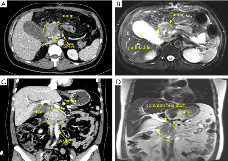
A 42-year-old man was admitted due to postoperation of acute upper gastrointestinal hemorrhage. He had repeated episodes of vomiting blood and underwent emergency surgery at a local hospital, which included suture hemostasis, distal subtotal gastrectomy, gastrojejunostomy (Billroth II anastomosis), and biopsy of a pancreatic head tumor. Pathological examination confirmed pancreatic adenocarcinoma. Physical examination revealed two abdominal drainage tubes. Peripheral blood tests revealed low hemoglobin (98 g/L), obstructive jaundice (total bilirubin 248 µmol/L, direct bilirubin 141.6 µmol/L), and elevated carbohydrate antigen 19-9 (CA19-9) (2,635.31 U/mL, normal range <37 U/mL). Pathogenetic examination identified Klebsiella in the drainage fluid. Enhanced computed tomography (CT) and magnetic resonance cholangiopancreatography (MRCP) revealed a mass (approximately 5.1 cm × 4.9 cm × 5.1 cm) in the pancreatic head encroaching on the wall of portal vein. The gallbladder, biliary ducts, and pancreatic duct were dilated, and the tumor’s blood supply involved the gastroduodenal artery (Figure 1). After antibiotic treatment and replacement of the drainage catheter, abdominal infection was well controlled. Cultures from the drainage were negative. On the 11th day of admission, the patient developed persistent upper abdominal distension, active bleeding from the abdominal drainage tube, and decreased hemoglobin levels, leading to a diagnosis of gastrointestinal hemorrhage and hemorrhagic shock. EPD was planned.
Figure 1.
Preoperative abdominal CT and MRI findings. (A) Transverse CT image showed a mass measuring approximately 5.1 cm × 4.9 cm × 5.1 cm invading the right wall of the portal vein. (B) Transverse MRI image. (C) Coronal CT image. (D) Coronal MRI image. SMV, superior mesenteric vein; SMA, superior mesenteric artery; PV, portal vein; CT, computed tomography; MRI, magnetic resonance imaging.
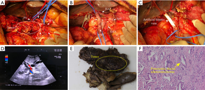
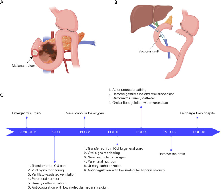
The patient was positioned supine for the laparotomy, and a midline incision was made. The abdominal cavity was explored, and peritoneal and liver metastases were excluded. After dissecting adherent intestinal segments, the omental mass was resected. Cholecystectomy was performed. Hepatic bile duct was divided and clamped. Portal vein and hepatic artery were carefully dissected and isolated. Superior mesenteric artery was dissected, and pancreatic neck was transected. A tumor in the head of the pancreas, approximately 5 cm in length, had encroached upon the portal vein (Figure 2A). An 8-mm PTFE graft with internal rings (Terumo, Camberley, UK) was used for portal vein reconstruction. Vascular clamps were applied to occlude the portal vein. The specimen was removed. Subsequently, the PTFE graft was sutured to the ends of the portal vein and superior mesenteric vein using a continuous 5-0 Prolene stitch (Figure 2B,2C). Portal vein was occluded for 33 minutes. After reconstruction, intraoperative ultrasound confirmed the patency of portal vein (Figure 2D). Pathological examination confirmed a moderately differentiated ductal adenocarcinoma (T3N1M1, Stage IV) (Figure 2E,2F). Pancreatic-jejunal anastomosis, biliary-enteric anastomosis, and entero-enteric anastomosis were performed sequentially (Figure 3A,3B). The operative time was 614 minutes, with intraoperative bleeding amounting to 1,700 mL. The patient recovered uneventfully and was discharged 16 days after surgery. The following figure presents the timeline of care events (Figure 3C).
Figure 2.
Intraoperative photos (A-D) and postoperative pathologic images (E,F). (A) Pancreatic mass invading the portal vein (white circle and white arrows). (B) Removal of the specimen (white dotted line). (C) Artificially revascularized portal vein (white arrows). (D) Intraoperative ultrasound confirmed the patency of portal vein. (E) Gross pathology. The yellow oval is the tumor, measuring about 5.1 cm × 4.9 cm × 5.1 cm. (F) HE pathologic staining picture (yellow arrow; magnification: ×100). PV, portal vein; HAP, hepatic artery proper; SpV, splenic vein; SMV, superior mesenteric vein; SMA, superior mesenteric artery; HE, hematoxylin and eosin.
Figure 3.
Schematic diagram of the operation. (A) Preoperative. (B) Postoperative. (C) The timeline of care events. POD, postoperative day; ICU, intensive care unit.
The PTFE graft showed good patency during the follow-up period (Figure 4). The patient underwent 8 cycles of chemotherapy (FOLFIRINOX regimen, folinic acid, fluorouracil, irinotecan, oxaliplatin) combined with immunotherapy (PD-1 monoclonal antibody, anti-programmed cell death-1). The patient passed away on August 7, 2021, due to multiple postoperative abdominal metastases and cachexia, with a postoperative survival time of 10 months.
Figure 4.
Postoperative evaluation of PTFE graft patency. (A) CT image at 3 months postoperatively. The PTFE graft was patency (yellow arrows). (B) Postoperative CT 3D reconstruction image (yellow arrows). PTFE, polytetrafluoroethylene; CT, computed tomography.
Previous studies have identified three main causes of bleeding associated with pancreatic cancer: gastric variceal bleeding secondary to splenic vein occlusion, bleeding from cancerous ulcers due to tumor invasion of the stomach or duodenum, and direct tumor bleeding via the pancreatic duct (10). Another mechanism involves the pancreas invading surrounding blood vessels and forming a fistula leading to the digestive tract (11,12). Ulcerative bleeding from malignant tumors of the gastrointestinal tract occurs when the tumor erodes the mucosal layer, leading to ulcer formation and subsequent damage or rupture of blood vessels within or near the ulcerated area. This condition typically results from a combination of tumor invasion, tissue ulceration, and vascular disruption, leading to gastrointestinal bleeding.
Currently, there are three methods for treating duodenal cancer ulcer bleeding: EH, TAE, and EPD. EH is generally preferred but has limitations, especially in cases where pancreatic tumors invade the duodenum, causing lumen stenosis or occlusion (13). TAE is feasible and effective for bleeding that cannot be controlled by EH but serves as only a temporary measure. EPD can achieve both hemostasis and tumor resection in cases of pancreaticoduodenal injury, perforation, and bleeding caused by tumor invasion. We have collected relevant case reports from PubMed and compiled them in Table 1. Comparison of the success rates across EH, TAE, and EPD revealed rates of 22.22%, 57.14%, and 87.50%, respectively. Given the limited clinical sample size, statistical validation is not feasible, and only large-scale cohort studies or randomized clinical trials can definitively establish EPD as the most effective approach. A literature search on PubMed did not identify any reported cases of EPD with portal vein reconstruction using PTFE grafts. These findings suggest that, in certain cases, EPD may be considered the precise surgical strategy for bleeding control.
Table 1. Summary of cases of gastrointestinal bleeding associated with pancreatic cancer collected by PubMed.
| No. | Author | Year | Bleeding site | Age (years) |
Gender | Hemostasis | Outcome | Survival time | Pathological |
|---|---|---|---|---|---|---|---|---|---|
| 1 | Ghaphery | 1966 | Aorta | 55 | Female | EPD | F | 0 hour | Unknown |
| 2 | Thorstad | 1987 | Superior mesenteric artery | 57 | Male | EH-TAE | F | 0 hour | PDAC |
| 3 | Mullan | 1990 | Gastric fundic vein | 63 | Female | EPD | S | 5 months | PDAC |
| 4 | Horiguchi | 1994 | Gastric fundic vein | 63 | Male | TAE | F | 21 days | Unknown |
| 5 | McDermott | 1995 | Gastric fundic vein | 46 | Male | TAE | S | 6 months | Unknown |
| 6 | Ferguson | 1997 | Gastric fundic vein | 71 | Male | EH-TAE | S | 18 weeks | Unknown |
| 7 | Lin | 2005 | Duodenal wall | 77 | Male | EH | S | 6 months | PDAC |
| 8 | Tomita | 2006 | Duodenal wall | 67 | Male | EH-EPD | S | 2 years | PDAC |
| 9 | Maeda | 2006 | Pancreaticoduodenal artery | 54 | Female | EH-TAE-PD | S | 37 days | SCA |
| 10 | Nikola Nikolov | 2007 | Vater | 43 | Female | EPD | S | Unknown | Unknown |
| 11 | Hidalgo | 2010 | Duodenal wall | 56 | Male | EH-PD | S | 3 months | Unknown |
| 12 | Ryoji Takada | 2014 | Duodenal wall | 43 | Female | Unprocessed | S | 7 months | Unknown |
| 13 | Higashiyama | 2015 | Gastroduodenal artery | 69 | Female | EH-TAE | S | 10 hours | Unknown |
| 14 | Yoshifumi Morita | 2018 | Superior mesenteric artery | 68 | Male | EH | S | 2 hours | Unknown |
| 15 | Zhe Jin | 2021 | Splenic artery | 49 | Male | EH-TAE | S | Unknown | Unknown |
| 16 | Yoshito Wada | 2022 | Duodenal wall | 77 | Female | EPD | S | 12 months | PDAC |
| 17 | Present case | 2023 | Duodenal wall | 42 | Male | EPD + PVR | S | 10 months | PDAC |
EPD, emergency pancreaticoduodenectomy; F, failure to stop bleeding; EH, endoscopic hemostasis; TAE, transcatheter arterial embolization; PDAC, pancreatic ductal adenocarcinoma; S, successful hemostasis; PD, pancreaticoduodenectomy; SCA, serous cystadenoma; PVR, portal vein reconstruction.
Portal vein reconstruction is common in pancreaticoduodenectomy with vascular resection, typically achieved through lateral venorrhaphy, primary end-to-end anastomosis, or the interposition of autologous or synthetic grafts (14,15). Autologous grafts, such as the saphenous vein, offer superior biocompatibility and minimal immunogenicity but are often limited by availability and donor site morbidity (16,17). Additionally, autologous replacement may not be feasible due to prior harvesting or the patient’s compromised health. Over time, the quality of autologous blood vessels can be difficult to guarantee, and the incidence of postoperative complications is higher (18). Synthetic alternatives, particularly PTFE, provide consistent quality and reduced operative time. Wang et al. highlight their benefits, including light weight and facilitation of tissue integration (19). However, artificial grafts carry inherent risks, such as thrombosis and infection (20). A comprehensive review on the history, progress, and future challenges of artificial blood vessels emphasizes emerging polymers and fabrication techniques, focusing on innovations designed to improve biocompatibility and mitigate complications (21). These advancements underscore the need for continued development in biomaterial science to refine vascular reconstruction strategies.
Because the patient was in an emergency condition of acute hemorrhagic shock and required reoperation, an artificial graft was utilized to reduce operative time and expedite the surgery while minimizing surgical trauma. The postoperative outcome demonstrated that the PTFE graft was not infected and maintained good patency, suggesting that under emergency conditions, the use of artificial grafts remains a viable and safe option.
This report underscores the complexity and high-risk nature of EPD combined with portal vein reconstruction using PTFE grafts. Despite the challenges, this procedure can be life-saving and provides a precise approach for patients with severe bleeding due to pancreatic cancer.
Supplementary
The article’s supplementary files as
Acknowledgments
Funding: None.
Ethical Statement: The authors are accountable for all aspects of the work in ensuring that questions related to the accuracy or integrity of any part of the work are appropriately investigated and resolved. All procedures performed in this article were in accordance with the ethical standards of the institutional and/or national research committee(s) and with the Declaration of Helsinki (as revised in 2013). Written informed consent was obtained from the patient’s guardians for the publication of this article and accompanying images. A copy of the written consent is available for review by the editorial office of this journal. This report was approved by the Ethics Committee of the First Affiliated Hospital of Guangxi Medical University (Approval No. 2023-E284-01).
Footnotes
Provenance and Peer Review: This article was a standard submission to the journal. The article has undergone external peer review.
Conflicts of Interest: All authors have completed the ICMJE uniform disclosure form (available at https://hbsn.amegroups.com/article/view/10.21037/hbsn-24-355/coif). The authors have no conflicts of interest to declare.
References
- 1.Tempero MA. NCCN Guidelines Updates: Pancreatic Cancer. J Natl Compr Canc Netw 2019;17:603-5. 10.6004/jnccn.2019.5007 [DOI] [PubMed] [Google Scholar]
- 2.Jiang R, Gao CC, Bai J, et al. Progress in the diagnosis and treatment of pancreatic cancer with acute pancreatitis as the initial symptom. Zhonghua Wai Ke Za Zhi 2024;62:971-5. 10.3760/cma.j.cn112139-20240418-00192 [DOI] [PubMed] [Google Scholar]
- 3.Iwai Y, Fujita Y, Takamoto T, et al. A case of R0 resection in conversion surgery after duodenal stent placement for locally advanced unresectable pancreatic cancer with duodenal stenosis. Nihon Shokakibyo Gakkai Zasshi 2024;121:407-14. 10.11405/nisshoshi.121.407 [DOI] [PubMed] [Google Scholar]
- 4.Vincent A, Herman J, Schulick R, et al. Pancreatic cancer. Lancet 2011;378:607-20. 10.1016/S0140-6736(10)62307-0 [DOI] [PMC free article] [PubMed] [Google Scholar]
- 5.Sharon P, Stalnikovicz R, Rachmilewitz D. Endoscopic diagnosis of duodenal neoplasms causing upper gastrointestinal bleeding. J Clin Gastroenterol 1982;4:35-8. 10.1097/00004836-198202000-00006 [DOI] [PubMed] [Google Scholar]
- 6.Loffroy R, Rao P, Ota S, et al. Embolization of acute nonvariceal upper gastrointestinal hemorrhage resistant to endoscopic treatment: results and predictors of recurrent bleeding. Cardiovasc Intervent Radiol 2010;33:1088-100. 10.1007/s00270-010-9829-7 [DOI] [PubMed] [Google Scholar]
- 7.Elmokadem AH, Abdelsalam H, El-Morsy A, et al. Trans-arterial embolization of malignant tumor-related gastrointestinal bleeding: technical and clinical efficacy. Egyptian Journal of Radiology and Nuclear Medicine 2019;50:45. [Google Scholar]
- 8.Popa C, Schlanger D, Chirică M, et al. Emergency pancreaticoduodenectomy for non-traumatic indications-a systematic review. Langenbecks Arch Surg 2022;407:3169-92. 10.1007/s00423-022-02702-6 [DOI] [PubMed] [Google Scholar]
- 9.Liao K, Wang H, Chen Q, et al. Prosthetic graft for superior mesenteric-portal vein reconstruction in pancreaticoduodenectomy: a retrospective, multicenter study. J Gastrointest Surg 2014;18:1452-61. 10.1007/s11605-014-2549-6 [DOI] [PubMed] [Google Scholar]
- 10.Tomita H, Osada S, Matsuo M, et al. Pancreatic cancer presenting with hematemesis from directly invading the duodenum: report of an unusual manifestation and review. Am Surg 2006;72:363-6. [PubMed] [Google Scholar]
- 11.Han B, Song ZF, Sun B. Hemosuccus pancreaticus: a rare cause of gastrointestinal bleeding. Hepatobiliary Pancreat Dis Int 2012;11:479-88. 10.1016/s1499-3872(12)60211-2 [DOI] [PubMed] [Google Scholar]
- 12.Thorstad BL, Keller FS. Fistula from the superior mesenteric artery to duodenum: a rare cause of death from pancreatic carcinoma. Gastrointest Radiol 1987;12:200-2. 10.1007/BF01885141 [DOI] [PubMed] [Google Scholar]
- 13.Ghaphery AD, Gupta R, Currie RA. Carcinoma of the head of the pancreas with aortoduodenal fistula. Am J Surg 1966;111:580-3. 10.1016/0002-9610(66)90289-3 [DOI] [PubMed] [Google Scholar]
- 14.Ouyang G, Zhong X, Cai Z, et al. The short- and long-term outcomes of laparoscopic pancreaticoduodenectomy combining with different type of mesentericoportal vein resection and reconstruction for pancreatic head adenocarcinoma: a Chinese multicenter retrospective cohort study. Surg Endosc 2023;37:4381-95. 10.1007/s00464-023-09901-2 [DOI] [PubMed] [Google Scholar]
- 15.Ma MJ, Cheng H, Chen YS, et al. Laparoscopic pancreaticoduodenectomy with portal or superior mesenteric vein resection and reconstruction for pancreatic cancer: A single-center experience. Hepatobiliary Pancreat Dis Int 2023;22:147-53. 10.1016/j.hbpd.2023.01.004 [DOI] [PubMed] [Google Scholar]
- 16.Yoshioka M, Uchinami H, Watanabe G, et al. Domino Reconstruction of the Portal Vein Using the External Iliac Vein and an ePTFE Graft in Pancreatic Surgery. J Gastrointest Surg 2017;21:1278-86. 10.1007/s11605-017-3413-2 [DOI] [PubMed] [Google Scholar]
- 17.Cai Y, Gao P, Li Y, et al. Laparoscopic pancreaticoduodenectomy with major venous resection and reconstruction: anterior superior mesenteric artery first approach. Surg Endosc 2018;32:4209-15. 10.1007/s00464-018-6167-3 [DOI] [PubMed] [Google Scholar]
- 18.Chu CK, Farnell MB, Nguyen JH, et al. Prosthetic graft reconstruction after portal vein resection in pancreaticoduodenectomy: a multicenter analysis. J Am Coll Surg 2010;211:316-24. 10.1016/j.jamcollsurg.2010.04.005 [DOI] [PubMed] [Google Scholar]
- 19.Wang D, Xu Y, Li Q, et al. Artificial small-diameter blood vessels: materials, fabrication, surface modification, mechanical properties, and bioactive functionalities. J Mater Chem B 2020;8:1801-22. 10.1039/c9tb01849b [DOI] [PMC free article] [PubMed] [Google Scholar]
- 20.Eftimie MA, Lungu V, Tudoroiu M, et al. Emergency Pancreatico-Duodenectomy with Superior Mesenteric and Portal Vein Resection and Reconstruction Using a Gore-Tex Vascular Graft. Chirurgia (Bucur) 2017;112:50-7. 10.21614/chirurgia.112.1.50 [DOI] [PubMed] [Google Scholar]
- 21.Cheung TT, Poon RT, Chok KS, et al. Pancreaticoduodenectomy with vascular reconstruction for adenocarcinoma of the pancreas with borderline resectability. World J Gastroenterol 2014;20:17448-55. 10.3748/wjg.v20.i46.17448 [DOI] [PMC free article] [PubMed] [Google Scholar]


