Abstract
Background
Kirschner wires or pins were widely used for osteosynthesis in trauma surgery. Breakage of osteosynthesis material and intra-thoracic migration is a complication that has occasionally been described. We reviewed the literature to study the frequency and pathophysiology of such migrations.
Methods
PubMed and Embase databases were searched for reports of intrathoracic osteosynthesis material migration. Cases were divided according to specific anatomic regions. We studied the time interval between initial operation, types of osteosynthesis material, intactness and trajectory of the material. Operative techniques and the outcome of material retrieval were analyzed.
Results
Of 3,592 potential articles, 102 manuscripts met all inclusion criteria describing 112 individual cases for a total of 124 different migrations. Risk of reporting bias was high. Osteosynthesis material predominately migrated into lung (29.0%), mediastinum (24.2%), major vessels/heart (18.5%), pleural space (9.7%) or spinal canal (13.7%). Migration occurred from four anatomical regions but predominantly the shoulder girdle (73.2%). The migration trajectory was not always predicable. We found that migration was linear in 83.8% (odds ratio 4.8, P=0.002) of reported cases if the origin was the clavicle compared to other regions. Intrapulmonary migrations were associated with a linear trajectory of intact material, while intrapleural migration were associated with non-linear migration of broken material. More than half of all reported migrations (51.8%) occurred later than one year after osteosynthesis, ranging from three days to 360 months. Major open surgery was performed for extraction in 66.9% of cases, video-assisted thoracoscopic surgery (VATS) 14.4% and local shoulder/neck incisions in 12.7%. Intra-thoracic migration was fatal in 4.5%. For osteosynthesis material retrieval from pulmonary parenchyma, VATS was used in only 25% and resulted in shorter hospital stays (T=−1.542, P=0.07), 3.2 days (W=0.890, P=0.47) compared to 6.2 days (W=0.879, P=0.056) for open surgery.
Conclusions
Intrathoracic migration of intact or broken Kirschner wires is not rare and potentially fatal. Migration trajectories and destination are difficult to predict. Systematic long-term radiological follow-up of such osteosynthesis material seems warranted. This review suggests that all intrathoracically migrated osteosynthesis material should be surgically removed. Minimally invasive approaches (VATS) should be considered whenever anatomy and clinical presentation allow this.
Keywords: Osteosynthesis, migration, thoracic, video-assisted thoracoscopic surgery (VATS), systematic review
Highlight box.
Key findings
• Despite more restrictive indications for their use, Kirschner wire migrations into the thorax are still encountered.
• 70% of patients were symptomatic prior to diagnosis of intrathoracic migration of osteosynthesis material.
• Pin migrations into every intrathoracic organ have been described, requiring major open surgery or even fatal (4.5%).
• Time to diagnosis was highly variable, ranging from three days to 30 years after osteosynthesis.
• Surgical access for removal was based on experience, site of migration and degree of emergency.
What is known and what is new?
• Intrathoracic migration is not rare and potentially fatal.
• Intact and broken wires can migrate. Migration trajectories and destination are poorly predictable.
• We found that mechanisms and trajectory of migration might be determined by the origin of osteosynthesis material and the anatomic region it migrated to. Migration might also be influenced by initial orientation of the osteosynthesis material, inadequate fixation (end bending) or breakage of the Kirschner, regional bone resorption and capillary action.
What is the implication, and what should change now?
• All Kirschner wires should be removed upon fracture healing or systematically followed-up especially if broken. Once intrathoracic migration of a K-wire is recognized, removal is necessary to avoid further migration into vital structures.
• Patient awareness and education about the material in their body needs to be promoted.
• Minimally invasive extraction (VATS) should be considered whenever anatomy and clinical presentation allows it.
Introduction
Rationale
Kirschner wires (or K-wires) are sterilized, sharpened, smooth stainless-steel pins. These and other wires (e.g., Steinmann pins) are widely used for osteosynthesis in orthopedics and trauma surgery (1). Although indications are now mostly restricted to pediatric traumatology and hand surgery, K-wires still seem to be commonly used for internal fixation of mid-clavicle fractures in current practice (2,3). Breakage and migration of such osteosynthesis material (OM) has been described various times in the literature, sometimes with fatal issue (3,4). Migrations have been described into lung parenchyma, pleural space and even into the heart or major mediastinal blood vessels (5). The physiology of these migrations into the thorax remains uncertain and no study has systematically analyzed their possible mechanisms. A systematic review of treatment for intra-thoracically migrated OM seems yet to be lacking.
Objectives
The aim of this systematic review of the literature was to better understand this complication, its prevalence and possibly the mechanisms and pathophysiology of such migrations. Our secondary aim was to analyze the feasibility and safety of video-assisted thoracoscopic surgery (VATS) for repair of these severe and potentially late complications. We present this article in accordance with the PRISMA reporting checklist (available at https://jtd.amegroups.com/article/view/10.21037/jtd-24-943/rc).
Methods
Search strategy
PubMed and Embase databases were searched. The following search terms: “intrathoracic”, “migration” and “osteosynthesis material” were used. The complete search strategy can be found in Table S1.
Study selection, eligibility criteria, and study outcomes
One author (R.V.D.) assessed eligibility of the articles based on title and abstract, conducted full-text analysis and extracted data. In case of doubt, a second experienced researcher (G.D.) was consulted. Studies were included from database inception with final searches carried out on May 21st, 2024. Only case reports, observational studies and case series describing intrathoracic migration of OM used for fracture related osteosynthesis in humans were included. Articles were excluded when the intrathoracic migration of material was caused by an immediate perioperative complication since this could not be defined as a migration. All reports of isolated neck and intraabdominal migrations of OM were excluded except if those articles described passage through the intrathoracic region or when multiple pins or fragments migrated to several anatomic regions, within the thorax, in one same patient. Furthermore, we excluded all articles reporting complications of other materials used for chest reconstruction (e.g., pectus bars, plates…). These topics have their specific literature. Articles written in a language other than English, Dutch, German or French; articles full text available; letters, editorials, or conference abstracts were also excluded. The complete inclusion and exclusion criteria can be found in Table S2.
Data extraction
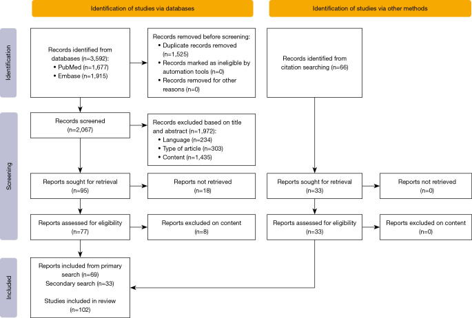
The results of the search were imported into Endnote (Version X9, Clearview Analytics, Philadelphia, PA, USA), where they were screened for duplicates. The duplicates were removed using the “Find duplicates” tool in Endnote. The remaining articles were imported to Rayyan (6) and screened according to the prespecified inclusion and exclusion criteria (Table S2). The following data were extracted from all case reports: title, authors, year of publication, age/gender of subject, related clinical symptoms, origin of OM, type of OM, trajectory, migrated regions, time between initial operation and diagnosis, operative technique, intraoperative findings, complications and duration of hospitalisation. When details on origin of OM or trajectory of the material were not specifically mentioned, we attempted to retrieve information from available imagery in the manuscript. Results were visually displayed using tables. Migrations were defined as antegrade if occurring from a peripheral position towards the chest. Migrations were defined as linear if the migration of OM followed a trajectory in line with the original position of the material. Migration was defined as non-linear if the trajectory deviated from this line. If OM migrated first into a vascular structure and then into the thorax via the vessel’s flow, the trajectory was considered unpredictable and called “intravascular embolization”. A PRISMA (7) flow diagram was used (Figure 1). Additional data were extracted from the selected manuscripts and summarized in Tables S3-37.
Figure 1.
PRISMA flow diagram of search results after data extraction.
Statistical analysis
The analysis was done in a descriptive manner mostly. Where possible, we used the Shapiro-Wilk test to test for normal distribution. When the P value exceeded 0.05, we accepted the null hypothesis and assumed that the data was normally distributed. The W statistic was provided and represents the difference between the estimated model and the observations (8). When testing our findings for significance, data were accompanied by a confidence interval (CI) of 95% and a P value showing significance if P<0.05. When comparing the averages of two separate groups, data was accompanied by a T-value showing the size of the difference relative to the variation in our sample data and a P value considered significant if P<0.05. The literature on intra-thoracically migrated OM is composed almost exclusively of individual case reports. Heterogeneity and reporting bias may prevent any complex statistical analysis.
Results
Search results
The search initially found 3,592 articles (PubMed: 1,677, Embase: 1,915). Duplicates were removed and 2,067 articles remained of which 1,990 articles were excluded (234 based on language, 303 based on type of publication, 1,435 based on content and 18 because no abstract or full text was available) at time of Title and Abstract screening. Another eight articles were excluded on content at time of full text screening, leaving 69 articles that were included. A secondary search was performed by snowballing the references of the articles included from primary search, leading to 33 additional inclusions (Figure 1). In total, 102 publications were included in this literature review, describing a total of 112 individual cases. In 12 cases, there were several pins or fragments that had migrated, for a total of 124 migration events.
Since only case reports, observational studies and case series were found, we assessed that all included cases had a large risk of bias.
General findings
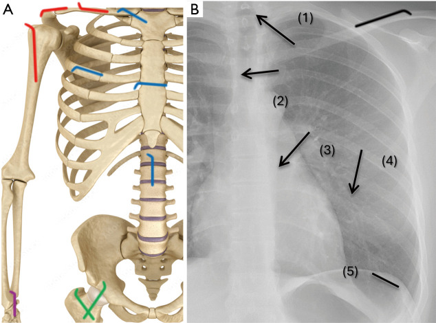
The cases could be divided according to the origin of the OM and according to the specific anatomic regions it migrated into, as seen in Figure 2. We found 36 cases of migration into the lung parenchyma, 30 into the mediastinum, 23 intracardiac, 12 into the pleural space, 17 into the spinal canal, three intraabdominal, and three to the neck. Some cases reported migration of the same OM into multiple regions as consequence of fragmentation or travel over long distance. Six cases described an intraabdominal and neck migration of OM that originated from the shoulder region. Although these cases matched our inclusion criteria, these six cases will not be discussed further, because they were considered to be outside the scope of our review. The origin of migration could be divided into four groups (Table 1). In 93 cases, the OM was described as K-wire. The remaining cases were labeled as Steinmann pins in 14 cases, sternal wire in three, Schanz screw in one and AO fixation pin in one case. In 85 cases (76%) there was a migration of an intact pin, while in 27 cases (24%) it was described as a migration of a broken part of the wire. In 67 cases (60%) the trajectory was described as linear, in 35 cases (31%) it was nonlinear and in 10 cases (9%) there was an intravascular embolization of the material.
Figure 2.
Overview of regions of origin and destinations of final migration. (A) Origins of migration, divided into four anatomic regions: shoulder region in 82 cases (73.2%), thoracic cage in 21 (18.8%), lower limb in three (2.7%) and distal upper limb in six (5.4%). (B) Intrathoracic areas of migration, divided into five anatomic regions, represented by numbered arrows—1: spinal canal in 17 cases (13.7%), 2: mediastinum in 30 (24.2%), 3: intracardiac in 23 (18.5%), 4: intrapulmonary in 36 (29.0%), and 5: into the pleural cavity in 12 (9.7%).
Table 1. Origin of migration, divided into four anatomic regions.
| Group | Cases (n=112) | % |
|---|---|---|
| Shoulder region | ||
| Total shoulder region | 82 | 73.2 |
| Clavicle | 34 | 30.4 |
| Proximal humerus | 27 | 24.1 |
| Acromioclavicular joint | 13 | 11.6 |
| Glenohumeral joint | 3 | 2.7 |
| Shoulder luxation fixation | 3 | 2.7 |
| Humeroscapular joint | 1 | 0.9 |
| Scapula | 1 | 0.9 |
| Thoracic cage | ||
| Total thoracic cage | 21 | 18.8 |
| Sternoclavicular joint | 14 | 12.5 |
| Sternum | 5 | 4.5 |
| Ribcage | 1 | 0.9 |
| Spine | 1 | 0.9 |
| Distal upper limb | ||
| Total distal upper limb | 3 | 2.7 |
| Distal radius | 2 | 1.8 |
| Index metacarpophalangeal joint | 1 | 0.9 |
| Lower limb | ||
| Total lower limb | 6 | 5.4 |
| Femur | 4 | 3.6 |
| Patella | 1 | 0.9 |
| Pelvic bone | 1 | 0.9 |
For migrations originating from the clavicula, the migration trajectory was linear in 83.8% (odds ratio 4.8, 95% CI: 1.81–15.1, P=0.002 compared to the trajectory of OM originating from other regions). This was numerically higher than for any other region. Figure 3 displays a non-linear migration of broken material into the pulmonary parenchyma to illustrate our subdivisions based on trajectory and intactness of the material.
Figure 3.
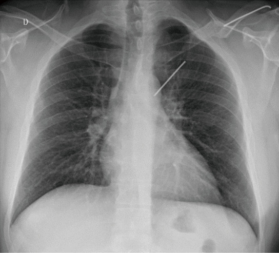
A chest radiograph confirming the position of a K-wire in the left hemithorax. The lateral half of the wire, measuring 6.4 cm, located in the distal third of the left clavicle. The medial half, measuring 6.2 cm, located in the right lung. Since the wire is broken and the migration is not in line with the originally placed position of the material, this migration is considered a “non-linear migration of broken material”.
In several cases, pin migrations occurred despite the authors describing preventive measures at the original osteosynthesis such as bending the end of the pin (9-11). In multiple cases, there was a migration of residual material, mostly broken pins, that were left behind after planned pin extraction (12,13). Most case reports gave no detailed info about the initial fracture fixation.
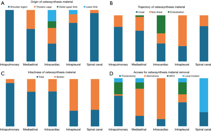
The mean age of the patients was 48.8 years (W=0.983, P=0.15), with ages ranging from 5 to 90 years old. In 76 cases the patient was male (68%) and in 36 female (32%). Mean time interval between initial operation and diagnosis of migration was 13.5 months (W=0.643, P<0.001), ranging from three days to 30 years. The mean time to diagnosis for linear migration was eight months (W=0.600, P<0.001) while it was 12.5 months (W=0.635, P<0.001) for non-linear migrations. Since the data in both groups were not normally distributed, statistical differences between both groups could not be calculated. Table 2 showed that when comparing the different time periods (divided by decades of publication) there was no significant difference in mean patient age, nor was there a difference in the mean delays between initial intervention to diagnosis (Table 2). In 34 cases (30%), patients were asymptomatic, and the migration was incidentally found on unrelated imaging. Thus, the majority (70%) were symptomatic with symptoms ranging from cough, dyspnea or localized pain to hemoptysis. Death due to migration or immediate postoperative period of removal was reported in five cases (detailed later) for an overall mortality of 4.5% (14-18). In 15 cases the diagnosis was made within the first month. In 38 cases the diagnosis was made between the first month and one year. In 45 cases the diagnosis was between the first year and 10 years. In 12 cases the diagnosis was later than 10 years after osteosynthesis. In two cases the interval was not mentioned. For all cases reporting the duration of hospital stay, the median was 5 days, but did not follow a normal distribution (W=0.897, P<0.001). However, the majority (70 cases) did not mention hospital stay duration. Major open surgery was performed for extraction in 66.9% of all cases, VATS in 14.4% and local shoulder/neck incision in 12.7%. Other reported treatment options were follow-up in three cases (19-21), bronchoscopic extraction (22), pericardiocentesis (18) and extraction during autopsy each in one case (15). Figure 4 displays the number of interventions per period and distributed according to surgical approach. We will further discuss the anatomic regions in more detail. Figure 5 shows how these groups differed among themselves in terms of origin, trajectory, intactness and surgical technique for OM retrieval.
Table 2. All 112 individual cases split into time periods.
| Period | No. of cases |
Mean patient age (years) |
Mean interval (months) |
|---|---|---|---|
| 1961–1970 | 6 | 31.2 | 16.2 |
| 1971–1980 | 8 | 38.0 | 63.2 |
| 1981–1990 | 7 | 36.8 | 7.7 |
| 1991–2000 | 15 | 42.8 | 37.3 |
| 2001–2010 | 35 | 49.1 | 31.1 |
| 2011–2020 | 35 | 56.7 | 68.2 |
| 2021–2030 | 6 | 56.3 | 81.3 |
The mean patient age and mean time from initial intervention to diagnosis were calculated for each period.
Figure 4.
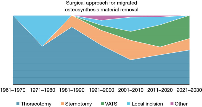
Graph showing the number of reported interventions per time period, distributed by surgical approach. “Local incision” includes laparotomy, neck or shoulder incision, supraclavicular incision or laminectomy. All cases of conservative treatment or autopsy findings were included into “other”. VATS, video-assisted thoracoscopic surgery.
Figure 5.
Comparison of the specific anatomic regions the OM migrated to, based on origin of OM (A), trajectory of OM (B), intactness of OM (C) and surgical access (D). VATS, video-assisted thoracoscopic surgery; OM, osteosynthesis material.
Intrapulmonary migration (Table 3)
Table 3. Intrapulmonary migration (36 cases).
| Author | Year | Age (y), gender | Origin | Trajectory | State | Interval | Operative technique |
|---|---|---|---|---|---|---|---|
| Dutta (23) | 2018 | 60 M | AC joint | Nonlinear | Total | 9 m | Thoracoscopy |
| Gopalakrishnan (24) | 2018 | 42 F | AC joint | Linear | Broken | 180 m | Thoracotomy |
| Retief (25) | 1978 | 27 M | AC joint | Linear | Total | 6 w | Laparotomy |
| Rey-Baltar (26) | 1964 | 29 M | Clavicle | Linear | Total | 7 m | Thoracotomy |
| Cabañero (27) | 2018 | 64 F | Clavicle | Linear | Total | 6 w | Thoracoscopy thoracotomy |
| Cortés-Julián (28) | 2015 | 56 M | Clavicle | Linear | Total | 360 m | Thoracoscopy |
| Hegemann (29) | 2005 | 59 M | Clavicle | Linear | Total | 6 w | Thoracoscopy |
| Irianto (30) | 2018 | 34 F | Clavicle | Linear | Total | 36 m | Thoracotomy |
| Nakayama (31) | 2009 | 30 M | Clavicle | Linear | Broken | 8 m | Thoracotomy |
| Amara (32) | 2024 | 61 M | Clavicle | Nonlinear | Total | 300 m | Thoracotomy |
| Reghine (33) | 2018 | 65 F | Clavicle | Nonlinear | Total | 240 m | Thoracotomy |
| Grauthoff (34) | 1978 | 50 M | Clavicle | Linear | Total | 49 m | Thoracotomy |
| Grauthoff (34) | 1978 | 60 M | Clavicle | Linear | Total | 228 m | Shoulder incision |
| Grauthoff (34) | 1978 | 18 M | Clavicle | Linear | Total | 96 m | Thoracotomy |
| Marchi (35) | 2008 | 26 M | Clavicle | Linear | Total | 156 m | Thoracoscopy thoracotomy |
| Suzuki (36) | 2017 | 69 F | GH joint | Linear | Total | 1 m | Shoulder incision |
| Tenconi (37) | 2014 | 79 F | GH joint | Linear | Total | N/A | Thoracotomy |
| Bleetman (38) | 2015 | 89 F | GH joint | Linear | Total | 4 w | Thoracoscopy |
| Schwartz (5) | 2011 | 78 F | HS joint | Linear | Total | 60 m | Sternotomy |
| Calkins (39) | 2001 | 48 M | Proximal humerus | Nonlinear | Total | 7 w | Thoracoscopy |
| Cameliere (12) | 2013 | 62 F | Proximal humerus | Nonlinear | Total | 48 m | Thoracotomy |
| Cerruti (10) | 2016 | 69 F | Proximal humerus | Linear | Total | 4 m | Thoracoscopy |
| Fuster (13) | 1990 | 42 M | Proximal humerus | Linear | Total | 8 m | Thoracotomy |
| Khan (40) | 2003 | 42 F | Proximal humerus | Nonlinear | Total | 4 m | Thoracotomy |
| Kim (41) | 2000 | 55 F | Proximal humerus | Linear | Total | 16 m | Thoracotomy |
| McCaughan (42) | 1969 | 12 M | Proximal humerus | Linear | Total | 39 m | Thoracotomy |
| Mellado (43) | 2004 | 85 F | Proximal humerus | Linear | Total | 10 d | Thoracotomy |
| Pientka (44) | 2016 | 78 M | Proximal humerus | Linear | Total | 6 w | Thoracotomy |
| Sergides (45) | 2009 | 59 M | Proximal humerus | Linear | Total | 3 m | Thoracotomy |
| Tristan (46) | 1964 | 12 F | Proximal humerus | Linear | Total | 3 m | Thoracotomy |
| van Hasselt (47) | 2021 | 90 F | Proximal humerus | Nonlinear | Total | 3 d | Thoracoscopy |
| Zacharia (48) | 2016 | 63 M | Proximal humerus | Nonlinear | Total | 36 m | Thoracoscopy |
| Chaurasia (49) | 2015 | 80 F | Proximal humerus | Linear | Total | N/A | Thoracoscopy |
| Bang (50) | 2017 | 26 F | Proximal humerus | Linear | Total | 6 w | Thoracotomy |
| Bedi (51) | 1997 | 8 M | Proximal humerus | Nonlinear | Broken | 1 w | Thoracotomy |
| Sarper (52) | 2003 | 46 M | Scapula | Linear | Total | 4 m | Thoracotomy |
Table S3 describing detailed anatomic regions for each case of migration was made available. M, male; F, female; AC, acromioclavicular; GH, glenohumeral; HS, humeroscapular; d, day; w, week; m, month; y, year; N/A, data not available.
Intrapulmonary migration occurred in 36 cases. In most cases, the trajectory was linear (27 cases, 75%). In nine cases (25%) the migration trajectory was nonlinear. In 33 cases (92%) there was a complete migration of intact material, while in three cases (8%) it was broken and only the distal fragment migrated. The period between initial placement and diagnosis of migration of OM ranged from three days to 30 years. The origin was always the shoulder region: in 16 cases the OM had been used for proximal humerus fractures, in 12 cases for clavicular fractures, acromioclavicular joint fixations in three, glenohumeral joint fixation in three, humeroscapular joint fixation in one and scapula fracture in one case. The OM originated from the right shoulder in 20 cases (56%) and the left shoulder in 16 cases (44%). Figure 6 depicts the exact anatomic locations of all reported cases with intra-pulmonary migration. In 25 cases (69%) migration occurred into the right lung and in seven cases (20%) into the left lung. In four cases (11%) the OM was embedded in multiple regions of the lung at the time of diagnosis. In 10 cases (28%), the OM had migrated to the lung opposite of the limb it had been placed. In all these 10 cases, the origin was the clavicle, the migration was linear and in 90% of them, the migration was from the left clavicle to the right lung. Cases of opposite side migration included a 78-year-old woman with a penetrating injury of the ascending thoracic aorta and superior vena cava caused by migration of a Steinman wire used for percutaneous fixation of a left-shoulder dislocation 5 years earlier (5). Four weeks after her initial operation when she was hospitalized for removal of the wires, the shoulder X-ray showed that one of the wires had already migrated into the chest wall. The migrated wire was left in place but later crossed the mediastinum to migrate into the opposite lung. The patient was treated with an emergent median sternotomy with a favorable outcome. Cameliere et al. (12) described a case where a K-wire made a non-linear migration from the right proximal humerus through the right upper and lower lobes, passing the scissure. They saw strong adherences of the lung at the top and to the diaphragm above the vena cava, adherent to the pulmonary artery. Concerning clinical presentations, the migration caused a hemothorax in five patients and a pneumothorax in three patients. In 28 cases minimal or no clinical repercussions of the migration were mentioned.
Figure 6.
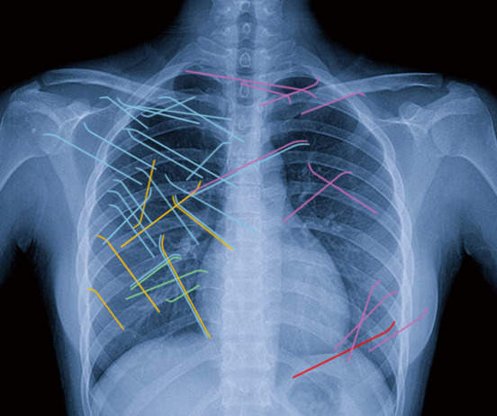
Cases of intrapulmonary migration into specific pulmonary anatomic regions, based on information and imaging provided in the original manuscripts. Right upper lobe (blue) 20 cases (56%), middle lobe (green) three cases (8%), right lower lobe (yellow) six cases (17%), left upper lobe (purple) 10 cases (27%), left lower lobe (red) one case (3%). In four cases (11%), the OM crossed multiple regions of pulmonary parenchyma during migration (shown in the image by two adjacent pins with different colors). OM, osteosynthesis material.
Treatment
The migrated wires were retrieved by thoracotomy in 23 cases (64%) and by VATS in nine cases (25%). Two wires were retrieved by local shoulder incision (34,36), one by laparotomy (25) and one by sternotomy (5). Cabañero et al. (27) performed a VATS through the left hemithorax via a 12-mm port and they converted non-urgently to an anterior thoracotomy for better visualization and extraction of the material. Marchi et al. (35) described a case where the patient was initially subjected to a right thoracoscopy that showed scattered pleural adhesions and no visual identification of the wire. Conversion from VATS to thoracotomy was deemed necessary and the wire was retrieved by a small thoracotomy. The mean duration of hospitalization for intrapulmonary migrations was 5.9 days (W=0.859, P=0.008). It was shorter for thoracoscopic removal [3.2 days (W=0.890, P=0.47)] than for thoracotomy cases [6.2 days (W=0.879, P=0.056)]; (T=−1.542, P=0.07).
Mediastinal migration (Table 4)
Table 4. Mediastinal migration (30 cases).
| Author | Year | Age (y), gender | Origin | Trajectory | Sate | Interval | Operative technique |
|---|---|---|---|---|---|---|---|
| Dutta (23) | 2018 | 60 M | AC Joint | Nonlinear | Total | 9 m | Thoracoscopy |
| Foster (11) | 2001 | 50 M | AC Joint | Nonlinear | Broken | 60 m | Neck incision |
| Richardson (22) | 1987 | 72 M | Cervical spine | Linear | Total | 4 d | Bronchoscopy |
| Cabañero (27) | 2018 | 64 F | Clavicle | Linear | Total | 6 w | Thoracoscopy thoracotomy |
| Orsini (53) | 2012 | 58 M | Clavicle | Linear | Total | 72 m | Sternotomy |
| Harmouchi (54) | 2021 | 41 M | Clavicle | Linear | Total | 24 m | Cervicotomy sternotomy |
| Harmouchi (54) | 2021 | 41 M | Clavicle | Linear | Total | 24 m | Cervicotomy sternotomy |
| Hegemann (29) | 2005 | 59 M | Clavicle | Linear | Total | 6 w | Thoracoscopy |
| Nakayama (31) | 2009 | 30 M | Clavicle | Linear | Broken | 8 m | Thoracotomy |
| Venissac (55) | 2000 | 26 M | Clavicle | Nonlinear | Broken | 4 m | Thoracoscopy |
| Venissac (55) | 2000 | 20 M | Clavicle | Nonlinear | Total | 1 m | Thoracotomy sternotomy |
| Wu (56) | 2009 | 48 M | Clavicle | Linear | Total | 108 m | Sternotomy |
| Wada (57) | 2005 | 84 M | Clavicle | Linear | Total | 24 m | Shoulder incision |
| Bezer (58) | 2009 | 26 M | Clavicle | Linear | Total | 24 m | Thoracoscopy sternotomy |
| Schwartz (5) | 2011 | 78 F | HS joint | Linear | Total | 60 m | Sternotomy |
| Harle (59) | 1967 | 70 F | Proximal humerus | Linear | Total | 15 m | N/A |
| Saxena (19) | 2014 | 31 F | Proximal humerus | Linear | Total | 240 m | Follow-up |
| Bang (50) | 2017 | 26 F | Proximal humerus | Linear | Total | 6 w | Thoracotomy |
| Mozaffari (60) | 2014 | 87 M | Shoulder | Nonlinear | Total | 8 d | Thoracoscopy |
| Sharma (61) | 2011 | 31 M | Shoulder | Nonlinear | Broken | 84 m | Thoracotomy |
| Kumar (62) | 2002 | 36 M | SC joint | Linear | Total | 4 w | Sternotomy |
| Özbey (63) | 2023 | 26 M | SC joint | Linear | Total | 156 m | Sternotomy |
| Leonard (18) | 1965 | 33 M | SC joint | Linear | Total | 17 m | Pericardio- centesis |
| Grauthoff (34) | 1978 | 55 M | SC joint | Nonlinear | Total | 4 w | Sternotomy |
| Leppilahti (64) | 1999 | 48 M | SC joint | Linear | Total | 26 d | Mediastinotomy |
| Daus (65) | 1993 | 45 F | SC joint | Linear | Total | 5 w | Sternotomy |
| Ballas (9) | 2012 | 56 M | SC joint | Nonlinear | Broken | 60 m | Laparotomy |
| Janssens de Varebeke (66) | 1994 | 62 F | SC joint | Linear | Broken | 7 m | Sternotomy |
| Hazelrigg (67) | 1994 | 64 M | Sternum | Nonlinear | Broken | 108 m | Sternotomy |
| Schreffler (68) | 2001 | 53 M | Sternum | Embolization | Broken | 24 m | Sternotomy |
Table S4 describing detailed anatomic regions for each case of migration was made available. M, male; F, female; AC, acromioclavicular; HS, humeroscapular; SC, sternoclavicular; d, day; w, week; m, month; y, year; N/A, data not available.
Mediastinal migration occurred in 30 cases. In 20 such cases, the migration was nonlinear, while in 9 cases it was linear. In one case there was a vascular embolization of OM. Schreffler et al. (68) described a case of a 53-year-old man who presented with massive, painless hemoptysis. A chest roentgenogram showed a segment of fractured sternal wire in the hilum of the right lung. In fact, an embolization of a broken K-wire from the sternum into the right ventricle, then embolization to the right middle pulmonary artery, with finally an erosion into an adjacent bronchus had occurred. In 22 cases (73%) there was a complete migration of the material, while in eight cases (27%) there was migration of broken material. The period between initial placement and diagnosis of migration of the OM ranged between four days and 20 years. The origin was shoulder region in 20 cases, sternoclavicular joint in eight cases and sternum in two cases.
Treatment
The migrated wires were retrieved by sternotomy in 14 cases, thoracotomy in four, thoracoscopy in four, laparotomy in one, mediastinotomy in one, neck incision in one and shoulder incision in one case. Harle et al. (59) provided no information about operative techniques. The remaining cases are described in more detail hereafter. Richardson et al. (22) described a case of early migration of a Steinman pin from the cervical vertebra into the left main bronchus. After resection of a vertebral chordoma the pin and methyl-acrylate had been used to fuse the vertebral bodies of C5 to C7. Although the postoperative radiography demonstrated the OM in satisfactory position, only three days later the patient complained of a mild sore throat without difficulties to breath or swallow. However, the next day radiography demonstrated the pin migrated into the left main bronchus and the authors were able to retrieve this intact pin by rigid bronchoscopy.
Leonard et al. (18) described the case of a 33-year-old man who was admitted because of a sharp stabbing chest pain present for three days. Imaging showed a K-wire in the anterior part of the mediastinum with its point near the base of the left main pulmonary artery. In their case, the K-wire caused a perforation of the right pulmonary artery with an organized hemopericardium of 300 mL. Resuscitative measures and emergency pericardiocentesis were of no avail and death ensued.
Saxena et al. (19) decided not to intervene in an asymptomatic patient and followed up a K-wire that had migrated to the posterior mediastinum across the midline into the prevertebral space behind the esophagus and aortic arch. Unfortunately, no late results were reported and outcome remains unknown.
Overall, the complications of migration into the mediastinum were mostly related to injury of great vessels and trachea. K-wires were found penetrating the trachea (11,22,31,54,56), brachiocephalic artery (56,57), brachiocephalic vein (61), esophagus (57) or piercing the ascending aorta and the superior vena cava (5). Orsini et al. (53) even described an aorto-esophageal fistula with hemorrhagic shock because of a K-wire migration through a thrombosed left subclavian artery into the esophagus of a patient with an arteria lusoria. After retrieval of OM via a median sternotomy extended into the left first intercostal space, the authors treated the fistula with a covered aortic stent and a right subclavian artery transposition without left subclavian revascularization. Postoperative difficulties with respiratory weaning required a tracheotomy and subsequent left pleural decortication. After one year of follow-up, the patient was reported to have regained his preoperative physical state. In the remaining 19 cases, no clinical repercussions of the migration were mentioned.
Intracardiac migration (Table 5)
Table 5. Intracardiac migration (23 cases).
| Author | Year | Age (y), gender | Origin | Trajectory | State | Interval | Operative technique |
|---|---|---|---|---|---|---|---|
| Wirth (20) | 2000 | 37 M | AC joint | Embolization | Broken | 240 m | Follow-up |
| Lenard (69) | 2009 | 45 M | AC joint | Embolization | Total | 60 m | Sternotomy |
| Tan (17) | 2016 | 5 M | Clavicle | Linear | Total | 7 d | Sternotomy |
| Nishizaki (70) | 2007 | 82 M | Clavicle | Nonlinear | Total | 36 m | Sternotomy |
| Goodsett (71) | 1999 | 50 M | Distal radius | Embolization | Broken | 42 m | Thoracotomy |
| Seipel (72) | 2001 | 50 M | Distal radius | Embolization | Total | 36 m | Sternotomy |
| Ono (73) | 2010 | 14 M | Epiphysis of femur | Embolization | Total | 6 m | Sternotomy |
| Leonardi (21) | 2014 | 71 F | Femur | Embolization | Broken | 252 m | Follow-up |
| Haapaniemi (74) | 1997 | 45 F | Index MCP joint | Embolization | Total | 31 m | Sternotomy |
| Biddau (75) | 2006 | 47 M | Patella | Embolization | Broken | 156 m | Sternotomy atriotomy |
| Park (76) | 2011 | 60 M | Pelvic bone | Nonlinear | Total | 240 m | Sternotomy |
| Hédon (77) | 2015 | 79 F | Proximal humerus | Linear | Total | 24 m | Sternotomy |
| Georges (78) | 2024 | 65 F | Proximal humerus | Nonlinear | Total | 3 m | Thoracotomy |
| Medved (16) | 2006 | 67 F | Proximal humerus | Linear | Total | 24 m | Sternotomy |
| Stemberga (15) | 2006 | 67 F | Proximal humerus | Nonlinear | Total | 24 m | Autopsy |
| Zhang (79) | 2014 | 50 M | Ribcage | Linear | Total | 32 m | Sternotomy |
| Clark (14) | 1974 | 18 M | SC joint | Linear | Total | 3 m | Thoracotomy pericardiotomy |
| Gulcan (80) | 2005 | 44 F | SC joint | Linear | Total | 6 m | Sternotomy |
| Liu (81) | 1993 | 35 M | SC joint | Linear | Total | 2 m | Sternotomy |
| Tubbax (82) | 1989 | 55 M | SC joint | Linear | Total | 8 d | Sternotomy |
| Wang (83) | 2021 | 55 M | SC joint | Linear | Broken | 5 m | Sternotomy |
| Levisman (84) | 2010 | 64 M | Sternum | Nonlinear | Broken | 84 m | Sternotomy |
| Anić (85) | 1997 | 48 F | Trochanter major | Embolization | Total | 66 m | Sternotomy, right atriotomy |
Table S5 describing detailed anatomic regions for each case of migration was made available. M, male; F, female; AC, acromioclavicular; MCP, metacarpophalangeal; SC, sternoclavicular; d, day; m, month; y, year.
Intracardiac migration occurred in 23 cases. The migration appeared linear in nine cases, nonlinear in five cases and the OM migrated by embolization into the heart in nine cases. In 17 cases (74%) there was a migration of the intact material, while in six cases (26%) there was migration of a broken part. The period between initial placement and diagnosis of OM migration ranged from seven days to 21 years. The OM originated from the shoulder region in eight cases, the thoracic cage in seven, the distal upper limbs in three and lower limbs in five cases. In nine cases there was confirmation of pericardial effusion or tamponade (14,70-72,77,78,80-82). Other complications ranged from tricuspid regurgitation (70,79), pulmonary artery perforation (82,86), cardioembolic stroke (77) or ventricle perforation (80). In 11 cases, no clinical repercussions of the migration were mentioned.
Treatment
The migrated wires were retrieved by sternotomy in 17 cases, thoracotomy in three cases. In two cases it was decided to perform follow-up without operation. No further information about long term follow-up of these patients was mentioned. In one case, the patient was only diagnosed at autopsy. Wirth et al. (20) described embolization of a broken K-wire with imaging suggesting the material overlying the ventricular portion of the cardiac silhouette. Leonardi et al. (21) described embolization of a broken K-wire into the anterior left ventricular myocardial wall. Their patient underwent close follow-up with clinical and radiographic examination every two months. In both cases, this conservative treatment had no short-term clinical repercussions but long-term outcome is unknown. Four cases (17%) of intracardiac migration resulted in death (14-17). Tan et al. (17) presented a case of a 5-year-old boy with intra-aortic migration of a K-wire used for the treatment of a right clavicle fracture 7 days previously. The CT-scan showed the wire to be partially inside the ascending aorta, which was associated with massive hemopericardium and cardiac tamponade. The patient developed cardiac arrest during induction of anesthesia for emergency surgery. Nevertheless, the K-wire was successfully retrieved via thoracotomy but the patient died 10 days postoperatively. Medved et al. (16) described the case of a 67-year-old woman who was admitted 24 months after intramedullary nailing of a right humerus fracture with three K-wires. Imaging revealed one of the K-wires in the right ventricle near the tricuspid valve. The patient died from multiorgan failure 36 hours post-admission and autopsy showed a 13.5 cm wire transpiercing the ventricle and left pericardium. Clark et al. (14) described a case of an 18-year-old man who presented with severe upper abdominal pain 3 months after sternoclavicular fixation with two K-wires. Imaging showed a K-wire in the middle mediastinum. Despite an emergent thoracotomy and pericardiotomy with successful removal of the K-wires, the patient died several hours after admission. Stemberga et al. (15) reported an autopsy discovery of a cardiac perforation by an intact pin used for humerus fracture.
Intrapleural migration (Table 6)
Table 6. Pleural space migration (12 cases).
| Author | Year | Age (y), gender | Origin | Trajectory | State | Interval | Operative technique |
|---|---|---|---|---|---|---|---|
| Sananta (87) | 2020 | 40 M | AC Joint | Nonlinear | Broken | 24 m | Thoracoscopy |
| Abbas (88) | 2009 | 27 M | AC Joint | Nonlinear | Broken | 8 m | Thoracoscopy |
| Aalders (89) | 1985 | 19 M | AC Joint | Linear | Total | 18 m | Laminectomy thoracotomy |
| Bezer (58) | 2009 | 26 M | Clavicle | Linear | Total | 24 m | Mediastinoscopy |
| Mamane (90) | 2009 | 34 M | Clavicle | Linear | Total | 2 m | Supraclavicular incision |
| Ozarslan (91) | 2014 | 49 F | Hip | Nonlinear | Total | 96 m | Thoracotomy |
| Emiroğullari (92) | 1999 | 45 M | Proximal humerus | Nonlinear | Total | 2 m | Thoracotomy |
| Kim (41) | 2000 | 55 F | Proximal humerus | Nonlinear | Total | 16 m | Thoracotomy |
| Ullmer (93) | 1998 | 80 F | Proximal humerus | Nonlinear | Broken | 36 m | Thoracoscopy |
| Julià (94) | 2012 | 83 F | Proximal humerus | Linear | Total | 1 m | Thoracotomy |
| Fowler (95) | 1981 | 14 M | SC joint | Nonlinear | Total | 2 m | Thoracotomy |
| Hamid (96) | 2012 | 67 M | Sternum | Nonlinear | Broken | 4 w | Sternotomy |
Table S6 describing detailed anatomic regions for each case of migration was made available. M, male; F, female; AC, acromioclavicular; w, week; m, month; y, year; SC, sternoclavicular.
Intrapleural migration was reported in 12 cases, including one case of intrapericardial migration. In eight cases (67%) the trajectory appeared nonlinear, while in four cases (33%) it seemed linear. In four cases (33%) there was a migration of the intact material, while in eight cases (67%) some broken parts migrated. Time interval between osteosynthesis and diagnosis of migration of the OM ranged from four weeks to eight years. The origin of OM was the shoulder girdle in nine cases, the thoracic cage in two and lower limb in one case.
In six patients (50%) the migration caused a pneumothorax (87,88,90,91,93). In five cases, no clinical repercussions were mentioned. In the case of Fowler (95), the K-wire, used for sternoclavicular fixation was found lying at the bottom of the pericardial cavity with fibrinous pericarditis.
Treatment
A thoracotomy was performed in six cases, a sternotomy in one case, VATS in three cases and through a supraclavicular incision in one. Bezer et al. (58) removed a K-wire through mediastinoscopy with a thoracoscope.
Spinal canal migration (Table 7)
Table 7. Spinal canal migration (17 cases).
| Author | Year | Age (y), gender | Origin | Trajectory | State | Interval | Operative technique |
|---|---|---|---|---|---|---|---|
| Aalders (89) | 1985 | 19 M | AC joint | Linear | Total | 18 m | Laminectomy thoracotomy |
| Li (86) | 2013 | 35 M | AC joint | Nonlinear | Total | 2 m | Thoracoscopy |
| Mankowski (97) | 2016 | 34 M | AC joint | Linear | Broken | 84 m | Neck incision |
| Mian (98) | 2012 | 41 M | Clavicle | Linear | Total | 24 m | Neck incision |
| Loncán (99) | 1998 | 22 M | Clavicle | Linear | Total | 2 m | Supraclavicular incision |
| Wang (100) | 2010 | 28 F | Clavicle | Linear | Broken | 48 m | Shoulder incision |
| Bennis (101) | 2008 | 57 M | Clavicle | Linear | Total | 4 m | Shoulder incision |
| Fransen (102) | 2007 | 30 M | Clavicle | Linear | Total | 12 m | Laminectomy |
| Regel (103) | 2002 | 50 M | Clavicle | Nonlinear | Broken | 3 m | Shoulder incision |
| Tsai (104) | 2009 | 49 M | Clavicle | Linear | Broken | 98 m | Shoulder incision |
| Gonsales (105) | 2017 | 42 M | Clavicle | Linear | Total | 3 m | Thoracotomy |
| Leppilahti (64) | 1999 | 56 M | Clavicle | Linear | Total | 11 d | Shoulder incision |
| Mamane (90) | 2009 | 34 M | Clavicle | Linear | Total | 2 m | Supraclavicular incision |
| Bedi (51) | 1997 | 8 M | Proximal humerus | Nonlinear | Broken | 1 w | Thoracotomy |
| Mellado (43) | 2004 | 85 F | Proximal humerus | Linear | Total | 10 d | Thoracotomy |
| Sarper (52) | 2003 | 55 F | Shoulder | Nonlinear | Total | 1 m | Thoracotomy |
| Furuhata (106) | 2020 | 68 M | Sternum | Linear | Broken | 84 m | Neck incision |
Table S7 describing detailed anatomic regions for each case of migration was made available. M, male; F, female; AC, acromioclavicular; d, day; w, week; m, month; y, year.
Spinal canal migration was reported in 17 cases. In four cases the trajectory seemed nonlinear while in 13 cases it was linear. In six cases (35%) there was a migration of the intact material, while in 11 cases (65%) broken material migrated. The interval between the initial operation and diagnosis of migration ranged from one week to eight years. The origin was the shoulder region in 16 cases and thoracic cage in one case.
In three cases (51,86,106) the migration caused leakage of cerebrospinal fluid. Loncán et al. (99) presented a case of Brown-Sequard syndrome because of a K-wire piercing the spinal canal through the T2–T3 left intervertebral foramen reaching the center of the spinal canal. Regel et al. (103) presented a case of tetraparesis with loss of bladder function because of a broken K-wire that had migrated into the right intervertebral foramen of C5/6, lying ventrally to the myelon in the epidural space. In 12 cases, no spine related complications of the migration were mentioned.
Treatment
A thoracotomy was performed in five cases, VATS in one, shoulder incision in five, neck incision in three, supraclavicular incision in two and laminectomy in one case. Aalders et al. (89) used a combined thoracotomy and laminectomy to extract a K-wire that migrated from the acromioclavicular joint to the second thoracic vertebra after 18 months.
Discussion
As confirmed by the large number of published cases, intra-thoracic migration of K-wires is not an exceptional event. Undoubtedly many more unpublished cases must have occurred. We found a large variety of clinical scenarios and various patterns and destinations of pin migrations. To give a more structured overview on the vast amount of data, we divided the cases according to the different thoracic regions or organs of migration. The pathophysiology of all these centripetal migrations towards the thorax remains uncertain. The fact that in most cases (73.2%), the origin of OM was the shoulder girdle could possibly be explained by the great freedom of movement of the shoulder and high muscle action in this region frequently leading to breakage of the wires (44). In comparison with the humerus, the clavicle remains relatively stable during respiration and shoulder movement. This could maybe explain why linear migration was the most prevalent pattern when the origin was the clavicle, compared to other regions of origin (odds ratio 4.8, 95% CI: 1.81–15.1, P=0.002). Migration of OM after proximal humerus fractures fixation might be predominantly nonlinear because of the higher local mobility and range of motion of the humerus. In some cases, the material even crossed multiple anatomic regions. Some K-wires were described to cross multiple lung lobes or even reach the contralateral lung. Specifically for intrapulmonary migrations, the side of migration is intriguing as most occurred into the right lung (25 cases or 69%) whereas the origin of OM was the right side in only 20 cases (56%). The left shoulder girdle was the origin in 16 cases (46%) but migration into the left lung occurred in only seven cases (20%) with the other 9 cases migrating to the right lung. This apparent side repartition imbalance is hard to explain. One might hypothesize that the right lung volume is larger than the left lung, contributing to 53.9% of the total lung volume and thus offering a larger destination space (107). Interestingly, in all 10 cases describing a migration to the opposite lung, this migration was linear originating from the clavicle and 9 of 10 times from the left clavicle to the right lung. These findings suggest that the fixity of the clavicle and large negative pressure pleural space on the right might cause favorable conditions for the wires to cross the mediastinum into the opposite lung parenchyma. Figure 6 visually confirms this right sided predominance of intra-pulmonary migrations. Although hypothetical, the clinical relevance of this finding lies in the anatomical dangers of a migration trajectory from left to right. As right to left migrations are rare, possibly the risk for injury of heart and major vessels could be less for OM implanted on the right side.
We confirm that mechanisms of antegrade OM migration are complex and seem influenced by multiple factors. Others have suggested the influence of regional bone resorption, capillary action but also surgical errors with insufficient measures taken to secure the fixating devices (e.g., by bending its end) (15,51,55). Patient age and the duration of presence of the material in the body might also have an influence. Use of K-wires in pediatric cases increases lifelong exposure risk to migration, whereas elderly patients might present with bone fragility and thus potentially increased risk of migration. However, we could not make any conclusions based on the patient’s profile, age, race or gender, because of the limited medical information provided in most case reports. In some cases, even a simultaneous migration of several wires or fragments was found (43,54), however, it remains unclear whether this was due to patient composition or due to inadequate surgical technique.
Intactness of the OM and their trajectory may also play a role. Intact wire migrations were more often seen in intrapulmonary migrations in comparison to intrapleural migrations (odds ratio 22.0, 95% CI: 4.1–118.6, P<0.001) Intrapulmonary migration also more often followed a linear trajectory (odds ratio 6.0, 95% CI: 1.5–24.8, P=0.013) comparison to intrapleural migrations. These findings could possibly be explained by a mechanism based on slow but progressive aspiration towards the lung parenchyma from respiratory movements and negative intra-pleural pressure combined with gravitational force. We suspect a potential additional role of apical lung adhesions. Apical pleural adhesions can be caused by traumas, major surgery or previous infections, as in the case described by Cameliere et al. (12). However, such adhesions could also be secondary to the migration itself and independently of their cause, they could possibly facilitate and even guide linear migration straight into the lung parenchyma. In comparison to intrapulmonary migration, we found that intrapleural migrations were more likely when the OM was broken. A pneumothorax might be due to puncture of the lung parenchyma by the OM, as described by Retief et al. (25). We found that the likelihood of an associated pneumothorax was significantly higher in the intrapleural migration group compared to intrapulmonary migrations (odds ratio 11.0, 95% CI: 2.1–56.5, P=0.004).
Even if migration patterns and their complications may differ, an adequate radiologic follow-up seems necessary for all groups for as long as any K-wire remains inside a patient. Intravascular migration of OM remains more difficult to predict. Similar migration patterns may be seen with other medical devices such as subcutaneously placed contraception devices that can migrate from forearms to various chest organs (108). Pellegrino et al. (109) highlight the importance of local joint mobility by finding that placement of such contraceptive devices close to joints may predispose to secondary migration into the pulmonary artery.
Migration of OM has been reported as early as the first day (most likely due to surgical errors at the time of placement) and as late as 30 years after osteosynthesis (28). Altogether, reported mortality was significant (4.5% of all cases) and was probably still underestimated because of lack of follow-up in most cases with a conservative attitude. As described above, the four cases (14-17) (17%) of deadly intracardiac migration make this the most lethal anatomic group. Nevertheless, migration into any of the thoracic compartments is potentially dangerous. Therefore, once intrathoracic migration of a K-wire is recognized, swift removal seems recommendable to avoid further migration towards other more vital intrathoracic structures. Considering that migration of OM is probably a rare event compared to the frequent use of such material, it may be debatable how follow-up should be organized but our study confirms the conclusions of orthopedic reviews that systematic removal of K-wires is recommended to avoid later migration (1). Ozarslan et al. (91) stated that the possibility of intrathoracic K-wire migration should be considered in any patient with a medical history of such osteosynthesis in any part of the body presenting with respiratory or cardiac complaints, pneumothorax or hemothorax at any later time. Considering that more than half of all migrations (52%) occurred later than one year after osteosynthesis, our review might suggest that systematic K-wire removal or strict follow-up is the only way to avoid these complications. Such systematic follow-up would carry an important burden in terms of costs and radiation exposure and systematical wire removal would come with a burden of costs, postoperative morbidity or even logistical difficulties. Specific studies on these issues seem to be lacking but belong to the fields of Traumatology and Orthopedics.
In the literature, minimally invasive techniques for removal of OM were used for only 25% of all intrapulmonary migrations, 25% of intrapleural migrations and 17% of mediastinal migrations. As seen in Figure 4, more recently reported cases were more likely to have been performed by minimally invasive techniques. Also, asymptomatic case presentations with stable patients favored minimally invasive approaches. A VATS approach has been proven to reduce tissue injury, reduce postoperative pain and shorten hospital stays compared to thoracotomy for many indications (110). If necessary, conversion to thoracotomy or sternotomy could always be performed (27,35). The reported conversions were all performed in order to have better exposure for safe OM extraction or when adhesions made VATS impossible. Among the few cases available, there was no mention of conversion-related complications. Our findings thus support that VATS should be considered whenever anatomy, clinical presentation and position of the OM allows a minimally invasive approach.
The greatest limitation of our study is that the real prevalence of OM migration remains unknown as we ignore the denominator of such K-wire osteosynthesis. Most of the 102 publications were single cases reports and therefore one may assume the existence of a large grey number of unreported cases both via a reporting bias for negative outcome and via journal rejection of redundant case reports adding little to previous knowledge. Available information must be handled with caution since often only the final position of OM during diagnosis is reported and the reader does not know what happened during the period of migration.
Our analysis of Table 2 shows increasing numbers of reported cases and increasing patient age by decades. Time intervals between osteosynthesis and diagnosis were fluctuant over the decades and do not suggest any clear impact of potentially more restrictive K-wire usage lately (Table 2). As use of K-wires is increasingly restricted to pediatric and hand surgery, the indications for their usage in the shoulder region may be narrowing. Nevertheless, the treatment of late complications remains relevant and many reports are recent (32). Furthermore, K-wires may remain the cheapest or the only material available in some places worldwide. Their continued use is illustrated by recent studies which did not find significant differences between plating and Kirschner wire internal fixation for clavicle fractures (111).
Finally, the danger of intrathoracic migration of OM lies in the element of unpredictability. Although most studies support the systematic removal of all K-wires, in clinical practice this has not always been possible, sometimes because of patients being lost to follow-up or failure to remove all parts of broken wires (11). Maybe better patient education might have a positive effect on patient awareness about materials inside their body and thus might help to avoid these complications (112).
Conclusions
This literature review may serve as a guide when confronted with cases of intra-thoracic migration of OM. Our findings could guide future care by helping to correctly interpret clinical findings, imaging, intraoperative findings and to compare similar cases. Once intrathoracic migration of a K-wire is recognized, we recommend removal to avoid further migration towards vital intrathoracic structures. We suggest a systematic follow-up and discussion of removal of all broken pins or K-wires. Patient awareness and education about potential risks of OM material needs to be promoted. Minimally invasive extraction by VATS should be considered whenever anatomy and clinical presentation allows it.
Supplementary
The article’s supplementary files as
Acknowledgments
Funding: The study was funded by the “Fondation des Hopitaux Robert Schuman Luxembourg”.
Ethical Statement: The authors are accountable for all aspects of the work in ensuring that questions related to the accuracy or integrity of any part of the work are appropriately investigated and resolved.
Footnotes
Reporting Checklist: The authors have completed the PRISMA reporting checklist. Available at https://jtd.amegroups.com/article/view/10.21037/jtd-24-943/rc
Conflicts of Interest: Both authors have completed the ICMJE uniform disclosure form (Available at https://jtd.amegroups.com/article/view/10.21037/jtd-24-943/coif). The authors report the funding from the “Fondation des Hopitaux Robert Schuman Luxembourg”. G.D. is the Associate Editor of European Journal of Cardio-Thoracic Surgery (EJCTS). The authors have no other conflicts of interest to declare.
References
- 1.Franssen BB, Schuurman AH, Van der Molen AM, et al. One century of Kirschner wires and Kirschner wire insertion techniques: a historical review. Acta Orthop Belg 2010;76:1-6. [PubMed] [Google Scholar]
- 2.Yang D, Zhou J, Wang L. Comparative clinical efficacy of anatomic plate and Kirschner wire internal fixation in midshaft clavicle fractures: A meta-analysis. Med Int (Lond) 2021;1:21. 10.3892/mi.2021.21 [DOI] [PMC free article] [PubMed] [Google Scholar]
- 3.Wijdicks FJ, Houwert RM, Millett PJ, et al. Systematic review of complications after intramedullary fixation for displaced midshaft clavicle fractures. Can J Surg 2013;56:58-64. 10.1503/cjs.029511 [DOI] [PMC free article] [PubMed] [Google Scholar]
- 4.Freund E, Nachman R, Gips H, et al. Migration of a Kirschner wire used in the fixation of a subcapital humeral fracture, causing cardiac tamponade: case report and review of literature. Am J Forensic Med Pathol 2007;28:155-6. 10.1097/PAF.0b013e31806195a1 [DOI] [PubMed] [Google Scholar]
- 5.Schwartz A, Thumerel M, Delcambre F, et al. Transaortic migration of a Steinman wire from the shoulder. Eur J Cardiothorac Surg 2011;40:517-9. 10.1016/j.ejcts.2010.12.015 [DOI] [PubMed] [Google Scholar]
- 6.Ouzzani M, Hammady H, Fedorowicz Z, et al. Rayyan-a web and mobile app for systematic reviews. Syst Rev 2016;5:210. 10.1186/s13643-016-0384-4 [DOI] [PMC free article] [PubMed] [Google Scholar]
- 7.Page MJ, McKenzie JE, Bossuyt PM, et al. The PRISMA 2020 statement: an updated guideline for reporting systematic reviews. BMJ 2021;372: n71. 10.1136/bmj.n71 [DOI] [PMC free article] [PubMed] [Google Scholar]
- 8.Ghasemi A, Zahediasl S. Normality tests for statistical analysis: a guide for non-statisticians. Int J Endocrinol Metab 2012;10:486-9. 10.5812/ijem.3505 [DOI] [PMC free article] [PubMed] [Google Scholar]
- 9.Ballas R, Bonnel F. Endopelvic migration of a sternoclavicular K-wire. Case report and review of literature. Orthop Traumatol Surg Res 2012;98:118-21. 10.1016/j.otsr.2011.09.015 [DOI] [PubMed] [Google Scholar]
- 10.Cerruti P, Mangano T, Giovale M, et al. Early asymptomatic intrathoracic migration of a threaded pin after proximal humeral osteosynthesis. Int J Shoulder Surg 2016;10:41-3. 10.4103/0973-6042.174520 [DOI] [PMC free article] [PubMed] [Google Scholar]
- 11.Foster GT, Chetty KG, Mahutte K, et al. Hemoptysis due to migration of a fractured Kirschner wire. Chest 2001;119:1285-6. 10.1378/chest.119.4.1285 [DOI] [PubMed] [Google Scholar]
- 12.Cameliere L, Rosat P, Heyndrickx M, et al. Migration of a Kirschner pin from the shoulder to the lung, requiring surgery. Asian Cardiovasc Thorac Ann 2013;21:222-3. [DOI] [PubMed] [Google Scholar]
- 13.Fuster S, Palliso F, Combalia A, et al. Intrathoracic migration of a Kirschner wire. Injury 1990;21:124-6. 10.1016/0020-1383(90)90074-5 [DOI] [PubMed] [Google Scholar]
- 14.Clark RL, Milgram JW, Yawn DH. Fatal aortic perforation and cardiac tamponade due to a Kirschner wire migrating from the right sternoclavicular joint. South Med J 1974;67:316-8. 10.1097/00007611-197403000-00017 [DOI] [PubMed] [Google Scholar]
- 15.Stemberga V, Bosnar A, Bralic M, et al. Heart embolization with the Kirschner wire without cardiac tamponade. Forensic Sci Int 2006;163:138-40. 10.1016/j.forsciint.2005.09.014 [DOI] [PubMed] [Google Scholar]
- 16.Medved I, Simic O, Bralic M, et al. Chronic heart perforation with 13.5 cm long Kirschner wire without pericardial tamponade: an unusual sequelae after shoulder fracture. Ann Thorac Surg 2006;81:1895-7. 10.1016/j.athoracsur.2005.06.061 [DOI] [PubMed] [Google Scholar]
- 17.Tan L, Sun DH, Yu T, et al. Death Due to Intra-aortic Migration of Kirschner Wire From the Clavicle: A Case Report and Review of the Literature. Medicine (Baltimore) 2016;95:e3741. 10.1097/MD.0000000000003741 [DOI] [PMC free article] [PubMed] [Google Scholar]
- 18.Leonard JW, Gifford RW, Jr. Migration of a Kirschner wire from the clavicle into the pulmonary artery. Am J Cardiol 1965;16:598-600. 10.1016/0002-9149(65)90040-8 [DOI] [PubMed] [Google Scholar]
- 19.Saxena R, Muthukkumaran S, Kumar S, et al. Migrated Kirschner wire in the posterior mediastinum. Heart Lung Circ 2014;23:e109-10. 10.1016/j.hlc.2013.09.011 [DOI] [PubMed] [Google Scholar]
- 20.Wirth MA, Lakoski SG, Rockwood CA, Jr. Migration of broken cerclage wire from the shoulder girdle into the heart: a case report. J Shoulder Elbow Surg 2000;9:543-4. 10.1067/mse.2000.109413 [DOI] [PubMed] [Google Scholar]
- 21.Leonardi F, Rivera F. Intravascular migration of a broken cerclage wire into the left heart. Orthopedics 2014;37:e932-5. 10.3928/01477447-20140924-90 [DOI] [PubMed] [Google Scholar]
- 22.Richardson M, Gomes M, Tsou E. Transtracheal migration of an intravertebral Steinmann pin to the left bronchus. J Thorac Cardiovasc Surg 1987;93:939-41. [PubMed] [Google Scholar]
- 23.Dutta R, Pal H, Patel LM, Singh TK. Thoracoscopic removal of k-wire penetrating lung and mediastinum. Arch Trauma Res 2018;7:85-6. [Google Scholar]
- 24.Gopalakrishnan M, Naik A, Kamath M, et al. Interesting case of retrieval of intrathoracic Kirschner wire. Indian J Thorac Cardiovasc Surg 2018;34:384-7. 10.1007/s12055-017-0586-y [DOI] [PMC free article] [PubMed] [Google Scholar]
- 25.Retief PJ, Meintjes FA. Migration of a kirschner wire in the body. A case report. S Afr Med J 1978;53:557-8. [PubMed] [Google Scholar]
- 26.Rey-Baltar E, Errazu D. Unusual outcome of steinman wire. Case of fractured clavicle. Arch Surg 1964;89:1024-5. 10.1001/archsurg.1964.01320060092018 [DOI] [PubMed] [Google Scholar]
- 27.Cabañero A, Ovejero P, Gorospe L, et al. Trans-mediastinal Kirschner wire migration treated by video-assisted thoracoscopy. Asian Cardiovasc Thorac Ann 2018;26:482-4. 10.1177/0218492318785262 [DOI] [PubMed] [Google Scholar]
- 28.Cortés-Julián G, Mier JM, Briseño C. Thoracoscopic Excision of Migrated Kirschner Wire to Right Pulmonary Hilum. Ann Thorac Surg 2015;100:1461-3. 10.1016/j.athoracsur.2014.12.102 [DOI] [PubMed] [Google Scholar]
- 29.Hegemann S, Kleining R, Schindler HG, et al. Kirschner wire migration in the contralateral lung after osteosynthesis of a clavicular fracture. Unfallchirurg 2005;108:991-3. 10.1007/s00113-005-0946-8 [DOI] [PubMed] [Google Scholar]
- 30.Irianto KA, Edward M, Fiandana A. K-wire migration to unexpected site. Int J Surg Open 2018;11:18-21. [Google Scholar]
- 31.Nakayama M, Gika M, Fukuda H, et al. Migration of a Kirschner wire from the clavicle into the intrathoracic trachea. Ann Thorac Surg 2009;88:653-4. 10.1016/j.athoracsur.2008.12.093 [DOI] [PubMed] [Google Scholar]
- 32.Amara KB, Zairi S, Radhia BB, et al. Intra-pulmonary migration of a clavicle osteosynthesis pin: a case report. J Med Case Rep 2024;18:184. 10.1186/s13256-024-04369-7 [DOI] [PMC free article] [PubMed] [Google Scholar]
- 33.Reghine ÉL, Cirino CCI, Neto AA, et al. Clavicle Kirschner Wire Migration into Left Lung: A Case Report. Am J Case Rep 2018;19:325-8. 10.12659/ajcr.908014 [DOI] [PMC free article] [PubMed] [Google Scholar]
- 34.Grauthoff H, Klammer HL. Complications due to migration of a Kirschner wire from the clavicle (author's transl). Rofo 1978;128:591-4. 10.1055/s-0029-1230910 [DOI] [PubMed] [Google Scholar]
- 35.Marchi E, Reis MP, Carvalho MV. Transmediastinal migration of Kirschner wire. Interact Cardiovasc Thorac Surg 2008;7:869-70. 10.1510/icvts.2008.185850 [DOI] [PubMed] [Google Scholar]
- 36.Suzuki T, Matsumura N, Iwamoto T, et al. Migration of a Kirschner wire into the lung with shoulder dislocation. BMJ Case Rep 2017;2017:bcr2017221850. 10.1136/bcr-2017-221850 [DOI] [PMC free article] [PubMed] [Google Scholar]
- 37.Tenconi S, Lococo F, Rapicetta C, et al. Intra pulmonary migration of a Kirschner wire after glenohumeral fixation. Lung 2014;192:217-8. 10.1007/s00408-013-9524-y [DOI] [PubMed] [Google Scholar]
- 38.Bleetman D, Guida GA, Bonfield SH, et al. Migration of a Glenohumeral Steinmann Pin Into the Thoracic Cavity. Ann Thorac Surg 2015;100:e19. 10.1016/j.athoracsur.2015.03.044 [DOI] [PubMed] [Google Scholar]
- 39.Calkins CM, Moore EE, Johnson JL, et al. Removal of an intrathoracic migrated fixation pin by thoracoscopy. Ann Thorac Surg 2001;71:368-70. 10.1016/s0003-4975(00)01722-7 [DOI] [PubMed] [Google Scholar]
- 40.Khan AA, Khan SU, Hossain Z. Intrathoracic migration of a humeral orthopedic pin. J Cardiovasc Surg (Torino) 2003;44:275-7. [PubMed] [Google Scholar]
- 41.Kim JH, Kwon JH, Hwang ED, et al. Intrathoracic migration of Steinmann pins. J Thorac Imaging 2000;15:301-3. 10.1097/00005382-200010000-00013 [DOI] [PubMed] [Google Scholar]
- 42.McCaughan JS, Jr, Miller PR. Migration of Steinmann pin from shoulder to lung. JAMA 1969;207:1917. [PubMed] [Google Scholar]
- 43.Mellado JM, Calmet J, García Forcada IL, et al. Early intrathoracic migration of Kirschner wires used for percutaneous osteosynthesis of a two-part humeral neck fracture: a case report. Emerg Radiol 2004;11:49-52. 10.1007/s10140-004-0361-4 [DOI] [PubMed] [Google Scholar]
- 44.Pientka WF, 2nd, Bates CM, Webb BG. Asymptomatic Migration of a Kirschner Wire from the Proximal Aspect of the Humerus to the Thoracic Cavity: A Case Report. JBJS Case Connect 2016;6:e77. 10.2106/JBJS.CC.16.00032 [DOI] [PubMed] [Google Scholar]
- 45.Sergides NN, Nikolopoulos DD, Yfadopoulos DK, et al. Intrathoracic migration of a Steinman wire: a case report and review of the literature. Cases J 2009;2:8321. 10.4076/1757-1626-2-8321 [DOI] [PMC free article] [PubMed] [Google Scholar]
- 46.Tristan TA, Daughtridge TG. Migration of a metallic pin from the humerus into the lung. N Engl J Med 1964;270:987-9. 10.1056/NEJM196405072701905 [DOI] [PubMed] [Google Scholar]
- 47.van Hasselt AJ, Hooghof JT, Huizinga MR, et al. Intrathoracic migration of a K-wire after percutaneous fixation of a proximal humerus fracture. Trauma Case Rep 2021;32:100425. 10.1016/j.tcr.2021.100425 [DOI] [PMC free article] [PubMed] [Google Scholar]
- 48.Zacharia B, Puthezhath K, Varghees I. Kirschner wire migration from subcapital humeral fracture site, causing hydropneumothorax. Chin J Traumatol 2016;19:305-8. 10.1016/j.cjtee.2015.12.010 [DOI] [PMC free article] [PubMed] [Google Scholar]
- 49.Chaurasia IK, Dubey K, Rajiwate F, et al. Thoracoscopic removal of unusual migrated K-wire from thorax: A case report. IAIM 2015;2:78-81. [Google Scholar]
- 50.Bang GA, Nana Muluem A, Nana Oumarou B, et al. Kirschner Wire Migration from Proximal Humerus into the Lung: Brief Report. Trauma Cases Rev 2017. doi: 10.23937/2469-5777/1510050. [DOI] [Google Scholar]
- 51.Bedi GS, Gill SS, Singh M, et al. Intrathoracic migration of a Kirschner wire: case report. J Trauma 1997;43:865-6. 10.1097/00005373-199711000-00023 [DOI] [PubMed] [Google Scholar]
- 52.Sarper A, Urgüden M, Dertsiz L, et al. Intrathoracic migration of Steinman wire. Interact Cardiovasc Thorac Surg 2003;2:210-1. 10.1016/S1569-9293(03)00047-1 [DOI] [PubMed] [Google Scholar]
- 53.Orsini B, Amabile P, Bal L, et al. Management of an aortoesophageal fistula caused by Kirschner wire migration in a patient with arteria lusoria. J Thorac Cardiovasc Surg 2012;144:e25-7. 10.1016/j.jtcvs.2012.05.022 [DOI] [PubMed] [Google Scholar]
- 54.Harmouchi H, Bouazzaoui AE, Bensalah A, et al. Intratracheal migration of two Kirschner wires after surgery for a clavicle fracture. Asian Cardiovasc Thorac Ann 2021;29:428-30. 10.1177/0218492320987933 [DOI] [PubMed] [Google Scholar]
- 55.Venissac N, Alifano M, Dahan M, et al. Intrathoracic migration of Kirschner pins. Ann Thorac Surg 2000;69:1953-5. 10.1016/s0003-4975(00)01198-x [DOI] [PubMed] [Google Scholar]
- 56.Wu YH, Lai CH, Luo CY, et al. Tracheoinnominate artery fistula caused by migration of a Kirschner wire. Eur J Cardiothorac Surg 2009;36:214-6. 10.1016/j.ejcts.2009.03.043 [DOI] [PubMed] [Google Scholar]
- 57.Wada S, Noguchi T, Hashimoto T, et al. Successful treatment of a patient with penetrating injury of the esophagus and brachiocephalic artery due to migration of Kirschner wires. Ann Thorac Cardiovasc Surg 2005;11:313-5. [PubMed] [Google Scholar]
- 58.Bezer M, Aydin N, Erol B, et al. Unusual migration of K-wire following fixation of clavicle fracture: a case report. Ulus Travma Acil Cerrahi Derg 2009;15:298-300. [PubMed] [Google Scholar]
- 59.Harle TS, Beard EF. Migration of Steinmann pins. Am J Roentgenol Radium Ther Nucl Med 1967;100:542-5. 10.2214/ajr.100.3.542 [DOI] [PubMed] [Google Scholar]
- 60.Mozaffari M, Estfan R, Sarkar S. Intrathoracic migration of an unbent Steinmann pin. Ann R Coll Surg Engl 2014;96:e21-3. 10.1308/003588414X13814021678916 [DOI] [PMC free article] [PubMed] [Google Scholar]
- 61.Sharma R, Tam RK. Migrating foreign body in mediastinum--intravascular Steinman pin. Interact Cardiovasc Thorac Surg 2011;12:883-4. 10.1510/icvts.2010.256503 [DOI] [PubMed] [Google Scholar]
- 62.Kumar P, Godbole R, Rees GM, et al. Intrathoracic migration of a Kirschner wire. J R Soc Med 2002;95:198-9. 10.1177/014107680209500409 [DOI] [PMC free article] [PubMed] [Google Scholar]
- 63.Özbey M, Türkmen U, Şahin E. Thirteen years of migration of Kirschner wires: A mediastinal foreign body. Turk Gogus Kalp Damar Cerrahisi Derg 2023;31:412-5. 10.5606/tgkdc.dergisi.2023.21980 [DOI] [PMC free article] [PubMed] [Google Scholar]
- 64.Leppilahti J, Jalovaara P. Migration of Kirschner wires following fixation of the clavicle--a report of 2 cases. Acta Orthop Scand 1999;70:517-9. 10.3109/17453679909000992 [DOI] [PubMed] [Google Scholar]
- 65.Daus GP, Drez D, Jr, Newton BB, Jr, et al. Migration of a Kirschner wire from the sternum to the right ventricle. A case report. Am J Sports Med 1993;21:321-2. 10.1177/036354659302100225 [DOI] [PubMed] [Google Scholar]
- 66.Janssens de Varebeke B, Van Osselaer G. Migration of Kirschner's pin from the right sternoclavicular joint resulting in perforation of the pulmonary artery main trunk. Acta Chir Belg 1993;93:287-91. Erratum in: Acta Chir Belg 1994;94:IX. [PubMed] [Google Scholar]
- 67.Hazelrigg SR, Staller B. Migration of sternal wire into ascending aorta. Ann Thorac Surg 1994;57:1023-4. 10.1016/0003-4975(94)90232-1 [DOI] [PubMed] [Google Scholar]
- 68.Schreffler AJ, Rumisek JD. Intravascular migration of fractured sternal wire presenting with hemoptysis. Ann Thorac Surg 2001;71:1682-4. 10.1016/s0003-4975(00)02311-0 [DOI] [PubMed] [Google Scholar]
- 69.Lenard L, Aradi D, Donauer E. Migrating Kirschner wire in the heart mimics acute coronary syndrome. Eur Heart J 2009;30:754. 10.1093/eurheartj/ehn548 [DOI] [PubMed] [Google Scholar]
- 70.Nishizaki K, Seki T. Intracardiac migration of a Kirschner wire from the right clavicle. Asian Cardiovasc Thorac Ann 2007;15:272-3. 10.1177/021849230701500325 [DOI] [PubMed] [Google Scholar]
- 71.Goodsett JR, Pahl AC, Glaspy JN, et al. Kirschner wire embolization to the heart: an unusual cause of pericardial tamponade. Chest 1999;115:291-3. 10.1378/chest.115.1.291 [DOI] [PubMed] [Google Scholar]
- 72.Seipel RC, Schmeling GJ, Daley RA. Migration of a K-wire from the distal radius to the heart. Am J Orthop (Belle Mead NJ) 2001;30:147-51. [PubMed] [Google Scholar]
- 73.Ono M, Goerler H, Boethig D, et al. Surgical removal of Kirschner wire from the right ventricle, migrated from the femur. Eur J Cardiothorac Surg 2010;37:486. 10.1016/j.ejcts.2009.08.022 [DOI] [PubMed] [Google Scholar]
- 74.Haapaniemi TA, Hermansson US. Cardiac arrhythmia caused by a Kirschner wire inside the heart. An unusual complication of finger osteosynthesis. J Hand Surg Br 1997;22:402-4. 10.1016/s0266-7681(97)80412-3 [DOI] [PubMed] [Google Scholar]
- 75.Biddau F, Fioriti M, Benelli G. Migration of a broken cerclage wire from the patella into the heart. A case report. J Bone Joint Surg Am 2006;88:2057-9. 10.2106/JBJS.E.00968 [DOI] [PubMed] [Google Scholar]
- 76.Park SY, Kang JW, Yang DH, et al. Intracardiac migration of a Kirschner wire: case report and literature review. Int J Cardiovasc Imaging 2011;27 Suppl 1:85-8. 10.1007/s10554-011-9977-0 [DOI] [PubMed] [Google Scholar]
- 77.Hédon C, Khoueiry Z, Verges M, et al. Late intracardiac orthopedic wire migration presenting as tamponade and stroke. Eur Heart J 2015;36:1546. 10.1093/eurheartj/ehu416 [DOI] [PubMed] [Google Scholar]
- 78.Georges O, De Dominicis F, Rahal MB, et al. Successful removal of an intra-pericardic Kirschner wire via fluoroscopy-guided minimal invasive approach: a case report. Surg Case Rep 2024;10:57. 10.1186/s40792-023-01803-8 [DOI] [PMC free article] [PubMed] [Google Scholar]
- 79.Zhang W, Song F, Yang Y, et al. Asymptomatic intracardiac migration of a Kirschner wire from the right rib. Interact Cardiovasc Thorac Surg 2014;18:525-6. 10.1093/icvts/ivt539 [DOI] [PMC free article] [PubMed] [Google Scholar]
- 80.Gulcan O, Sezgin AT, Bolat B, et al. Right ventricular penetration and cardiac tamponade as a late complication of Kirschner wire placement in the sternoclavicular joint. Interact Cardiovasc Thorac Surg 2005;4:295-6. 10.1510/icvts.2005.107896 [DOI] [PubMed] [Google Scholar]
- 81.Liu HP, Chang CH, Lin PJ, et al. Pulmonary artery perforation after Kirschner wire migration: case report and review of the literature. J Trauma 1993;34:154-6. [PubMed] [Google Scholar]
- 82.Tubbax H, Hendzel P, Sergeant P. Cardiac perforation after Kirschner wire migration. Acta Chir Belg 1989;89:309-11. [PubMed] [Google Scholar]
- 83.Wang P, Chen C, Liu B, et al. Intracardic migration of Kirschner wire from the right sternoclavicular joint: a case report. BMC Surg 2021;21:294. 10.1186/s12893-021-01292-2 [DOI] [PMC free article] [PubMed] [Google Scholar]
- 84.Levisman J, Shemin RJ, Robertson JM, et al. Migrated sternal wire into the right ventricle: case report in cardiothoracic surgery. J Card Surg 2010;25:161-2. 10.1111/j.1540-8191.2009.00955.x [DOI] [PubMed] [Google Scholar]
- 85.Anić D, Brida V, Jelić I, et al. The cardiac migration of a Kirschner wire. A case report. Tex Heart Inst J 1997;24:359-61. [PMC free article] [PubMed] [Google Scholar]
- 86.Li Y, Wang B, Lv G, et al. Video-assisted thoracoscopic surgery for migration of a Kirschner wire in the spinal canal: a case report and literature review. Turk Neurosurg 2013;23:803-6. 10.5137/1019-5149.JTN.5300-11.1 [DOI] [PubMed] [Google Scholar]
- 87.Sananta P, Dradjat RS, Julana R, et al. Migration of K-wire into the cavum pleura after the reduction of acromioclavicular dislocation, a case report and review of literature. Int J Surg Case Rep 2020;74:192-5. 10.1016/j.ijscr.2020.08.004 [DOI] [PMC free article] [PubMed] [Google Scholar]
- 88.Abbas A, Richmond N, McCormack DJ, et al. A 27-year-old man presenting with acute chest pain and dyspnea. Chest 2009;135:1684-7. 10.1378/chest.08-2750 [DOI] [PubMed] [Google Scholar]
- 89.Aalders GJ, van Vroonhoven TJ, van der Werken C, et al. An exceptional case of pneumothorax--'a new adventure of the K wire'. Injury 1985;16:564-5. 10.1016/0020-1383(85)90087-7 [DOI] [PubMed] [Google Scholar]
- 90.Mamane W, Breitel D, Lenoir T, et al. Spinal migration of a Kirschner wire after surgery for clavicular nonunion. A case report and review of the literature. Chir Main 2009;28:367-9. 10.1016/j.main.2009.08.007 [DOI] [PubMed] [Google Scholar]
- 91.Ozarslan F, Arıkan O, Acat M, et al. Intrathoracic migration of a Kirschner wire. J Surg Case Rep 2014;2014:rjt132. 10.1093/jscr/rjt132 [DOI] [PMC free article] [PubMed] [Google Scholar]
- 92.Emiroğullari ON, Kahraman C, Taşdemir K, et al. Intrathoracic migration of a Kirschner wire. Asian Cardiovasc Thorac Ann 1999;7:319-20. [Google Scholar]
- 93.Ullmer E, Bolliger CT, Solèr M. Pneumothorax or the end of a wire's journey. Respiration 1998;65:151. 10.1159/000029247 [DOI] [PubMed] [Google Scholar]
- 94.Julià D, Baldó X, Gómez N, et al. Transthoracic migration of a Kirschner wire from the humerus to the abdomen. Eur J Cardiothorac Surg 2012;41:1197-8. 10.1093/ejcts/ezr138 [DOI] [PubMed] [Google Scholar]
- 95.Fowler AW. Migration of a wire from the sternoclavicular joint to the pericardial cavity. Injury 1981;13:261-2. 10.1016/0020-1383(81)90255-2 [DOI] [PubMed] [Google Scholar]
- 96.Imran Hamid U, Gillespie S, Lynchehaun C, et al. Traumatic Bilateral Pneumothoraces due to Sternal Wire Migration. Case Rep Med 2012;2012:438429. 10.1155/2012/438429 [DOI] [PMC free article] [PubMed] [Google Scholar]
- 97.Mankowski B, Polchlopek T, Strojny M, et al. Intraspinal migration of a Kirschner wire as a late complication of acromioclavicular joint repair: a case report. J Med Case Rep 2016;10:66. 10.1186/s13256-016-0844-4 [DOI] [PMC free article] [PubMed] [Google Scholar]
- 98.Mian MK, Nahed BV, Walcott BP, et al. Intraspinal migration of a clavicular Steinmann pin: case report and management strategy. J Clin Neurosci 2012;19:310-3. 10.1016/j.jocn.2011.05.018 [DOI] [PubMed] [Google Scholar]
- 99.Loncán LI, Sempere DF, Ajuria JE. Brown-Sequard syndrome caused by a Kirschner wire as a complication of clavicular osteosynthesis. Spinal Cord 1998;36:797-9. 10.1038/sj.sc.3100683 [DOI] [PubMed] [Google Scholar]
- 100.Wang SQ, Gao YS, Mei J, et al. Migration of a broken Kirschner pin into thoracic spinal canal 4 years following internal fixation of a clavicle fracture. Eur J Orthopaed Surg Traumol 2010;20:493-5. [Google Scholar]
- 101.Bennis S, Scarone P, Lepeintre JF, et al. Asymptomatic spinal canal migration of clavicular K-wire at the cervicothoracic junction. Orthopedics 2008;31: orthosupersite.com/view.asp?rID=32939. 10.3928/01477447-20081201-03 [DOI] [PubMed] [Google Scholar]
- 102.Fransen P, Bourgeois S, Rommens J. Kirschner wire migration causing spinal cord injury one year after internal fixation of a clavicle fracture. Acta Orthop Belg 2007;73:390-2. [PubMed] [Google Scholar]
- 103.Regel JP, Pospiech J, Aalders TA, et al. Intraspinal migration of a Kirschner wire 3 months after clavicular fracture fixation. Neurosurg Rev 2002;25:110-2. 10.1007/s101430100186 [DOI] [PubMed] [Google Scholar]
- 104.Tsai CH, Hsu HC, Huan CY, et al. Late migration of threaded wire (schanz screw) from right distal clavicle to the cervical spine. J Chin Med Assoc 2009;72:48-51. 10.1016/S1726-4901(09)70021-8 [DOI] [PubMed] [Google Scholar]
- 105.Gonsales D, Aguilar-Salinas P, Cavicchioli A, et al. The Migration of Kirschner Wire from Left Distal Clavicle to the IntraDural Anterior Thoracic Spine. Open Access J Neurol Neurosurg 2017;2:555595. [Google Scholar]
- 106.Furuhata R, Nishida M, Morishita M, et al. Migration of a Kirschner wire into the spinal cord: A case report and literature review. J Spinal Cord Med 2020;43:272-5. 10.1080/10790268.2017.1419915 [DOI] [PMC free article] [PubMed] [Google Scholar]
- 107.Fleming J, Conway J, Majoral C, et al. Determination of regional lung air volume distribution at mid-tidal breathing from computed tomography: a retrospective study of normal variability and reproducibility. BMC Med Imaging 2014;14:25. 10.1186/1471-2342-14-25 [DOI] [PMC free article] [PubMed] [Google Scholar]
- 108.Ohannessian A, Levy A, Jaillant N, et al. A French survey of contraceptive implant migration to the pulmonary artery. Contraception 2019;100:255-7. 10.1016/j.contraception.2019.05.016 [DOI] [PubMed] [Google Scholar]
- 109.Pellegrino A, Damiani GR, Loverro M, et al. Distal migration of contraceptive device in a sub-segmental branch of the pulmonary artery. Eur J Obstet Gynecol Reprod Biol 2017;215:260-1. 10.1016/j.ejogrb.2017.06.012 [DOI] [PubMed] [Google Scholar]
- 110.Cao C, Manganas C, Ang SC, et al. Video-assisted thoracic surgery versus open thoracotomy for non-small cell lung cancer: a meta-analysis of propensity score-matched patients. Interact Cardiovasc Thorac Surg 2013;16:244-9. 10.1093/icvts/ivs472 [DOI] [PMC free article] [PubMed] [Google Scholar]
- 111.Ngarmukos C, Parkpian V, Patradul A. Fixation of fractures of the midshaft of the clavicle with Kirschner wires. Results in 108 patients. J Bone Joint Surg Br 1998;80:106-8. 10.1302/0301-620x.80b1.7880 [DOI] [PubMed] [Google Scholar]
- 112.Johansson K, Nuutila L, Virtanen H, et al. Preoperative education for orthopaedic patients: systematic review. J Adv Nurs 2005;50:212-23. 10.1111/j.1365-2648.2005.03381.x [DOI] [PubMed] [Google Scholar]


