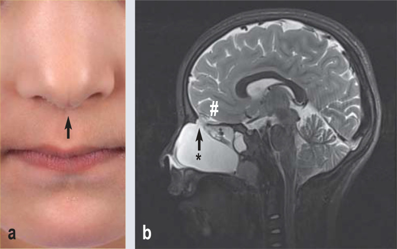
A 6-year-old girl presented to our department with impaired nasal breathing. Examination revealed a cleft nose (Figure a) and a smoothly demarcated endonasal mass displacing the nasal septum to the right. On the basis of cranial MRI, transethmoidal meningocele was diagnosed (Figure b). Following complementary computed tomography, we performed complication-free endonasal resection and duraplasty. At 3 months postoperatively, the patient was symptom-free. Meningoencephalocele (with brain parenchyma) or meningocele (without brain parenchyma) are classified according to localization as frontal, ethmoidal, parietal, and occipital. The skull base defects are usually congenital. Their incidence is approximately 1:35,000. They are a rare differential diagnosis of impaired nasal breathing in children, alongside adenoids and dermoid cysts. Biopsies should be avoided due to the risk of ascending infections in the case of rhinoliquorrhea. Spontaneous meningitis is possible and decisive for the prognosis. Forms of facial dysmorphism such as cleft nose are simple clinical indications of more complex clinical pictures.
Figure.
a) Cleft nose: divergence of the greater alar cartilage with indentation of the tip of the nose (arrow), broad nasal bridge.
b) Cranial MRI, 3 Tesla, sagittal T2W:
cystic formation (asterisk) in the nasal cavity, isointense to cerebrospinal fluid. Defect of the cribriform plate (arrow) connecting to the subarachnoid space. No herniation of brain parenchyma (hash).
Acknowledgments
Translated from the original German by Christine Rye.
Footnotes
Conflict of interest statement: The authors declare that no conflict of interest exists.


