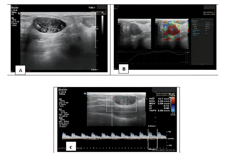
Figure 2. A 28-year-old female patient presented with a right breast lump.
A: Greyscale ultrasound showed a well-defined, wider-than-taller, oval-shaped hypoechoic lesion in the right upper outer quadrant.
B: Strain elastography measured a strain ratio of 2.48.
C: Doppler showed a PI of 0.8 and a RI of 0.75. FNAC confirmed the lesion as a fibroadenoma.
PI: pulsatility index; RI: resistance index; FNAC: fine needle aspiration cytology.

