


Abstract
Introduction and importance
Para-duodenal hernias make up 53 % of all internal hernias, with 40 % being left-sided and 13 % being right-sided. The occurrence of symptoms in adults is rare, ranging from 0.2 % to 0.5 %. The sex ratio for internal hernia often exhibits a male predominance, with around three males affected for every one female. Emergency operations typically result in the discovery of the right para-duodenal hernia, an uncommon internal hernia with aberrant anatomy. In this case report, we conducted a comprehensive analysis of right para-duodenal hernia. This condition poses a diagnostic challenge and requires prompt surgical management in emergency patients.
Case presentation
A 71-year-old male presented to the emergency department with a 24-h history of intermittent abdominal pain in the abdominal region. The patient described the pain as colicky and reported experiencing nausea and vomiting. All laparotorical investigations were normal. A CT scan showed that the jejunal bowel loops were positioned incorrectly, with one below and to the right of the second part of the duodenum and the other at an incorrect angle to the duodenum-jejunal junction. We diagnosed a suspected para-duodenal hernia. An emergency operation was scheduled, and instead a diagnosis of right para-duodenal hernia was made.
Following the surgery, the patient made a full recovery and received discharge within 5 days.
Clinical discussion
Internal hernias are a rare and unusual type of intestinal obstruction that are defined by the small bowel becoming trapped in an abnormal intra-abdominal cavity defect. Less than 1 % and up to 6 % of all related bowel obstruction, respectively, are roughly related.
In our cases, due to the late presentation of massive abdominal distention, peritonitis, and hemodynamic alterations, we preferred an explorative laparotomy, which revealed intestinal ischemia, which is trapped in the hernia sac and is not suitable for laparoscopic surgery.
Conclusion
Although right para-duodenal hernia is a very uncommon type of para-duodenal hernia, its anatomical features can vary significantly. It is critical to increase healthcare professionals' knowledge about the illness and ensure prompt diagnosis and surgical intervention. This intervention will effectively mitigate potential problems and significantly enhance patient outcomes.
Keywords: Bowel obstruction, Preduodenal hernia, Bowel ischemia, Resection and anastomosis
Highlights
-
•
Para-duodenal hernias make up 53% of all internal hernias, with 40% being left-sided and 13% being right-sided. The occurrence of symptoms in adults is rare, ranging from 0.2% to 0.5%. The sex ratio for internal hernia often exhibits a male predominance, with around three males affected for every one female.
-
•
PDH can be divided into left and right PDH according to completely different embryological and anatomical features. Right PDH is less common than left PDH, with a 1:3 ratio, The right para-duodenal hernia occurs due to an abnormality in the second stage of embryonic mid gut rotation.
-
•
The majority are asymptomatic and can be detected during surgery and clinical diagnosis of para-duodenal hernia is difficult due to its rarity and diverse symptoms.
-
•
If the suspect has ischemia and strangulation, visceral perforation, or significant bowel distension (bowel diameter > 4 cm), significant adhesions in the peritoneal fluid, hemodynamic instability, or a contraindication to pneumo-peritoneum, open explorative laparotomy is recommended. vi. Furthermore, laparotomy is a suitable treatment option in areas with limited equipment or where laparoscopy is not available.
1. Introduction and importance
Para-duodenal hernias (PDHs) are congenital internal hernias. The incidence of PDH is unclear because some patients remain asymptomatic throughout their lives [1]. According to autopsy results, the incidence of internal hernias is between 0.2 % and 0.9 %, while PDH accounts for approximately 53 % of internal hernias [2]. PDH can be divided into left and right PDH according to completely different embryological and anatomical features [3,4]. Right PDH is less common than left PDH, with a 1:3 ratio [5]. The right para-duodenal hernia occurs due to an abnormality in the second stage of embryonic midgut rotation [6].
Although congenital, most patients are entirely asymptomatic and diagnosed incidentally during abdominal surgery or abdominal imaging [7]. The clinical presentation in symptomatic patients can vary from recurrent episodes of abdominal pain to acute intestinal obstruction or strangulation [8,9]. It is impossible to make a clinical diagnosis of right para-duodenal hernia as there are no specific symptoms or signs. Small bowel contrast studies may be helpful, but a computed tomography (CT) scan with oral and intravenous contrast is the best modality to establish the diagnosis [10]. Although right para-duodenal hernia accounts for only 0.2 to 0.9 % of all intestinal obstruction cases, the morbidity and mortality are high in patients with an acute presentation [11]. In this case report, we present our experience in managing a patient of right para-duodenal hernia with small bowel strangulation.
The work has been reported in line with the SCARE 2023 criteria.
2. Case presentation
A 71-year-old male presented to the emergency department with a 24-h history of intermittent abdominal pain in the abdominal region. The pain was described as colicky and associated with nausea and vomiting. There were no recent changes in bowel habits or urinary symptoms. His medical history was notable for hypertension, and he developed a transient ischemic attack 1 year ago, and he was taking an angiotensin-converting enzyme inhibitor aspirin and atorvastatin. Upon initial examination, the patient appeared uncomfortable but not acutely distressed. Vital signs were within normal limits, and physical examination revealed tenderness in the epigastric region of the abdomen with diffuse distension of the abdomen, and there was no fecal or gas passage for the last 2 days; bowel sounds were active. The digital rectal examination was unremarkable. Given the persistent nature of the abdominal pain, further investigation was initiated. Laboratory investigations revealed a normal complete blood count, liver function tests, and pancreatic enzyme levels.
Serum electrolytes and renal function were within normal limits. The first done radiologic investigations were chest and erect abdominal x-rays (Figs. 1, 2), which show air fluid level, and again abdominal ultrasounds were warranted, and the result was slightly dilated bowel loops with air fluid levels and some fluid in between bowel loops. IV contrast abdominal CT were recommended, and the results were abnormal orientation of the jejunal bowel loops being interposed below and to the right of the second part of the duodenum associated with an abnormal position of the duodeno-jejunal junction. Distended small bowel seen at the level of distal jejunum with a twisted-like configuration of the mesentery and two transitional points opposite to each other along with multiple air-fluid levels, denoting closed loop obstruction (Figs. 3, 4, 5), and operative strategy were decided and the patient was emergently transferred to the operation theater, and under general anesthesia, midline laparotomies were performed, and upon entering the peritoneal cavity, there were dilated jejunal loops with ischemic changes.
Figs. 1, 2.

Chest and abdominal X-ray shows that there is no air diaphragm but demonstrates multiple air fluid level which shoes bowel obstruction.
Fig. 3.

Abdominal CT-scan shows gastric dilatation for proximal bowel obstruction.
Figs. 4, 5.

Abnormal orientations of the jejunal bowel loops being interposed below and to the right of the second part of the duodenum associated with an abnormal position of the duodeno-jejunal junction, with multiple air-fluid levels, denoting closed loop obstruction.
During surgery, we noted that the small bowel loops were ischemic. There were jejunal loops entering and trapped to the transverse mesocolon at the right upper quadrant, and jejunal loops were reduced without untwisting and we made resection and anastomosis of the ischemic bowel. The bowel 120 cm from the Treitz ligament and 90 cm of the bowel were resected with a linear stapler (side to side stapled anastomosis), and we closed the mesenteric defect with Vicryl 2.0 (Fig. 6, Fig. 7, Fig. 8).
Fig. 6.
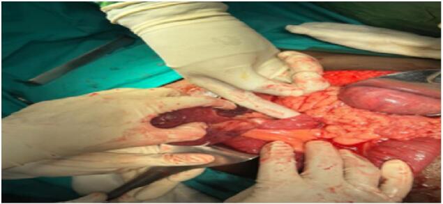
Identifying the defect of paraduodenal hernia were the bowel is trapped.
Fig. 7.
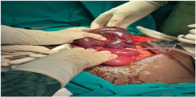
The ischemic part of the bowel.
Fig. 8.
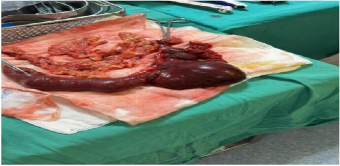
The resected part of the small bowel.
Postoperatively, the patient was transferred to the intensive care unit for further follow-up and closed monitoring, and after two days of taking a semi-solid diet at postoperative day 3, we started his full diet and tolerated and discharged successfully on postoperative day 5.
3. Clinical discussion
Internal hernias are a rare and unusual type of intestinal obstruction that are defined by the small bowel becoming trapped in an abnormal intra-abdominal cavity defect [12]. PDH is caused by an abnormal rotation of the primitive midgut during embryonic life, resulting in mesoscopic hernia [11]. Treitz and Jonnesco were the first to describe this phenomenon in 1857 and 1889, respectively [13]. According to where they are located on the Treitz ligament, para- duodenal hernias can be classified as either left- or right-sided (left-sided PDH: 75 % vs right-sided PDH: 25 %). Right PDH is a part of or the entire small intestine, entrapped in Waldeyer's fossa due to congenital factors [14]. It is located, behind the superior mesenteric artery and inferior to the third portion of the duodenum and the hernia extends inferolateral behind the ascending mesocolon from the right transverse mesocolon, also hernia is more common in males and mostly presents in the fourth to sixth decade [15]. The earliest age of presentation documented in published literature is in a 1-week-old neonate [16]. Research indicates that right-sided preduodenal hernias are larger and more fixed than left-sided hernias [17]. Non-rotation of the small bowel is related with them.
The majority are asymptomatic and can be detected during surgery and clinical diagnosis of para-duodenal hernia is difficult due to its rarity and diverse symptoms. Clinicians notice nothing specific [18]. Approximately 50 % of gangrene and perforation cases are discovered accidently during surgery or post-mortem, as an acute intestinal obstruction, staying asymptomatic and the symptoms include acute to chronic stomach pain, nausea, vomiting, and, rarely, fever [19]. Asymptomatic, recurrent ileus from childhood, or weight loss may occur. Most patients present with severe and diffuse abdominal pain, vomiting and nausea from acute small intestinal obstruction, and ileus from repeated cramps or torsion. The severity of symptoms depends on obstruction degree [20]. This illness may mirror other upper GI symptoms, causing doctors to misdiagnose the para-duodenal hernia. Patients may develop obstructive signs and complications due to delays in diagnosis, inappropriate medical treatments, and drugs.
Multidetector CT is best for internal hernia diagnosis and preoperative assessment. On CT imaging, right para-duodenal hernias appear as sac-like masses of dilated small bowel loops in Waldeyer's fossa in the right upper abdomen [21]. Due to intestinal malrotation, the superior mesenteric vein is on the left, and the third segment of the duodenum does not cross. When there is obstruction, one can observe bowel dilatation, elevated air-fluid levels, and mesenteric congestion with fat stranding. Moreover, CT has the advantage of ruling out the possibility of another related cause of the obstruction [22].
There are various surgical approaches, depending on the patient's condition and findings in the pre-op radiological assessment, but the standard surgical approach is laparoscopic repair due to better postoperative surgical outcomes, and this condition is becoming more common in the literature [23]. If the suspect has ischemia and strangulation, visceral perforation, or significant bowel distension (bowel diameter > 4 cm), significant adhesions in the peritoneal fluid, hemodynamic instability, or a contraindication to pneumo-peritoneum, open explorative laparotomy is recommended [24]. In our cases, due to the late presentation of massive abdominal distention, peritonitis, and hemodynamic alterations, we preferred an explorative laparotomy, which revealed intestinal ischemia, which is trapped in the hernia sac and is not suitable for laparoscopic surgery.
Furthermore, laparotomy is a suitable treatment option in areas with limited equipment or where laparoscopy is not available [25].
The surgical goal should be to return the bowel to its natural anatomical position, correct the hernia sac defect by closing a suture, to remove the sac is not mandatory and then the mesenteric and colonic arteries should not injury during repair [26].
Despite a mortality rate of over 50 % in cases of bowel ischemia requiring bowel resection, our patient, who had intestinal ischemia and >90 cm of intestine necrosis, had a resection anastomosis. Despite this, there was no fatality and the patient was discharged in excellent condition.
4. Conclusion
To summarize, strangulated para-duodenal hernias are uncommon sources of sudden abdominal pain that necessitate a strong level of suspicion, swift diagnosis, and surgical treatment. Chronic intermittent post-prandial stomach pain is a frequently occurring symptom that is crucial in signaling the need for more examination. The diagnosis of strangulated para- duodenal hernias might be difficult due to the diversity of clinical presentations.
It is crucial to increase knowledge about the illness among healthcare professionals and guarantee swift diagnosis and timely surgical intervention. Based on the patient's state and pre-op radiological assessment, laparoscopic repair is the conventional surgical method due to better postoperative surgical outcomes, and this condition is becoming more common in the literature. Open explorative procedures are recommended for suspects with ischemia, strangulation, visceral perforation, bowel distension (bowel diameter > 4 cm), peritoneal fluid adhesions, hemodynamic instability, or pneumo-peritoneum, laparotomy advised
This intervention will aid in mitigating potential problems and enhancing the overall health outcomes of patients.
CRediT authorship contribution statement
O.N.Ç., M.RA., and N.A.Y. wrote the manuscript and corrected the manuscript for its scientific basis.
O.N.Ç., M.RA., and N.A.Y. collected the data for the study. All authors have read and approved the final manuscript.
Consent
Written informed consent was obtained from the patient for publication of this case report and accompanying images. A copy of the written consent is available for review by the Editor-in-Chief of this journal on request.
Ethical approval
Not applicable.
Guarantor
Nor Abdi Yasin.
Research registration number
Not applicable.
Consent for publication
For the publication of this case report and any accompanying pictures, the patient provided written informed consent. A copy of the written consent is available for review by the Editor-in-Chief of this journal on request.
Provenance and peer review
Not commissioned, externally peer reviewed.
Funding
None.
Declaration of competing interest
The authors declare no conflict of interest and this study received no funding support.
Acknowledgements
Not applicable.
Data availability
The author declares that all data in this article are available within the article.
References
- 1.Wang X.L., Jin G.X., Xu J.F., Chen Z.R., Wu L.M., Jiang Z.L. Right paraduodenal hernia, classification, and selection of surgical methods: a case report and review of the literature. J. Med. Case Rep. Dec 30 2023;17(1):536. doi: 10.1186/s13256-023-04286-1. [DOI] [PMC free article] [PubMed] [Google Scholar]
- 2.Lamprou V., Krokou D., Karlafti E., Panidis S., Kougias L., Tzikos G., Ioannidis A., Netta S., Thomaidou E., Paramythiotis D. Right paraduodenal hernia as a cause of acute abdominal pain in the emergency department: a case report and review of the literature. Diagnostics. Nov 9 2022;12(11):2742. doi: 10.3390/diagnostics12112742. [DOI] [PMC free article] [PubMed] [Google Scholar]
- 3.Oshita K., Yoshimitsu M., Yunoki K., Imaoka K., Yano T., Idani H., Okajima M. Reduced-port surgery for right paraduodenal hernia in an adult patient: a case report and review of the literature. Asian Journal of Endoscopic Surgery. Jul 2021;14(3):598–601. doi: 10.1111/ases.12908. [DOI] [PubMed] [Google Scholar]
- 4.Sohrabi C., Mathew G., Maria N., Kerwan A., Franchi T., Agha R.A. The SCARE 2023 guideline: updating consensus Surgical CAse REport (SCARE) guidelines. Int J Surg Lond Engl. 2023;109(5):1136. doi: 10.1097/JS9.0000000000000373. [DOI] [PMC free article] [PubMed] [Google Scholar]
- 5.Manojlović D., Čekić N., Palinkaš M. Left paraduodenal hernia–a diagnostic challenge: case report. Int. J. Surg. Case Rep. Aug 1 2021;(85) doi: 10.1016/j.ijscr.2021.106138. [DOI] [PMC free article] [PubMed] [Google Scholar]
- 6.Tanaka H., Mitsutomoe S., Nagao N., Komori S., Suetsugu T., Iwata Y., Watanabe T., Tanaka C., Kawai M. Right paraduodenal hernia presenting with strangulated obstruction with intestinal malrotation: a case report. Journal of Surgical Case Reports. May 2024;2024(5) doi: 10.1093/jscr/rjae311. [DOI] [PMC free article] [PubMed] [Google Scholar]
- 7.Kadhem S., Ali M.H., Al-Dera F.H., Alzayer N.A., Alyagoub H.M. Left paraduodenal hernia: case report of rare cause of recurrent abdominal pain. Cureus. Mar 2020;12(3) doi: 10.7759/cureus.7156. [DOI] [PMC free article] [PubMed] [Google Scholar]
- 8.Atak T. A rare cause of intestinal obstruction: right paraduodenal hernia. Turkish Journal of Trauma & Emergency Surgery. Mar 2024;30(3):226. doi: 10.14744/tjtes.2023.20352. [DOI] [PMC free article] [PubMed] [Google Scholar]
- 9.Xu H., Nie N., Kong F., Zhong B. Large left paraduodenal hernia with intestinal ischemia: a case report and literature review. J. Int. Med. Res. Sep 2020;48(9) doi: 10.1177/0300060520955040. [DOI] [PMC free article] [PubMed] [Google Scholar]
- 10.Omarov N., Özata İ.H., Balık E. Right paraduodenal hernia accompanying superior mesenteric vein thrombosis: a rare case. BMJ Case Reports CP. Jun 1 2021;14(6) doi: 10.1136/bcr-2020-241324. [DOI] [PMC free article] [PubMed] [Google Scholar]
- 11.Joshi B.R., Gautam S., Yadav S.A., Gupta R.K. Case report: right paraduodenal hernia–a unique case of rare internal hernia presenting as acute small bowel obstruction. F1000Research. Dec 15 2021;(10):1282. [Google Scholar]
- 12.Patel N.J., Peddapati J.R., Barot S., Mareedu S., Erepally P., Bhanja S. Left para-duodenal hernia presenting with recurrent abdominal pain: a diagnostic challenge. Cureus. Aug 18 2024;16(8) doi: 10.7759/cureus.67107. [DOI] [PMC free article] [PubMed] [Google Scholar]
- 13.Vanmali A., Maharajh J., Haines M. Left paraduodenal hernia: embryological and radiological findings. SA Journal of Radiology. 2021;25(1) doi: 10.4102/sajr.v25i1.1979. [DOI] [PMC free article] [PubMed] [Google Scholar]
- 14.Moshref L.H., Alqahtani S.H., Majeed Z.A., Miro J. Left paraduodenal hernia: a rare cause of recurrent abdominal pain: case report. Pan Afr. Med. J. Nov 3 2021;40(1) doi: 10.11604/pamj.2021.40.135.32065. [DOI] [PMC free article] [PubMed] [Google Scholar]
- 15.Yamani Z., Budianto A. Total bowel obstruction with right paraduodenal hernia: a rare case report. Indonesian Journal of General Surgery. Jul 8 2020;1(1):1–4. [Google Scholar]
- 16.Carter M., Hughes B.D., Lim I.I., Kaul A., Farrell P.R., Garrison A.P. Laparoscopic Ladd’s procedure for the repair of a right paraduodenal hernia in pediatric patients–a case series. J Pediatr Surg Case Rep. Aug 1 2023;(95) [Google Scholar]
- 17.Brogna B., Urciuoli L., Musto L.A., Iovine L. A late presentation of a left paraduodenal hernia in an elderly patient admitted in emergency: a case report. Radiology Case Reports. Apr 1 2024;19(4):1356–1360. doi: 10.1016/j.radcr.2024.01.013. [DOI] [PMC free article] [PubMed] [Google Scholar]
- 18.Samanvitha H, Shivagange SP, Sajjan SS, Naveen SG, Harsha MT. When the gut took a wrong turn to the right: a case report of a rare complication of closed loop small bowel obstruction with small bowel volvulus in a right paraduodenal hernia with midgut malrotation. Cureus. 2024 Jan;16(1). [DOI] [PMC free article] [PubMed]
- 19.Kakkar D., Kochar S., Prasad S. Right para duodenal hernia presenting as small bowel obstruction: a rare case report. International Surgery Journal. Jun 25 2020;7(7):2410–2413. [Google Scholar]
- 20.John M., Michael H., Matthew S., Anthony U., David D., Pablo G., Christopher E. Identification and repair of left-sided paraduodenal hernia using both laparoscopic and robotic techniques. Case Reports in Surgery. 2020;2020(1) doi: 10.1155/2020/7569530. [DOI] [PMC free article] [PubMed] [Google Scholar]
- 21.Giri O.A., Bhat S.N., Kuppusamy B. Lessons learnt in the management of paraduodenal hernias: a case series. Surg. Endosc. Jan 2024;38(1):443–448. doi: 10.1007/s00464-023-10513-z. [DOI] [PubMed] [Google Scholar]
- 22.Muramatsu R., Nobuoka T., Ito T., Ogawa T., Korai T., Takemasa I. Laparoscopic mesh repair and Toupet fundoplication for parahiatal hernia complicated by sliding hiatal hernia: a case report. Int. J. Surg. Case Rep. Nov 1 2022;(100) doi: 10.1016/j.ijscr.2022.107664. [DOI] [PMC free article] [PubMed] [Google Scholar]
- 23.Kwan B., Theodore J.E., Wong J. Laparoscopic paraduodenal hernia repair with bioabsorbable mesh: a case of a novel technique for a rare cause of bowel obstruction. Int. J. Surg. Case Rep. Jan 1 2020;(70):1–4. doi: 10.1016/j.ijscr.2020.03.035. [DOI] [PMC free article] [PubMed] [Google Scholar]
- 24.Misheva B., Hajjar R., Sebajang H., Schwenter F. Minimally invasive management of a paraduodenal hernia with intestinal malrotation. Journal of Surgical Case Reports. Sep 2020;2020(9) doi: 10.1093/jscr/rjaa326. [DOI] [PMC free article] [PubMed] [Google Scholar]
- 25.Kollmann L., Lock J.F., Kollmann C., Vladimirov M., Germer C.T., Seyfried F. Surgical treatment of internal hernia after Roux–en-Y gastric bypass—impact of institutional standards and surgical approach. Langenbeck’s Arch. Surg. Aug 17 2023;408(1):318. doi: 10.1007/s00423-023-03049-2. [DOI] [PMC free article] [PubMed] [Google Scholar]
- 26.Lakmal M.A., Gamage B.D., Sanjeewa S.H., Varun S. Laparoscopic repair of a rare type of internal hernia: a case of left para-duodenal hernia. Sri Lanka. Journal of Surgery. Jul 31 2022;40(2) [Google Scholar]
Associated Data
This section collects any data citations, data availability statements, or supplementary materials included in this article.
Data Availability Statement
The author declares that all data in this article are available within the article.