Simple Summary
This study developed and evaluated Bayesian Network models for lung cancer risk prediction using a decade of data from 38,944 high-risk individuals in Denmark. The models were trained and validated on datasets with varying sizes and levels of missing data to reflect real-world screening scenarios. The results showed that a model trained on a small, complete dataset (AUC 0.78) performed similarly on a larger dataset with 21% missing data (AUC 0.78), but performance decreased when 39% of data were missing (AUC 0.67). The laboratory results and smoking data were the most informative variables, significantly outperforming models based only on age and smoking status (AUC 0.70). These findings suggest that BN models can maintain strong predictive performance despite incomplete data and highlight the value of including standard laboratory results in future LC screening programs.
Keywords: lung cancer, bayesian networks, prediction models, screening, early detection, missing data, risk stratification
Abstract
Background/Objectives: Lung cancer (LC) is the leading cause of cancer mortality, making early diagnosis essential. While LC screening trials are underway globally, optimal prediction models and inclusion criteria are still lacking. This study aimed to develop and evaluate Bayesian Network (BN) models for LC risk prediction using a decade of data from Denmark. The primary goal was to assess BN performance on datasets varying in size and completeness, simulate real-world screening scenarios, and identify the most valuable data sources for LC screening. Methods: The study included 38,944 patients evaluated for LC, with 11,284 (29%) diagnosed. Data on comorbidities, medications, and general practice were available for the entire cohort, while laboratory results, smoking habits, and other variables were only available for subsets. The cohort was divided into four subsets based on data availability, and BNs were trained and validated across these subsets using cross-validation and external validation. To determine the optimal combination of variables, all possible data combinations were evaluated on the samples that contained all the variables (n = 5587). Results: A model trained on the small, complete dataset (AUC 0.78) performed similarly on a larger dataset with 21% missing data (AUC 0.78). Performance dropped when 39% of data were missing (AUC 0.67), resulting in informative variables missing completely in the dataset. Laboratory results and smoking data were the most informative, significantly outperforming models based only on age and smoking status (AUC 0.70). Conclusions: BN models demonstrated moderate to strong predictive performance, even with incomplete data, highlighting the potential value of incorporating laboratory results in LC screening programs.
1. Introduction
Lung cancer (LC) is the most common cancer globally. It ranks as the most common cancer in men and the second most common in women worldwide, with approximately 2.5 million new cases and 1.8 million deaths in 2022 [1,2,3]. The potential to reduce mortality through early detection via low-dose computed tomography (LDCT) screening has been in focus since the pivotal results from the National Lung Screening Trial (NLST) in the US and the Dutch–Belgian Randomized Lung Cancer Screening Trial (NELSON) [4,5]. These landmark studies have driven numerous local pilot programs and the gradual implementation of LC screening across various countries [6,7,8,9,10,11,12,13,14,15,16]. Presently, the United States is the only country with nationwide LC screening, guided by the U.S. Preventive Services Task Force (USPSTF) recommendations. These guidelines advise LDCT screening for individuals aged 50–80 who are current smokers or have quit within the past 15 years and have a 20-pack–year smoking history [17]. Similar to the USPSTF criteria, most screening trials focus on dichotomized risk factors such as age and smoking intensity. While effective in identifying many high-risk individuals, this approach misses a considerable number of LC cases, particularly among those with atypical risk profiles. For instance, under the current USPSTF criteria, only 68% of LC cases in the United States are detected [18].
Several individualized risk models have shown superior performance over these traditional selection criteria. Among the most recognized and widely integrated are the Liverpool Lung Project (LLP) model [19] and the Prostate, Lung, Colorectal, and Ovarian (PLCOm2012) model [20,21,22]. These models have been employed to select participants based on individualized risk scores in the UK Lung Cancer Screening (UKLS) trial and the ongoing 4-in-the-Lung-Run trial [6,8].
While logistic regression models like LLP and PLCOm2012 are interpretable and well-suited for certain data structures, prediction models based on artificial intelligence (AI) and machine learning (ML) methods offer additional advantages. These models excel in handling complex, high-dimensional, and non-linear data, aligning with the growing use of electronic health record data in predictive modeling [23].
Bayesian networks (BNs), introduced in the 1980s as a subfield of AI, are graphical models that represent probabilistic dependencies among variables. They use directed acyclic graphs (DAGs) to represent the structure of these dependencies. Each variable in a BN is associated with a set of conditional probability distributions, which serve as the parameters that define how the variables interact and influence each other [24].
BNs offer several advantages over other ML methods. Unlike many ML models that depend on imputation or complete-case analysis, BNs handle missing data within their probabilistic framework, estimating the most likely values based on observed variables. A major advantage of BNs is their ability to manage missing data not only during the training phase but also during the inference or classification phase. This capability, along with their strength in modeling complex relationships, has led to growing interest in their application within medical healthcare, especially for disease diagnosis, treatment planning, and decision-making. The scope of BN applications is broad, with notable usage in areas such as cardiology, oncology, psychiatry, and pulmonology [25,26]. The intuitive nature of DAGs also allows for the incorporation of expert knowledge, making BNs particularly relevant in LC screening scenarios.
We previously investigated the risk of LC based on smoking and laboratory results from high-risk individuals suspected of having LC in Southern Denmark [27]. From this dataset, we developed ML methods and BN models capable of predicting LC risk [28,29]. In this study, we expand our analysis to incorporate additional data sources from a larger cohort, resulting in datasets with varying levels of missing data that better reflect real-world conditions. Our study aims to assess how data quantity and quality affect the resulting models and their validation in different combinations of data types from Danish electronic health records and registry data. Specifically, we aim to address the following objectives:
Can a model trained on high-quality, complete data still perform well when validated on data of lower quality?
How does model performance vary with different levels of data completeness and dataset sizes?
Which combination of risk factors yields the best performance?
2. Materials and Methods
2.1. Study Cohort and Data Sources
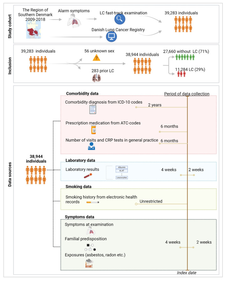
The study cohort comprised patients evaluated at the LC fast-track clinics in the Region of Southern Denmark from 1 January 2009 to 31 December 2018. Details on the definition of the study cohort and data sources are outlined in related work [27]. In the final cohort of 38,944 individuals examined for suspected LC, 29% were diagnosed with LC (Figure 1). A broad range of data was collected on this cohort, categorized into four datasets based on data availability:
Figure 1.
Study cohort, inclusion criteria, and data collection. The data sources included in the study were categorized into comorbidity data, laboratory results, smoking history, and symptoms data. Comorbidity data encompassed information on ICD-10 codes, prescription medications, the number of visits, and quick tests performed in general practice. Laboratory results consisted of 20 different analyses. Smoking history provided detailed records of smoking habits in binary format, while symptoms data included information on common symptoms, familial predispositions, and relevant exposures to LC. These data were collected for specific periods leading up to the date of inclusion, referred to as the index date, and are depicted by the bars on the right side of the image. Created with Biorender.com.
Comorbidity Dataset: This dataset contains binary information on the international classification of diseases—10th revision (ICD-10) codes obtained from a hospital level, the presence of prescribed medications indicated by anatomic therapeutic chemical (ATC) codes, and the number of consultations and C-reactive protein (CRP) rapid tests conducted in general practice. ICD-10 codes were registered if they appeared within two years prior to the LC fast-track examination, while the interval for ATC codes and general practice data was six months. These data were available for the entire population and were filtered to a subset of ICD-10 and ATC codes that field experts identified as being potentially associated with LC risk [27].
Laboratory Dataset: This dataset includes the results of 20 common laboratory analyses performed at the LC fast-track clinics. These analyses were included if conducted within the period of four weeks before the LC examination until two weeks after. This was performed in order to reflect the laboratory status at the time of the LC examination.
Smoking Dataset: This dataset provides binary information on smoking status, categorized as either never smoker or current/former smoker. This information was derived from free-text data in subfields of the electronic health records, which were manually annotated when available. The absence of data in this subfield limited this dataset to a subset of the entire population [30].
Symptom Dataset: This dataset contains manually annotated data from free text regarding the presence of the most common symptoms recorded in the primary journal of the LC examination. Additionally, it includes information on genetic predispositions to LC (parents or siblings with LC) and relevant exposures, such as radon or asbestos, based on the same records. The primary journals were filtered to include entries recorded within four weeks before to two weeks after the LC examination date. To reduce the burden of manual annotation, we focused on annotating data for a subset of individuals with complete information from the previously described datasets. This reduced cohort has previously been compared to the remaining individuals with missing data and showed overall similarity. However, the reduced cohort had a lower rate of comorbidities among LC patients and exhibited fewer differences in medication prescriptions compared to the remaining cohort [27].
2.2. Experimental Setup
2.2.1. Discretization
Before constructing the BN models, continuous variables were binned using the minimum description length (MDL) strategy [31]. This strategy seeks to find the optimal number of bins for the continuous laboratory results. The optimal number of bins is described by a model that offers the shortest overall description of the data, balancing model complexity (avoids overfitting) and accuracy (avoids underfitting). In initial exploratory experiments, we compared the MDL discretization method with the standard clinical reference intervals provided by the laboratory departments, which are typically based on 95% confidence intervals. We observed no significant differences between the two approaches and, for the sake of simplicity, chose to use the MDL strategy in the final experiments.
2.2.2. Model Development
The development of the BN models involved two key phases: structure learning and parameter learning. For structure learning, we employed the K2 algorithm introduced by Cooper et al. in 1992 [32]. This algorithm uses a greedy search strategy to identify the most suitable structure for the DAG. It iteratively adds one variable at a time, aiming to maximize the scoring function, which reflects the likelihood of the data given the network structure. This process continues until no further improvement in the score is observed or the maximum number of parent nodes per variable is reached, which was tested with 1–10 parent nodes in this case. The data used for structure learning needed to be complete, so missing continuous variables were imputed with the mean while missing discrete variables were imputed with the mode. The best structure was selected based on its area under the ROC curve (AUC), as estimated using 10-fold cross-validation.
In the parameter learning phase, we learned the conditional probability tables using the Expectation–Maximization (EM) algorithm [33]. The EM algorithm handles missing values in two steps: the expectation step, where it estimates the missing data based on the observed data, and the maximization step, where it adjusts the parameters to maximize the likelihood of the observed data. These steps are repeated until the parameter estimates stabilize.
We conducted preliminary exploratory experiments comparing the K2 algorithm with expert-drawn graphs and found no significant differences between them. For simplicity, we decided to use the K2 algorithm for the experiments described in this article.
2.2.3. Division of Study Cohort
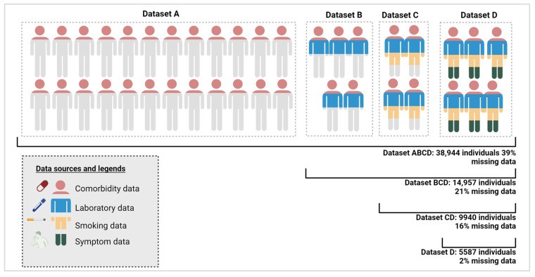
To investigate model performance with varying dataset sizes, completeness levels, and attributes, the study cohort was divided into subsets. Figure 2 displays these subsets, Datasets A to D, derived based on the four data categories: Dataset A includes individuals holding only comorbidity data; Dataset B includes individuals with comorbidity and laboratory results; Dataset C holds individuals with comorbidity, lab, and smoking data; and Dataset D individuals encompassing all four categories.
Figure 2.
Datasets A–D and the combination of these with increasing degree of missing data. The color code of the individuals reflects the type of data available on this subset from the four combinations of data: comorbidity, laboratory, smoking, and symptoms data. Created with Biorender.com.
Datasets A–D were combined to reflect a real-world distribution of missing data within datasets, where certain groups of individuals have complete data, while others lack information in some categories, such as smoking habits or symptoms. While Dataset D represents a small but nearly complete dataset with only 2% missing data, the other dataset combinations have higher rates of missing data: Dataset CD has 16% missing data, Dataset BCD has 21% missing data, and Dataset ABCD has 39% missing data. It should be noted that in the less complete datasets, the missing data levels include variables that are completely missing. General variables such as sex and age were included in all developed models.
2.3. Evaluation Setup
2.3.1. Evaluation 1
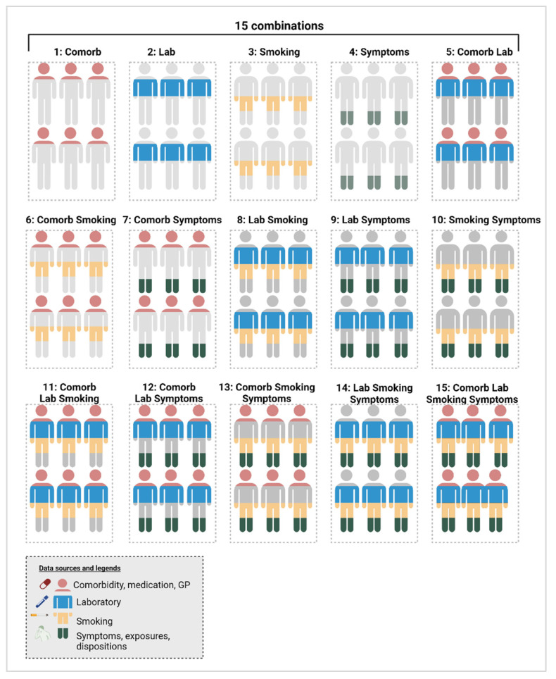
Models were trained on the records present in the four combinations of increasingly complete datasets (ABCD/BCD/CD/D), as described in Figure 2. Additionally, each of these four combinations was trained using all possible sub-combinations of the various data categories. This results in 15 different models, which can be found in Figure 3. One example is a model trained on dataset ABCD, using only the comorbidity-related variables.
Figure 3.
The 15 models trained and validated on dataset D, comparing different combinations of the four data types: comorbidity, laboratory results, smoking history, and symptoms data. Created with Biorender.com, https://www.biorender.com, accessed on 5 August 2024).
Validation involved a combination of 10-fold cross-validation on overlapping datasets and external validation on non-overlapping datasets. For example, a model trained on dataset D was evaluated using cross-validation within dataset D. In addition, the model trained on dataset D was externally validated on dataset A. When larger datasets were used for validation, the results from both validation methods were integrated. For instance, the performance of a model trained on dataset D and validated on dataset ABCD was assessed by combining the cross-validation results from dataset D with the external validation results from datasets A, B, and C.
2.3.2. Evaluation 2
To determine the optimal set of variables overall, we used the most complete dataset (dataset D) for both training and validation using 10-fold cross-validation. We evaluated the performance using their AUC score of the 15 potential combinations of the four data categories shown in Figure 3. By comparing the same dataset, the cohort size remained consistent, ensuring that any performance improvements were solely due to the combination of variables rather than an increase in cohort size.
2.4. Statistical Analyses
Baseline characteristics were described using the median and interquartile range (IQR) for continuous variables and number and percentage for categorical variables. The validation of the experiments was conducted using AUCs, along with 95% confidence intervals (CIs) calculated using a standard normal distribution. Discrimination was assessed through the AUCs, and the true positive rate (TPR/sensitivity) and true negative rate (TNR, specificity) were evaluated at the default probability cut-off of 0.5. All experiments were conducted using the WEKA framework version 3.8 [34].
3. Results
3.1. Baseline Characteristics
Table 1 presents the baseline characteristics for each dataset category. The LC group was older and had a higher proportion of females compared to the non-LC group, with ages of 70 years (IQR 63–77) versus 67 years (IQR 56–75) and 48.4% versus 45.3% females, respectively (p < 0.001 for both). Both groups commonly had comorbidities such as other malignancies (13.3%), chronic pulmonary disease (12.4%), and pneumonia (10.5%). However, LC patients were significantly more likely to have metastatic solid tumors, cerebrovascular disease, and peripheral vascular disease, whereas non-LC patients had a higher prevalence of other malignancies. Antibiotics were the most frequently prescribed medication in both groups, with no variation across groups (43.9%). Other medication categories were more frequently prescribed to LC patients.
Table 1.
Baseline characteristics for the non-LC and LC groups, including demographic data as well as information from the comorbidity, laboratory, smoking, and symptoms datasets. Categorical or binary data are expressed as fractions and compared using chi-squared tests, while continuous variables are shown as medians with interquartile ranges and p-values calculated using Wilcoxon signed-rank tests.
| Demography | Non-LC | LC | p-Value |
|---|---|---|---|
| Total, no. (%) | 27,660 (100) | 11,284 (100) | |
| Age, median (IQR) | 67 (56–75) | 70 (63–77) | <0.001 |
| Females, no. (%) | 12,515 (45.3) | 5461 (48.4) | <0.001 |
| LC stage, no. (%) | |||
| I | 2.001 (17.7) | ||
| II | 914 (8.1) | ||
| III | 2242 (19.9) | ||
| IV | 5440 (48.2) | ||
| Unknown | 687 (6.1) | ||
| Comorbidity dataset | Non-LC | LC | p-value |
| Total, no. (%) | 27,660 (100) | 11,284 (100) | |
| Myocardial infarction | 454 (1.6) | 225 (2.0) | 0.02 |
| Congestive cardiac failure | 198 (0.7) | 69 (0.61) | 0.26 |
| Peripheral vascular disease | 828 (3.0) | 555 (4.9) | <0.001 |
| Cerebrovascular disease | 915 (3.3) | 525 (4.7) | <0.001 |
| Dementia | 200 (0.7) | 77 (0.7) | 0.67 |
| Chronic pulmonary disease | 3379 (12.2) | 1429 (12.7) | 0.22 |
| Rheumatological disease | 533 (1.9) | 228 (2.0) | 0.54 |
| Liver disease | 198 (0.7) | 65 (0.6) | 0.13 |
| Diabetes mellitus | 1245 (4.5) | 566 (5.0) | 0.03 |
| Hemiplegia or paraplegia | 30 (0.1) | 18 (0.2) | 0.19 |
| Renal disease | 522 (1.9) | 179 (1.6) | 0.05 |
| Metastatic solid tumor | 772 (2.8) | 6.5 (5.4) | <0.001 |
| AIDS/HIV infection | 19 (0.1) | 9 (0.1) | 0.94 |
| Pulmonary tuberculosis | 48 (0.2) | 8 (0.1) | 0.02 |
| Sarcoidosis | 79 (0.3) | 18 (0.2) | 0.02 |
| Interstitial lung disease | 194 (0.7) | 74 (0.7) | 0.62 |
| Abscess | 157 (0.6) | 43 (0.4) | 0.02 |
| Pleural disease | 725 (2.6) | 297 (2.6) | 0.95 |
| Pneumonia | 2944 (10.6) | 1132 (10.0) | 0.07 |
| SumCCI, sum (median) | 0 (50) | 0 (50) | <0.001 |
| Other malignancies | 3859 (14.0) | 1321 (11.7) | <0.001 |
| Antibiotics | 12,130 (43.9) | 4954 (43.9) | 0.93 |
| COPD inhalations | 7028 (25.4) | 3490 (30.9) | <0.001 |
| Antihypertensives | 9560 (34.6) | 4660 (41.3) | <0.001 |
| Glucocorticoids | 2770 (10.0) | 1534 (13.6) | <0.001 |
| Metformin | 1694 (6.1) | 837 (7.4) | <0.001 |
| Antidepressants | 3960 (14.3) | 1838 (16.3) | <0.001 |
| Consultations at GP | 3093 (11.2) | 932 (8.3) | <0.001 |
| CRP rapid tests at GP | 13,053 (47.2) | 5275 (46.8) | |
| Laboratory data | Non-LC | LC | p-value |
| Total, no. (%) | 10,503 (100) | 4454 (100) | |
| B-Hemoglobin, mmol/L | 8.7 (8.0–9.3) | 8.40 (7.7–9.0) | <0.001 |
| B-Leucocytes, 109/L | 7.64 (6.20–9.46) | 9.12 (7.43–11.20) | <0.001 |
| B-Neutrophils, 109/L | 4.70 (3.58–6.20) | 6.10 (4.71–7.95) | <0.001 |
| B-Lymphocytes, 109/L | 1.81 (1.39–2.33) | 1.74 (1.30–2.27) | <0.001 |
| NLR | 2.6 (1.8–3.8) | 3.4 (2.4–5.2) | <0.001 |
| B-Monocytes, 109/L | 0.65 (0.51–0.84) | 0.76 (0.59–0.97) | <0.001 |
| B-Basophils, 109/L | 0.04 (0.02–0.06) | 0.04 (0.02–0.06) | <0.001 |
| B-Eosinophils, 109/L | 0.17 (0.10–0.27) | 0.14 (0.07–0.25) | <0.001 |
| B-Platelets, 109/L | 272 (223–334) | 311 (250–391) | <0.001 |
| P-Albumin, g/L | 43 (41–45) | 42 (39–44) | <0.001 |
| Total Calcium, mmol/L | 2.34 (2.27–2.41) | 2.36 (2.29–2.43) | <0.001 |
| P-CRP, mg/L | 3.7 (1.4–10.0) | 9.9 (3.0–32.0) | <0.001 |
| P-ALAT, U/L | 22 (16–31) | 18 (13–26) | <0.001 |
| P-LDH, U/L | 192 (169–221) | 214 (182–257) | <0.001 |
| P-Alkaline phosphatase, U/L | 75 (62–92) | 83 (68–102) | <0.001 |
| P-Bilirubin-total, μmol/L | 7 (6–10) | 7 (5–9) | <0.001 |
| P-Amylase (pancreatic), U/L | 25 (19–34) | 25 (18–34) | 0.79 |
| P-INR | 1.0 (0.9–1.1) | 1.0 (0.9–1.1) | <0.001 |
| P-Creatinine, mmol/L | 76 (64–89) | 72 (60–87) | <0.001 |
| P-Sodium, mmol/L | 140 (138–142) | 139 (136–141) | <0.001 |
| P-Potassium, mmol/L | 4.0 (3.8–4.3) | 4.0 (3.8–4.3) | 0.08 |
| Smoking status | Non-LC | LC | p -value |
| Total | 7435 (100) | 2505 (100) | |
| Never smoker | 2288 (30.8) | 196 (1.8) | <0.001 |
| Former or current smoker | 5147 (69.2) | 2309 (92.2) | |
| Symptoms, familial predispositions and exposures, no. (%) | Non-LC | LC | p -value |
| Total | 3733 (100) | 1854 (100) | |
| Predispositions | 253 (6.8) | 167 (9.0) | 0.00 |
| Exposures | 785 (21.0) | 354 (19.1) | 0.09 |
| Hemoptysis | 694 (18.6) | 212 (11.4) | <0.001 |
| Pneumonia | 671 (18.0) | 303 (16.3) | 0.13 |
| Cough | 2012 (53.9) | 969 (52.3) | 0.25 |
| Dyspnoea | 1365 (36.6) | 663 (35.8) | 0.56 |
| Fever | 286 (7.2) | 81 (4.4) | <0.001 |
| Weight loss | 822 (22.0) | 584 (31.5) | <0.001 |
| Fatigue | 684 (18.3) | 428 (23.1) | <0.001 |
| Hot flash | 402 (10.8) | 177 (9.6) | 0.16 |
| Hoarseness | 174 (4.7) | 92 (5.0) | 0.62 |
| Back pain | 133 (3.6) | 129 (7.0) | <0.001 |
| Other pain | 340 (9.1) | 250 (13.5) | <0.001 |
| Angina | 428 (11.5) | 256 (13.8) | 0.01 |
| Headache | 144 (3.1) | 65 (3.5) | 0.37 |
| Dizziness | 161 (4.3) | 96 (5.2) | 0.15 |
| Edema | 196 (5.3) | 108 (5.8) | 0.37 |
In laboratory results, most median values fell within clinical standard reference intervals, which were based on 95% CIs. Nevertheless, LC patients showed significantly elevated levels of white blood cells (leukocytes, neutrophils, monocytes), platelets, calcium, CRP, lactate dehydrogenase (LDH), and alkaline phosphatase compared to non-LC patients. Conversely, LC patients had lower levels of hemoglobin, eosinophils, lymphocytes, albumin, alanine aminotransferase (ALAT), creatinine, and sodium. The proportion of current or former smokers was significantly higher among LC patients (92%) compared to non-LC patients (69%, p < 0.001). The most common symptoms in both groups included cough (53.4%), dyspnea (36.3%), weight loss (25.2%), fatigue (19.9%), and hemoptysis (16.2%). Weight loss, fatigue, back pain, and other pain symptoms were more prevalent among LC patients, while hemoptysis and fever were more common in the non-LC cohort (p < 0.001 for all). For further details on baseline characteristics, please refer to the related literature [27].
3.2. Performance Assessment
3.2.1. Evaluation 1
Table 2 presents the AUC values for models trained on datasets ABCD, BCD, CD, and D, with validation conducted on datasets A, B, C, D, BCD, CD, and ABCD. For each combination, the 15 possible combinations of the four data types were evaluated, but for simplicity, only the best-performing combination is shown.
Table 2.
AUC measures and 95% CI obtained from various combinations of datasets A-D used for training and validation. For each dataset, the table includes the results of testing all 15 possible combinations of data types, with the optimal combination highlighted for each case. The two best performing models are highlighted in bold.
| Val. Data | Training Data ABCD | Training Data BCD | Training Data CD | Training Data D | ||||
|---|---|---|---|---|---|---|---|---|
| Variables | AUC (95% CI) | Variables | AUC (95% CI) | Variables | AUC (95% CI) | Variables | AUC (95% CI) | |
| A | Comorb Symptoms | 0.63 (0.62–0.64) |
Comorb Lab | 0.62 (0.60–0.63) |
Comorb | 0.60 (0.59–0.61) |
Comorb Smoking Symptoms | 0.60 (0.59–0.61) |
| B | Comorb Lab Symptoms | 0.78 (0.75–0.82) |
Comorb Lab Smoking |
0.79
(0.75–0.83) |
Comorb Lab | 0.78 (0.74–0.81) |
Comorb Lab | 0.77 (0.73–0.80) |
| C | Lab Smoking | 0.72 (0.67–0.76) |
Lab Smoking Symptoms | 0.72 (0.78–0.77) |
Lab Smoking Symptoms | 0.73 (0.69–0.78) |
Lab Smoking | 0.73 (0.58–0.80) |
| D | Comorb Lab Smoking Symptoms | 0.75 (0.72–0.79) |
Comorb Lab Smoking Symptoms | 0.77 (0.73–0.80) |
Comorb Lab Smoking | 0.77 (0.73–0.80) |
Comorb Lab Smoking Symptoms |
0.78
(0.75–0.82) |
| CD | Comorb Lab Smoking Symptoms | 0.76 (0.73–0.79) |
Comorb Lab Smoking Symptoms | 0.77 (0.74–0.80) |
Comorb Lab Smoking Symptoms | 0.78 (0.75–0.81) |
Comorb Lab Smoking Symptoms | 0.77 (0.74–0.80) |
| BCD | Comorb Lab Smoking Symptoms | 0.77 (0.75–0.79) |
Comorb Lab Smoking Symptoms | 0.78 (0.76–0.80) |
Comorb Lab Smoking | 0.78 (0.75–0.80) |
Comorb Lab Smoking Symptoms | 0.78 (0.75–0.80) |
| ABCD | Comorb Lab Smoking Symptoms | 0.69 (0.68–0.70) |
Comorb Lab Smoking | 0.68 (0.67–0.69) |
Comorb Lab Smoking | 0.66 (0.65–0.67) |
Comorb Lab Smoking Symptoms | 0.67 (0.66–0.68) |
Model performance varied significantly, ranging from an AUC of 0.60 (95% CI 0.59–0.61) for the worst-performing model to 0.79 (95% CI 0.75–0.83) for the best-performing model. The top-performing model was trained using dataset BCD and validated on dataset B using cross-validation, incorporating data on comorbidities, laboratory results, and smoking history. This dataset included 14,957 individuals and had 21% missing data. There was no significant difference between this model and the second-best model when considering the overlap in confidence intervals. The second-best model achieved an AUC of 0.78 (95% CI 0.75–0.82) and was trained and validated on the smaller, nearly complete dataset D, utilizing all four types of data variables. When tested on larger datasets with higher rates of missing data, its performance remained relatively stable, with AUC values slightly decreasing to 0.77 (95% CI 0.74–0.80) on dataset CD (16% missing data) and 0.78 (95% CI 0.75–0.80) on dataset BCD (21% missing data). However, its performance declined significantly to an AUC of 0.67 (95% CI 0.66–0.68) when validated on dataset ABCD, which had 39% missing data. Overall, all models performed poorly when validated on dataset ABCD, particularly on dataset A alone, regardless of the training set used.
3.2.2. Evaluation 2
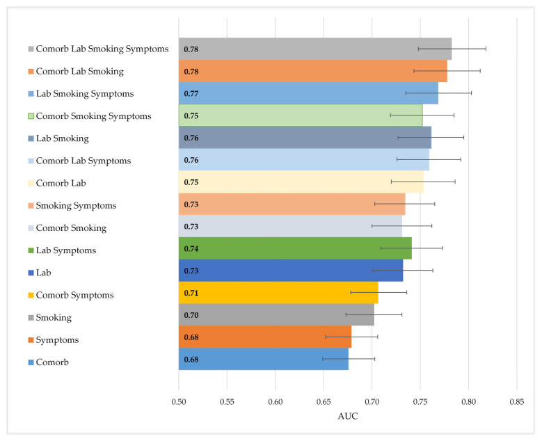
Figure 4 illustrates the performance comparison across the 15 dataset combinations, using dataset D for training and validation. The top-performing model, which utilized all four types of data, achieved an AUC of 0.783 (95% CI 0.748–0.818). This model, previously identified as the second-best overall, showed slightly better performance compared to the model that used only laboratory and smoking data (AUC 0.761, 95% CI 0.727–0.795), though there was considerable overlap in confidence intervals. Both of these models significantly outperformed the one based solely on smoking status, along with age and gender, which had an AUC of 0.702 (95% CI 0.673–0.731). Overall, models that included laboratory and smoking data achieved the best performance, while comorbidities and symptoms had the least impact on model performance.
Figure 4.
Comparison of AUCs when combining the four types of data in models trained and validated on dataset 4 (5587 individuals). The 95% confidence intervals are indicated with bars.
4. Discussion
4.1. Summary of Results
In this study, we developed BNs to predict the risk of lung cancer (LC) among a high-risk population in southern Denmark based on a decade of data. We analyzed data from 38,944 individuals, categorized into four types: comorbidity data, laboratory results, smoking history, and symptom-related data. To evaluate model performance across varying data completeness and sizes, the dataset was divided into smaller subsets based on the structure of the missing data.
We found that a relatively small high-quality training dataset could produce acceptable performance when validated on a dataset of lower quality. This was demonstrated by both the top-performing model, trained on dataset BCD, and the second-best model, trained on dataset D. Model performance remained largely stable until tested on a dataset containing 39% missing values (dataset ABCD). The best results were achieved by incorporating all four types of data (comorbidities, laboratory, smoking, and symptoms). Overall, laboratory and smoking data had the greatest impact on model performance, while symptom-related data had the least influence.
4.2. Interpretation and Comparison
We demonstrated that a model trained on high-quality data maintains strong performance even when validated against datasets with higher rates of missing information. This was true for both the top-performing model trained on dataset BCD and validated on dataset B, as well as the second-best model trained on dataset D, which yielded nearly identical results when validated on datasets CD, BCD, and B. This indicates that including lower-quality data does not significantly impair the model’s performance and can still offer valuable insights. For example, a model built from comorbidity, laboratory, smoking, and symptoms data on a small cohort (dataset D) remains consistently effective when applied to larger cohorts with partially missing data for smoking and symptoms (dataset BCD).
This ability to handle missing data is advantageous for screening, as it allows a model to be trained on a bigger cohort, which includes individuals with some missing data, without immediately harming model performance. This approach enables a larger proportion of individuals to be included in the training data without the need to exclude or impute missing data, which can introduce bias or errors, especially if the missing data are not missing at random. For instance, different subpopulations may have a different probability of having a specific laboratory test taken, resulting in different rates of missingness for this variable. Imputing this test based on the global population could introduce bias. BNs diminish these biases by using a probabilistic framework that accounts for the different subpopulations [24].
The model’s ability to handle missing data makes it scalable to various populations, including those in resource-limited settings where comprehensive data collection, such as smoking history or symptoms, may be difficult. For example, even when relying solely on data from ICD-10 comorbidity diagnoses and laboratory results due to recruitment challenges, the model still achieves an acceptable AUC of 0.77 when trained on dataset D and validated on dataset B.
This ability to handle missing values shows the advantages of using a BN during the training phase. In addition to this, it brings a similar benefit when the finalized model is utilized in clinical practice. Since the model can handle incomplete records, it becomes possible to use the model to screen a new patient without having to apply the full set of medical tests. This means that expensive, invasive, time-consuming, or otherwise inconvenient data to collect could potentially be skipped without compromising the accuracy of the model. This is especially relevant in screening scenarios as this reduces the barrier to entry, potentially allowing more individuals to be screened at a cheaper cost and with less discomfort.
However, when validated on dataset ABCD, which has 39% missing data, the model’s performance drops significantly, indicating a threshold for the amount of missing data the model can effectively handle. This effect may be compounded by the fact that dataset ABCD introduces a large number of records with missing lab values, which appear to be an important variable in our model. However, it should be noted that the drop in performance is present regardless of which set of attributes was used. It is well-known from the literature that although BNs are capable of managing missing data, their performance deteriorates as the quality of missing data decreases [35,36]. The exact rate or threshold of deterioration can vary depending on factors such as the dataset, model complexity, and the pattern of missing data. In cases where data are not missing at random—such as in this study, where entire categories of data were missing (e.g., all comorbidity or all lab values)—the impact on model performance is more pronounced compared to situations where data are missing completely at random [35,37].
Additionally, performance declines when validating on dataset A alone, suggesting that the comorbidity dataset by itself may not provide sufficient information for LC detection. This could be due to the fact that a significant proportion of patients in both groups had any of the included comorbidities: 62% of the LC cohort and 65% of the non-LC cohort. Furthermore, the comorbidity dataset included data on general practitioner visits and CRP measurements, but these variables did not significantly differentiate between LC and non-LC patients. Consequently, their presence likely diminished the dataset’s overall significance. Despite a higher overall rate of prescription medication among LC patients, this difference did not substantially enhance the predictive power when combined with variables related to ICD-10 codes and general practice.
The performance of four combinations of variables was compared, with the best results achieved by using all four data types. This combination of all four data types was comparable to the models that only used laboratory and smoking data, suggesting that adding comorbidity and symptoms data can only slightly enhance performance. Interestingly, both these combinations surpassed the conventionally used LC screening criteria, which rely solely on smoking status (along with age and sex). This underlines the potential of using other data sources in the selection of eligible individuals. Laboratory and smoking data have already been utilized by the members of the same study group to create ML and BN models [28,29]. In those models, dataset C, comprising 9940 individuals, was used with a focus on smoking and laboratory data, as initial exploration highlighted their significance in distinguishing LC from non-LC patients. The DES model developed by Flyckt et al. achieved a performance of 0.77, while the BN model by Henriksen et al. reached a similar performance of 0.76 on the same dataset. In this current study, dataset C was not used alone for model training, so a direct comparison cannot be made. However, the results indicate that the performance of both previous models can be matched or even surpassed by incorporating additional data sources (such as comorbidity and symptoms data) and/or expanding to a larger dataset despite its higher rate of missing data.
The PLCOm2012 model, which is frequently used, has demonstrated superior performance compared to the NLST eligibility criteria, with AUC values ranging from 0.76 to 0.81 in validation studies [38,39,40]. Although these results are similar to those found in this paper, a direct comparison is challenging. This is because the PLCOm2012 model was developed and validated using screening populations with a lower incidence of LC than in the current study. Additionally, the PLCOm2012 model is designed to predict 6-year risk, whereas this study focuses on predicting risk at the time of examination or diagnosis, which is more about detection. Despite these differences, our findings are noteworthy given that all individuals in this study are considered high-risk, and the control group is not as healthy as those in typical screening populations. This makes it more difficult for the model to discriminate LC patients from non-LC individuals.
Future research will involve analyzing data from COPD outpatients who are at moderate risk for LC compared to the high-risk patients in this study. Additionally, with a Danish LC screening trial in the planning phase, exploring the development or validation of BNs for this cohort could be valuable. This research could help assess whether these models can exceed the performance of the criteria used in the forthcoming Danish screening program.
4.3. Methodological Considerations
The models developed in this study utilized a large dataset spanning a decade and covering an entire region of Denmark. This dataset stands out due to its integration of various data sources, including manual annotations on symptoms, dispositions, exposures, and smoking, from relatively large cohorts—a data collection approach that is quite rare. The combination and diversity of these data sources offer a comprehensive view of the distributions and risk factors for LC within this high-risk population. The experimental design, which combined datasets to simulate varying degrees of missing data, mirrors real-life scenarios where some patients might lack information on smoking and symptoms or may not have undergone laboratory tests. This approach provides a more realistic depiction compared to earlier studies that used artificially introduced missing values that were completely random [29].
Despite its strengths, this study has several limitations that warrant consideration. The analyses rely exclusively on retrospective data, which introduces several biases due to the constraints of using variables collected at specific times. The most critical limitation is selection bias, as the dataset primarily includes detailed information on individuals who have visited hospital settings, with limited representation of the broader population eligible for screening. This focus on high-risk patients may result in a model that performs poorly when applied to less severe cases or healthier individuals. Consequently, while the dataset is valuable for certain analyses, it is suboptimal for LC screening, which requires a dataset that better represents the general population.
Comorbidity data is also biased toward hospital-recorded diagnoses, leading to a high proportion of patients being classified as having no comorbidities. Incorporating data from general practice, such as the International Classification of Primary Care (ICPC) codes that include symptoms and diagnostic information at the population level, could help address this limitation. Furthermore, using symptom data sourced from the general population might enhance the model’s ability to distinguish between LC and non-LC patients, as non-LC individuals would likely exhibit fewer symptoms. Other omitted variables, such as socioeconomic status, may also influence both LC risk and smoking status. Including such data could introduce more variation in risk and improve model discrimination and performance. However, we chose not to include socioeconomic data, as it is centralized at Statistics Denmark and unavailable for real-time use in clinical settings. Instead, we prioritized data sources that are accessible for everyday clinical practice.
Another limitation relates to information bias and the quality of particularly smoking status, which is often inconsistently recorded. A more detailed smoking dataset that includes packs–years and years since quitting could enable more precise predictions and a fairer comparison to current screening criteria. However, the aim of this study was to analyze real-world data, acknowledge its incompleteness and variability, and explore how model performance changes when validated across different subgroups within the dataset.
A more general limitation is linked to the experimental setup, where we analyzed pooled data without assessing individual variables within these datasets. For example, the optimal performance might involve combining COPD, three lab tests, smoking, and age, but our results suggest that the significance of individual variables like COPD could be reduced by the lack of significance from other variables in the comorbidity dataset. Certain medication prescriptions may become more prominent when assessed independently. This approach was chosen to simplify the experimental setup, given the complexity arising from multiple subpopulations with missing data and the inclusion of four different datasets, but will be explored further in future studies.
5. Conclusions
Our findings demonstrate that it is feasible to develop an LC prediction model using a high-quality dataset and achieve satisfactory performance even when validating with lower-quality data with up to 39% missing data. The most effective variable combination was achieved by integrating all types of data, with smoking and laboratory results proving to be particularly informative. These results are relevant for LC screening scenarios, where data are often disjointed and difficult to obtain. Future research should focus on validating the model in a broader population, such as COPD outpatients. Furthermore, its clinical potential to improve screening efficiency and optimize resource utilization should be assessed, emphasizing analyses of specific feature importance rather than limiting the focus to the broader data groupings explored in this study. Finally, further exploration of models that can handle missing data is crucial to identifying the most effective strategies for improving prediction accuracy in real-world settings.
Author Contributions
Conceptualization, M.H.B.H. and F.v.D.; methodology, F.v.D. and M.H.B.H.; software, F.v.D.; validation, F.v.D.; formal analysis, F.v.D.; investigation, M.H.B.H. and F.v.D.; resources, F.v.D. and M.H.B.H.; data curation, M.H.B.H., M.A.K.A. and E.H.; writing—original draft preparation, M.H.B.H. and F.v.D.; writing—review and editing, M.H.B.H., F.v.D., L.W., T.F.H., O.H., C.L.B., L.H.J. and I.B.; visualization, M.H.B.H. and F.v.D.; supervision, L.W., T.F.H. and I.B; project administration, M.H.B.H. and F.v.D.; funding acquisition, M.H.B.H. All authors have read and agreed to the published version of the manuscript.
Institutional Review Board Statement
The study was conducted in accordance with the Declaration of Helsinki (as revised in 2013) and approved by the Danish Data Protection Agency (19/30673, 12 June 2020) and the Danish Patient Safety Authority (3-3013-3132/1, 30 March 2020).
Informed Consent Statement
Patient consent was waived due to the retrospective nature of this study.
Data Availability Statement
Data may be shared upon reasonable request.
Conflicts of Interest
The authors declare no conflicts of interest. The funders had no role in the design of the study; in the collection, analyses, or interpretation of data; in the writing of the manuscript; or in the decision to publish the results.
Funding Statement
The project was funded by The Region of Southern Denmark, The University of Southern Denmark, The Dagmar Marshall Foundation, the Lilly and Herbert Hansen Foundation, the Hede Nielsen Family Foundation, The Beckett Foundation, and The Danish National Research Center for Lung Cancer, Danish Cancer Society (grant no. R198-A14299). The funding sources did not participate in the data collection, analyses, or writing of the manuscript.
Footnotes
Disclaimer/Publisher’s Note: The statements, opinions and data contained in all publications are solely those of the individual author(s) and contributor(s) and not of MDPI and/or the editor(s). MDPI and/or the editor(s) disclaim responsibility for any injury to people or property resulting from any ideas, methods, instructions or products referred to in the content.
References
- 1.World Health Organization Lung Cancer [Internet] 2023. [(accessed on 13 December 2023)]. Available online: https://www.who.int/news-room/fact-sheets/detail/lung-cancer.
- 2.Thandra K.C., Barsouk A., Saginala K., Aluru J.S., Barsouk A. Epidemiology of lung cancer. Contemp. Oncol. 2021;25:45–52. doi: 10.5114/wo.2021.103829. [DOI] [PMC free article] [PubMed] [Google Scholar]
- 3.Bray F., Laversanne M., Sung H., Ferlay J., Siegel R.L., Soerjomataram I., Jemal A. Global cancer statistics 2022: GLOBOCAN estimates of incidence and mortality worldwide for 36 cancers in 185 countries. CA A Cancer J. Clin. 2024;74:229–263. doi: 10.3322/caac.21834. [DOI] [PubMed] [Google Scholar]
- 4.Aberle D., Adams A., Berg C., Black W., Clapp J., Fagerstrom R., Gareen I.F., Gatsonis C., Marcus P.M., Sicks J.D. Reduced Lung-Cancer Mortality with Low-Dose Computed Tomographic Screening. N. Engl. J. Med. 2011;365:395–409. doi: 10.1056/NEJMoa1102873. [DOI] [PMC free article] [PubMed] [Google Scholar]
- 5.de Koning H.J., van der Aalst C.M., de Jong P.A., Scholten E.T., Nackaerts K., Heuvelmans M.A., Lammers J.-W.J., Weenink C., Yousaf-Khan U., Horeweg N., et al. Reduced Lung-Cancer Mortality with Volume CT Screening in a Randomized Trial. N. Engl. J. Med. 2020;382:503–513. doi: 10.1056/NEJMoa1911793. [DOI] [PubMed] [Google Scholar]
- 6.Field J.K., Vulkan D., Davies M.P.A., Baldwin D.R., Brain K.E., Devaraj A., Eisen T., Gosney J., Green B.A., Holemans J.A., et al. Lung cancer mortality reduction by LDCT screening: UKLS randomised trial results and international meta-analysis. Lancet Reg. Health. 2021;10:100179. doi: 10.1016/j.lanepe.2021.100179. [DOI] [PMC free article] [PubMed] [Google Scholar]
- 7.Paci E., Puliti D., Pegna A.L., Carrozzi L., Picozzi G., Falaschi F., Pistelli F., Aquilini F., Ocello C., Zappa M., et al. Mortality, survival and incidence rates in the ITALUNG randomised lung cancer screening trial. Thorax. 2017;72:825–831. doi: 10.1136/thoraxjnl-2016-209825. [DOI] [PubMed] [Google Scholar]
- 8.van der Aalst C., Vonder M., Hubert J., Moldovanu D., Schmitz A., Delorme S., Kaaks R., ten Haaf K., Oudkerk M., de Koning H. P1. 14-04 European lung cancer screening implementation: 4-IN-THE-LUNG-RUN trial. J. Thorac. Oncol. 2023;18:S217. doi: 10.1016/j.jtho.2023.09.352. [DOI] [Google Scholar]
- 9.McWilliams A.M., Mayo J.R., Im Ahn M., MacDonald S.L.S., Lam S.C. Lung cancer screening using multi-slice thin-section computed tomography and autofluorescence bronchoscopy. J. Thorac. Oncol. 2006;1:61–68. doi: 10.1016/S1556-0864(15)31514-8. [DOI] [PubMed] [Google Scholar]
- 10.dos Santos R.S., Franceschini J.P., Chate R.C., Ghefter M.C., Kay F., Trajano A.L.C., Pereira J.R., Succi J.E., Fernando H.C., Júnior R.S. Do current lung cancer screening guidelines apply for populations with high prevalence of granulomatous disease? Results from the First Brazilian Lung Cancer Screening Trial (BRELT1) Ann. Thorac. Surg. 2016;101:481–488. doi: 10.1016/j.athoracsur.2015.07.013. [DOI] [PubMed] [Google Scholar]
- 11.Blanchon T., Bréchot J.-M., Grenier P.A., Ferretti G.R., Lemarié E., Milleron B., Chagué D., Laurent F., Martinet Y., Beigelman-Aubry C., et al. Baseline results of the Depiscan study: A French randomized pilot trial of lung cancer screening comparing low dose CT scan (LDCT) and chest X-ray (CXR) Lung Cancer. 2007;58:50–58. doi: 10.1016/j.lungcan.2007.05.009. [DOI] [PubMed] [Google Scholar]
- 12.Becker N., Motsch E., Gross M.-L., Eigentopf A., Heussel C.P., Dienemann H., Schnabel P.A., Eichinger M., Optazaite D.-E., Puderbach M., et al. Randomized study on early detection of lung cancer with MSCT in Germany: Results of the first 3 years of follow-up after randomization. J. Thorac. Oncol. 2015;10:890–896. doi: 10.1097/JTO.0000000000000530. [DOI] [PubMed] [Google Scholar]
- 13.Kreftforeningen Lung Cancer Screenings to Begin in Norway [Internet] [(accessed on 27 June 2024)]. Available online: https://kreftforeningen.no/forebygging/screening-og-masseundersokelser/?gad_source=1&gclid=Cj0KCQiAo5u6BhDJARIsAAVoDWvb6pfV9_cs9bw5nBXUc5JvXYNlLAvWWByrea7eNuftzaq9aTj4-3oaAjlqEALw_wcB.
- 14.Kaneko M., Eguchi K., Ohmatsu H., Kakinuma R., Naruke T., Suemasu K., Moriyama N. Peripheral lung cancer: Screening and detection with low-dose spiral CT versus radiography. Radiology. 1996;201:798–802. doi: 10.1148/radiology.201.3.8939234. [DOI] [PubMed] [Google Scholar]
- 15.Lu L., Tan Y., Schwartz L.H., Zhao B. Hybrid detection of lung nodules on CT scan images. Med. Phys. 2015;42:5042–5054. doi: 10.1118/1.4927573. [DOI] [PubMed] [Google Scholar]
- 16.Hu P., Dai M., Shi J., Ren J., Li J., Liao X., Du L., Liu Y., Chen Z., Wu N., et al. The feasibility study of a randomized cancer screening trial in China. Cancer Res. 2016;76:1795. doi: 10.1158/1538-7445.AM2016-1795. [DOI] [Google Scholar]
- 17.U.S. Preventive Services Task Force Final Recommendation Statement. Lung Cancer: Screening [Internet] 2021. [(accessed on 22 May 2024)]. Available online: https://www.uspreventiveservicestaskforce.org/uspstf/recommendation/lung-cancer-screening.
- 18.Pinsky P.F., Berg C.D. Applying the National Lung Screening Trial eligibility criteria to the US population: What percent of the population and of incident lung cancers would be covered? J. Med. Screen. 2012;19:154–156. doi: 10.1258/jms.2012.012010. [DOI] [PubMed] [Google Scholar]
- 19.Cassidy A., Myles J.P., van Tongeren M., Page R.D., Liloglou T., Duffy S.W., Field J. The LLP risk model: An individual risk prediction model for lung cancer. Br. J. Cancer. 2008;98:270–276. doi: 10.1038/sj.bjc.6604158. [DOI] [PMC free article] [PubMed] [Google Scholar]
- 20.Tammemägi M.C., Katki H.A., Hocking W.G., Church T.R., Caporaso N., Kvale P.A., Chaturvedi A.K., Silvestri G.A., Riley T.L., Commins J., et al. Selection criteria for lung-cancer screening. N. Engl. J. Med. 2013;368:728–736. doi: 10.1056/NEJMoa1211776. [DOI] [PMC free article] [PubMed] [Google Scholar]
- 21.Lebrett M.B., Balata H., Evison M., Colligan D., Duerden R., Elton P., Greaves M., Howells J., Irion K., Karunaratne D., et al. Analysis of lung cancer risk model (PLCO(M2012) and LLP(v2)) performance in a community-based lung cancer screening programme. Thorax. 2020;75:661–668. doi: 10.1136/thoraxjnl-2020-214626. [DOI] [PMC free article] [PubMed] [Google Scholar]
- 22.Tammemägi M.C., Church T.R., Hocking W.G., Silvestri G.A., Kvale P.A., Riley T.L., Commins J., Berg C.D. Evaluation of the lung cancer risks at which to screen ever- and never-smokers: Screening rules applied to the PLCO and NLST cohorts. PLoS Med. 2014;11:e1001764. doi: 10.1371/journal.pmed.1001764. [DOI] [PMC free article] [PubMed] [Google Scholar]
- 23.Chiu H.-Y., Chao H.-S., Chen Y.-M. Application of Artificial Intelligence in Lung Cancer. Cancers. 2022;14:1370. doi: 10.3390/cancers14061370. [DOI] [PMC free article] [PubMed] [Google Scholar]
- 24.Wüthrich M.V., Merz M. Statistical Foundations of Actuarial Learning and Its Applications. Springer; Cham, Switzerland: 2022. Bayesian Methods, Regularization and Expectation-Maximization; pp. 207–266. [Google Scholar]
- 25.McLachlan S., Dube K., Hitman G.A., Fenton N.E., Kyrimi E. Bayesian networks in healthcare: Distribution by medical condition. Artif. Intell. Med. 2020;107:101912. doi: 10.1016/j.artmed.2020.101912. [DOI] [PubMed] [Google Scholar]
- 26.Polotskaya K., Muñoz-Valencia C.S., Rabasa A., Quesada-Rico J.A., Orozco-Beltrán D., Barber X. Bayesian Networks for the Diagnosis and Prognosis of Diseases: A Scoping Review. Mach. Learn. Knowl. Extr. 2024;6:1243–1262. doi: 10.3390/make6020058. [DOI] [Google Scholar]
- 27.Henriksen M.B., Hansen T.F., Jensen L.H., Brasen C.L., Peimankar A., Ebrahimi A., Wii U.K., Hilberg O. A Collection of Multiregistry Data on Patients at High Risk of Lung Cancer. Transl. Lung Cancer Res. 2023;12:2392–2411. doi: 10.21037/tlcr-23-495. [DOI] [PMC free article] [PubMed] [Google Scholar]
- 28.Flyckt R.N.H., Sjodsholm L., Henriksen M.H.B., Brasen C.L., Ebrahimi A., Hilberg O., Hansen T.F., Wiil U.K., Jensen L.H., Peimankar A. Pulmonologists-Level lung cancer detection based on standard blood test results and smoking status using an explainable machine learning approach. Sci. Rep. 2024. Accepted for publication .
- 29.Henriksen M.B., van Daalen F., Wee L., Hansen T.F., Jensen L.H., Brasen C.L., Hilberg O.B.I. Lung cancer detection using Bayesian networks: A retrospective development and validation study on a Danish population of high-risk individuals. Cancer Med. 2024. Accepted for publication .
- 30.Ebrahimi A., Henriksen M.B.H., Brasen C.L., Hilberg O., Hansen T.F., Jensen L.H., Peimankar A., Wiil U.K. Identification of patients’ smoking status using an explainable AI approach: A Danish electronic health records case study. BMC Med. Res. Methodol. 2024;24:114. doi: 10.1186/s12874-024-02231-4. [DOI] [PMC free article] [PubMed] [Google Scholar]
- 31.Fayyad U.M., Irani K.B. Multi-interval discretization of continuous-valued attributes for classification learning. IJCAI. 1993;93:1022–1029. [Google Scholar]
- 32.Cooper G.F., Herskovits E. A Bayesian method for the induction of probabilistic networks from data. Mach. Learn. 1992;9:309–347. doi: 10.1007/BF00994110. [DOI] [Google Scholar]
- 33.Dempster A., Laird N., Rubin D., Dempster A.P., Laird N.M., Rubin D. Likelihood from incomplete data via the em algorithm. JR Stat. Soc. B. 1977;39:1–38. doi: 10.1111/j.2517-6161.1977.tb01600.x. [DOI] [Google Scholar]
- 34.Frank E., Hall M.A., Witten I.H. The WEKA Workbench. Morgan Kaufmann; Burlington, MA, USA: 2016. [Google Scholar]
- 35.Ke X., Keenan K., Smith V.A. Treatment of missing data in Bayesian network structure learning: An application to linked biomedical and social survey data. BMC Med. Res. Methodol. 2022;22:326. doi: 10.1186/s12874-022-01781-9. [DOI] [PMC free article] [PubMed] [Google Scholar]
- 36.Lin J.-H., Haug P.J. Exploiting missing clinical data in Bayesian network modeling for predicting medical problems. J. Biomed. Inform. 2008;41:1–14. doi: 10.1016/j.jbi.2007.06.001. [DOI] [PubMed] [Google Scholar]
- 37.Friedman N., Geiger D., Goldszmidt M. Bayesian network classifiers. Mach. Learn. 1997;29:131–163. doi: 10.1023/A:1007465528199. [DOI] [Google Scholar]
- 38.Tammemägi M.C. Selecting lung cancer screenees using risk prediction models-where do we go from here. Transl. Lung Cancer Res. 2018;7:243–253. doi: 10.21037/tlcr.2018.06.03. [DOI] [PMC free article] [PubMed] [Google Scholar]
- 39.Jantzen R., Ezer N., Camilleri-Broët S., Tammemägi M.C., Broët P. Evaluation of the accuracy of the PLCO(m2012) 6-year lung cancer risk prediction model among smokers in the CARTaGENE population-based cohort. C. Open. 2023;11:E314–E322. doi: 10.9778/cmajo.20210335. [DOI] [PMC free article] [PubMed] [Google Scholar]
- 40.Robbins H.A., Alcala K., Swerdlow A.J., Schoemaker M.J., Wareham N., Travis R.C., Crosbie P.A.J., Callister M., Baldwin D.R., Landy R., et al. Comparative performance of lung cancer risk models to define lung screening eligibility in the United Kingdom. Br. J. Cancer. 2021;124:2026–2034. doi: 10.1038/s41416-021-01278-0. [DOI] [PMC free article] [PubMed] [Google Scholar]
Associated Data
This section collects any data citations, data availability statements, or supplementary materials included in this article.
Data Availability Statement
Data may be shared upon reasonable request.


