Abstract
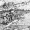
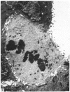
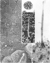
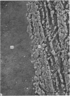
The teat and lactiferous sinus epithelium from the mammary glands of 23 lactating ewes was examined by light and electron microscopy. Most of the sinus epithelium consisted of two layers of non-secretory cells but, in the lactiferous sinus, cells with the same ultrastructural features as alveolar secretory cells were also found. Secretory cells sometimes occupied more than 50% of the total area of the sinus. Many non-secretory cells in the lactiferous sinus possessed a single cilium but they were less common in the teat sinus. 'Accessory glands', which opened directly into the lumen of the gland, were found beneath the epithelium in both the teat and the lactiferous sinuses. From their ultrastructure it was clear that these glands consisted of normal secretory alveoli and that they produced normal milk components. It is suggested that the mixed population of secretory and non-secretory cells in the lactiferous sinus provides unique material for the experimental study of many aspects of mammary gland physiology.
Full text
PDF
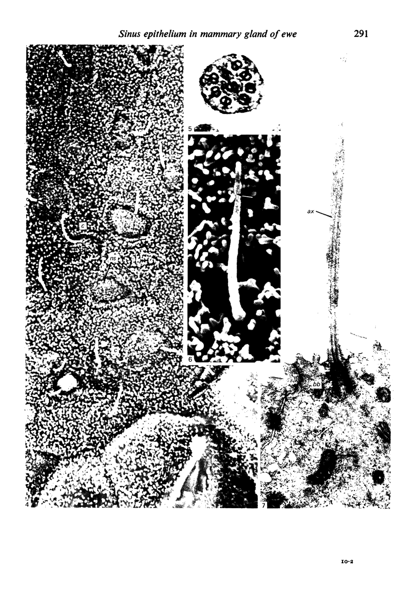
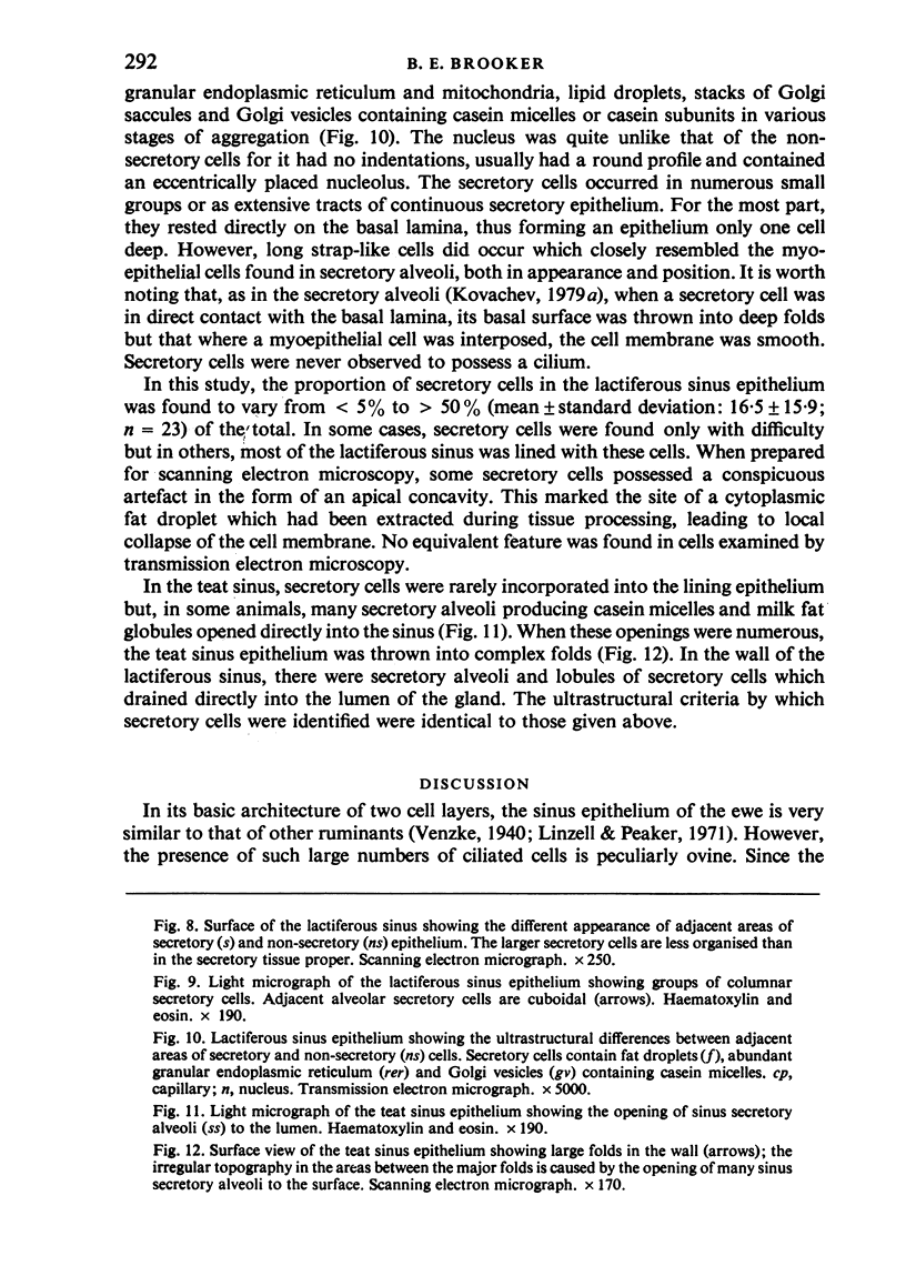
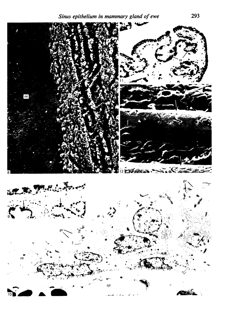
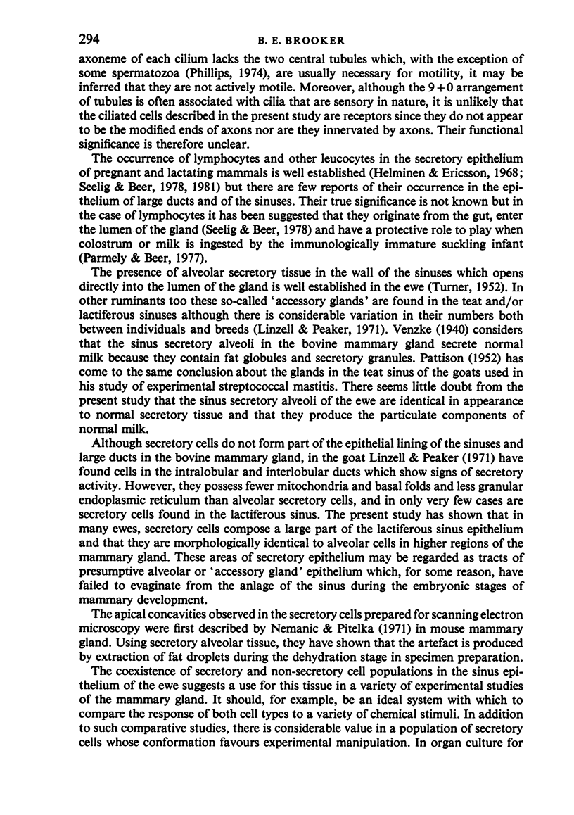
Images in this article
Selected References
These references are in PubMed. This may not be the complete list of references from this article.
- Brooker B. E. Pseudopod formation and phagocytosis of milk components by epithelial cells of the bovine mammary gland. Cell Tissue Res. 1983;229(3):639–650. doi: 10.1007/BF00207703. [DOI] [PubMed] [Google Scholar]
- FARQUHAR M. G., PALADE G. E. Junctional complexes in various epithelia. J Cell Biol. 1963 May;17:375–412. doi: 10.1083/jcb.17.2.375. [DOI] [PMC free article] [PubMed] [Google Scholar]
- Helminen H. J., Ericsson J. L. Studies on mammary gland involution. I. On the ultrastructure of the lactating mammary gland. J Ultrastruct Res. 1968 Nov;25(3):193–213. doi: 10.1016/s0022-5320(68)80069-3. [DOI] [PubMed] [Google Scholar]
- Lee C. S., Lascelles A. K. The histological changes in involuting mammary glands of ewes in relation to the local allergic response. Aust J Exp Biol Med Sci. 1969 Oct;47(5):613–623. doi: 10.1038/icb.1969.155. [DOI] [PubMed] [Google Scholar]
- Linzell J. L., Peaker M. The permeability of mammary ducts. J Physiol. 1971 Aug;216(3):701–716. doi: 10.1113/jphysiol.1971.sp009548. [DOI] [PMC free article] [PubMed] [Google Scholar]
- PATTISON I. H. Studies on experimental streptococcal mastitis. VI. Histological examination of the teats of affected goats. J Comp Pathol. 1952 Jan;62(1):1–5. [PubMed] [Google Scholar]
- Parmely M. J., Beer A. E. Colostral cell-mediated immunity and the concept of a common secretory immune system. J Dairy Sci. 1977 Apr;60(4):655–665. doi: 10.3168/jds.S0022-0302(77)83915-5. [DOI] [PubMed] [Google Scholar]
- Seelig L. L., Jr, Beer A. E. Intraepithelial leukocytes in the human mammary gland. Biol Reprod. 1981 Jun;24(5):1157–1163. [PubMed] [Google Scholar]
- Seelig L. L., Jr, Beer A. E. Transepithelial migration of leukocytes in the mammary gland of lactating rats. Biol Reprod. 1978 Jun;18(5):736–744. doi: 10.1095/biolreprod18.5.736. [DOI] [PubMed] [Google Scholar]


