Abstract
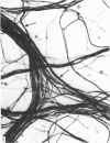
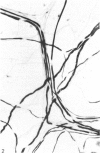
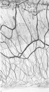
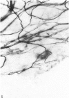
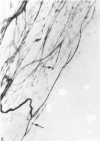
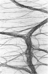
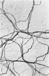
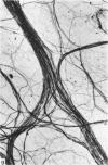
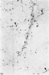
A silver impregnation technique (Linder, 1978) has been applied to whole mounts of the rat iris. The results suggest that only sensory fibres, both myelinated and non-myelinated, are stained. They disappear only after a trigeminal lesion and their distribution is different from that of catecholaminergic intrinsic fibres. Staining of the iris reveals a conspicuous pattern of innervation, characterised by a circular bundle and a thin plexus in the ciliary body, and by prominent bundles of fibres with a loose network of thin smooth fibres in the external part of the dilator plate and with a denser network in the central area. Nerve endings are seen on the dilator plate, in the sphincter as well as in the ciliary body. It is possible by a slight modification of the technique to stain myelinated and non-myelinated fibres separately. It results in a deep staining of the myelin while thin fibres are relatively clear. This method provides clear and reproducible staining of the nerves of the iris. It can be combined with various histochemical and immunocytochemical techniques. This will permit further studies to be made on the development of sensory and central nervous tissues, when grafted to the normal or the selectively denervated iris.
Full text
PDF


Images in this article
Selected References
These references are in PubMed. This may not be the complete list of references from this article.
- BEATIE J. C., STILWELL D. L., Jr Innervation of the eye. Anat Rec. 1961 Sep;141:45–61. doi: 10.1002/ar.1091410107. [DOI] [PubMed] [Google Scholar]
- Clarke P. G. Labelling of dying neurones by peroxidase injected intravascularly in chick embryos. Neurosci Lett. 1982 Jun 30;30(3):223–228. doi: 10.1016/0304-3940(82)90403-7. [DOI] [PubMed] [Google Scholar]
- Huhtala A. Origin of myelinated nerves in the rat iris. Exp Eye Res. 1976 Mar;22(3):259–265. doi: 10.1016/0014-4835(76)90053-1. [DOI] [PubMed] [Google Scholar]
- Hökfelt T., Kellerth J. O., Nilsson G., Pernow B. Substance p: localization in the central nervous system and in some primary sensory neurons. Science. 1975 Nov 28;190(4217):889–890. doi: 10.1126/science.242075. [DOI] [PubMed] [Google Scholar]
- Linder J. E. A simple and reliable method for the silver impregnation of nerves in paraffin sections of soft and mineralized tissues. J Anat. 1978 Dec;127(Pt 3):543–551. [PMC free article] [PubMed] [Google Scholar]
- MALMFORS T., NILSSON O. PARASYMPATHETIC POST-GANGLIONIC DENERVATION OF THE IRIS AND THE PAROTID GLAND IN THE RAT. Acta Morphol Neerl Scand. 1964;6:81–85. [PubMed] [Google Scholar]
- Miller A., Costa M., Furness J. B., Chubb I. W. Substance P immunoreactive sensory nerves supply the rat iris and cornea. Neurosci Lett. 1981 May 29;23(3):243–249. doi: 10.1016/0304-3940(81)90005-7. [DOI] [PubMed] [Google Scholar]
- Olson L. Fluorescence histochemical evidence for axonal growth and secretion from transplanted adrenal medullary tissue. Histochemie. 1970;22(1):1–7. doi: 10.1007/BF00310543. [DOI] [PubMed] [Google Scholar]
- Olson L., Malmfors T. Growth characteristics of adrenergic nerves in the adult rat. Fluorescence histochemical and 3H-noradrenaline uptake studies using tissue transplantations to the anterior chamber of the eye. Acta Physiol Scand Suppl. 1970;348:1–112. [PubMed] [Google Scholar]
- Olson L., Seiger A. A system of atypical catecholamine-containing nerve fibres in the rat iris present after total superior cervical ganglionectomy. Med Biol. 1980 Apr;58(2):94–100. [PubMed] [Google Scholar]
- Olson L., Seiger A. Brain tissue transplanted to the anterior chamber of the eye. 1. Fluorescence histochemistry of immature catecholamine and 5-hydroxytryptamine neurons reinnervating the rat iris. Z Zellforsch Mikrosk Anat. 1972;135(2):175–194. doi: 10.1007/BF00315125. [DOI] [PubMed] [Google Scholar]
- Richardson K. C. Electron microscopic identification of autonomic nerve endings. Nature. 1966 May 14;210(5037):756–756. doi: 10.1038/210756a0. [DOI] [PubMed] [Google Scholar]
- Saari M., Johansson G. Myelinated nerves of the rat iris. Acta Anat (Basel) 1974;89(1):139–144. doi: 10.1159/000144278. [DOI] [PubMed] [Google Scholar]
- Seiger A. Growth interaction between locus coeruleus and trigeminal ganglion after intraocular double grafting. Med Biol. 1980 Jun;58(3):149–157. [PubMed] [Google Scholar]
- Seiger A., Olson L. Reinitiation of directed nerve fiber growth in central monoamine neurons after intraocular maturation. Exp Brain Res. 1977 Aug 8;29(1):15–44. doi: 10.1007/BF00236873. [DOI] [PubMed] [Google Scholar]
- Skagerberg G., Emson P. C., Björklund A. Transplantation of irides or sensory ganglia to the anterior eye chamber of the rat: survival and sprouting of substance P-containing neurones. Neurosci Lett. 1982 Jan 22;28(1):29–34. doi: 10.1016/0304-3940(82)90203-8. [DOI] [PubMed] [Google Scholar]
- Stefanini M., De Martino C., Zamboni L. Fixation of ejaculated spermatozoa for electron microscopy. Nature. 1967 Oct 14;216(5111):173–174. doi: 10.1038/216173a0. [DOI] [PubMed] [Google Scholar]


