Abstract
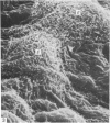
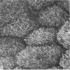
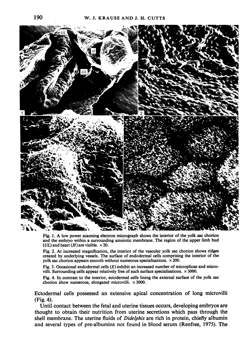
The 10 day opossum embryo is enclosed by a yolk sac chorion in which distinct vascular and non-vascular regions are established before attachment to the uterine epithelium. The embryo is closely associated with the vascular region which becomes adherent to the uterine lining. Cells of the external layer bear abundant, closely packed, long microvilli, which may serve to absorb nutrients from the uterine secretions. Cells of the inner surface show only scattered microvilli and microplicae on their apical surfaces.
Full text
PDF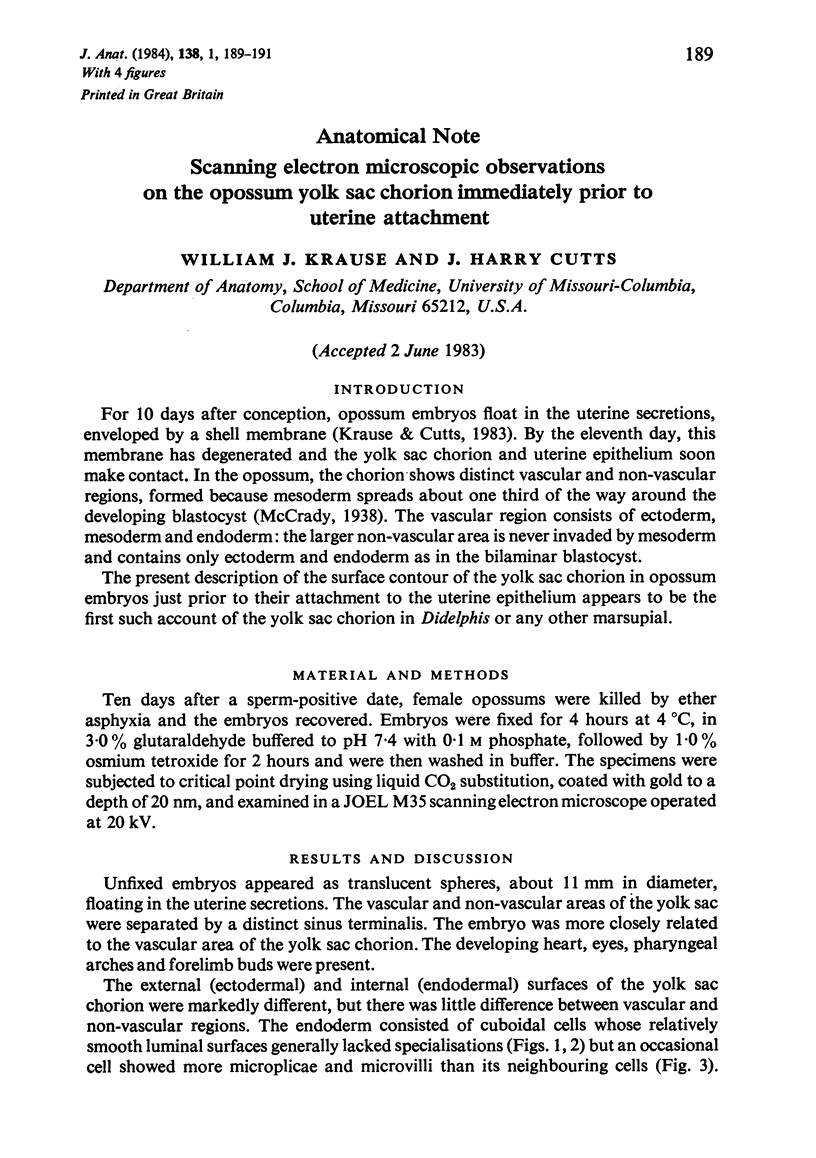
Images in this article
Selected References
These references are in PubMed. This may not be the complete list of references from this article.
- Renfree M. B. Uterine proteins in the marsupial, Didelphis Marsupialis virginiana, during gestation. J Reprod Fertil. 1975 Jan;42(1):163–166. doi: 10.1530/jrf.0.0420163. [DOI] [PubMed] [Google Scholar]


