Abstract
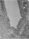
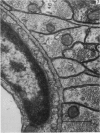
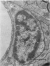
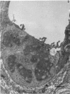
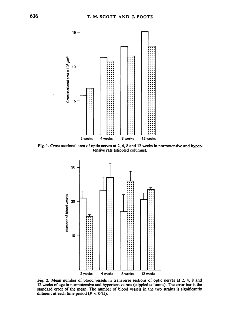
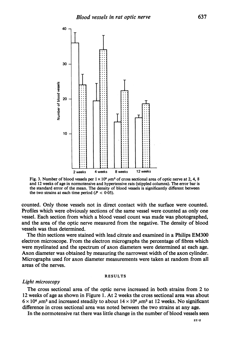
The blood vessels in the optic nerve of normotensive and hypertensive rats have been examined at 2, 4, 8 and 12 weeks of age. The pattern of development was found to be different in the two strains, with the number of blood vessels in the hypertensive rat optic nerve being lower at 2 weeks, but greater at 12 weeks than the normotensive rat. There appeared to be no correlation between vascularity and either myelination or changes in the fibre diameter spectrum at the ages studied. It is concluded that while the cause of the increased vascularity of the optic nerve in hypertensive rats is not known, it appears to be without effect in the structural development of the optic nerve.
Full text
PDF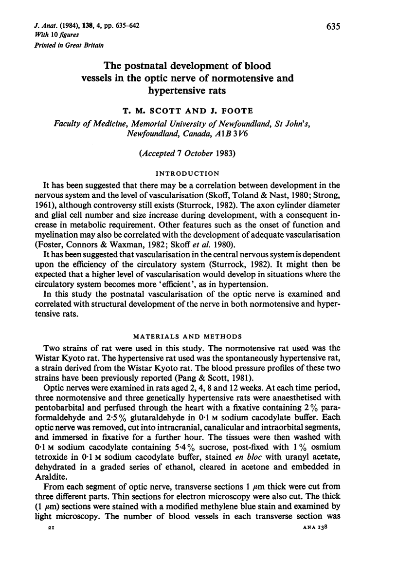
Images in this article
Selected References
These references are in PubMed. This may not be the complete list of references from this article.
- Bohlen H. G., Gore R. W., Hutchins P. M. Comparison of microvascular pressures in normal and spontaneously hypertensive rats. Microvasc Res. 1977 Jan;13(1):125–130. doi: 10.1016/0026-2862(77)90121-2. [DOI] [PubMed] [Google Scholar]
- Foster R. E., Connors B. W., Waxman S. G. Rat optic nerve: electrophysiological, pharmacological and anatomical studies during development. Brain Res. 1982 Mar;255(3):371–386. doi: 10.1016/0165-3806(82)90005-0. [DOI] [PubMed] [Google Scholar]
- Pang S. C., Scott T. M. Stereological analysis of the tunica media of the aorta and renal artery during the development of hypertension in the spontaneously hypertensive rat. J Anat. 1981 Dec;133(Pt 4):513–526. [PMC free article] [PubMed] [Google Scholar]
- Skoff R. P., Toland D., Nast E. Pattern of myelination and distribution of neuroglial cells along the developing optic system of the rat and rabbit. J Comp Neurol. 1980 May 15;191(2):237–253. doi: 10.1002/cne.901910207. [DOI] [PubMed] [Google Scholar]
- Sturrock R. R. A quantitative study of vascularisation of the prenatal rabbit spinal cord. J Anat. 1982 Aug;135(Pt 1):89–96. [PMC free article] [PubMed] [Google Scholar]


