

Abstract
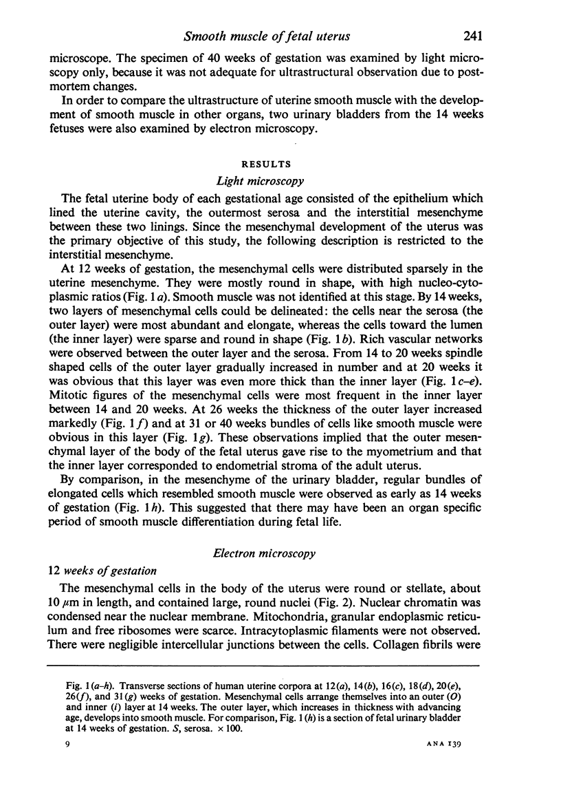
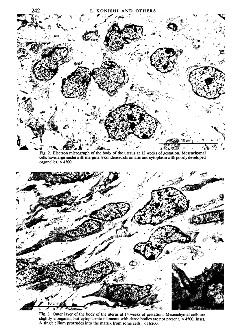
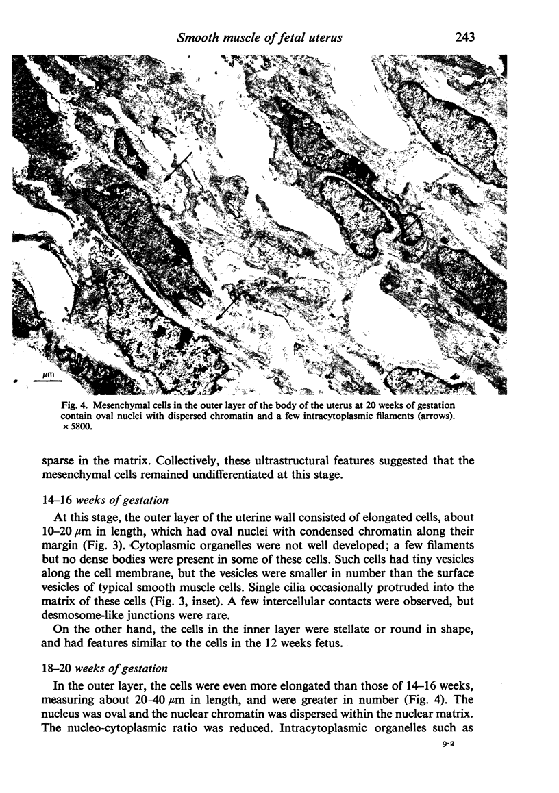
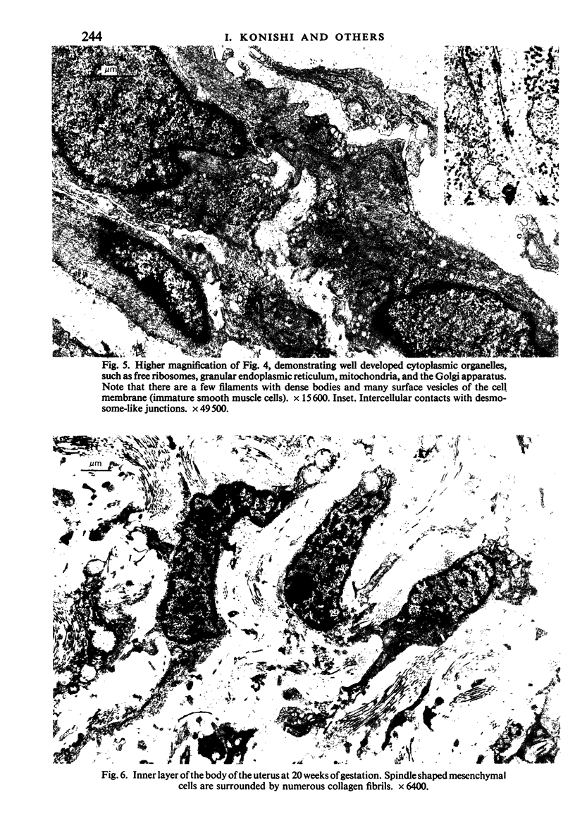
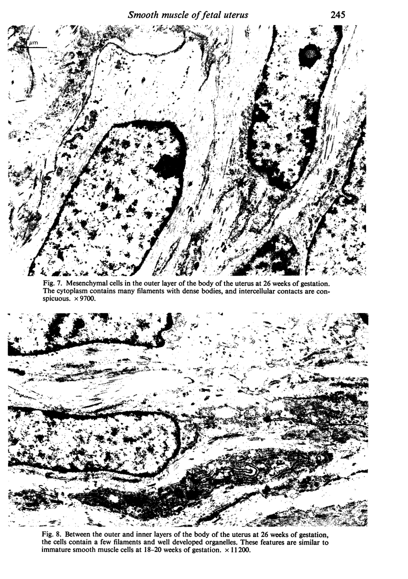
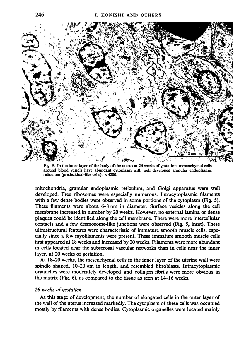
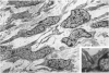
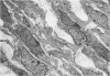
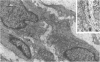
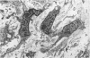
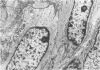
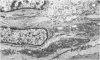
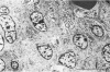
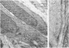
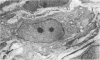
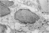
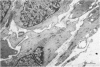
Prenatal development of uterine smooth muscle was studied by light and electron microscopy in specimens obtained from ten human fetuses between 12 and 40 weeks gestation. Light microscopical observation of transverse sections of the body of the uterus revealed that the outer, subserosal layer of elongated cells was distinguishable from 14 weeks. This layer was more cellular than the inner layer and increased its thickness with advancing age. Ultrastructurally, the mesenchymal cells of the uterus did not contain myofilaments until 16 weeks. At 18 weeks, however, spindle shaped cells in the outer layer had a few filaments with dense bodies and well developed organelles and were identified as immature smooth muscle cells. By 31 weeks, the cells in this layer developed into almost mature smooth muscle cells, which contained abundant cytoplasmic filaments, dense plaques and surface vesicles along the cell membrane, and an external lamina. Therefore, in the human fetal uterus, smooth muscle differentiation begins at about 18 weeks and myometrium is formed in the outer layer of the wall by 31 weeks gestation. In the inner layer of the uterus that corresponds to the endometrial stroma, in addition to fibroblast-like cells, there were cells with plump cytoplasm which contained well developed granular endoplasmic reticulum and Golgi apparatus and which were identified around blood vessels by 26 weeks. These features resembled those of predecidual cells, and suggested the influence of sex steroids on the human fetal uterus.
Full text
PDF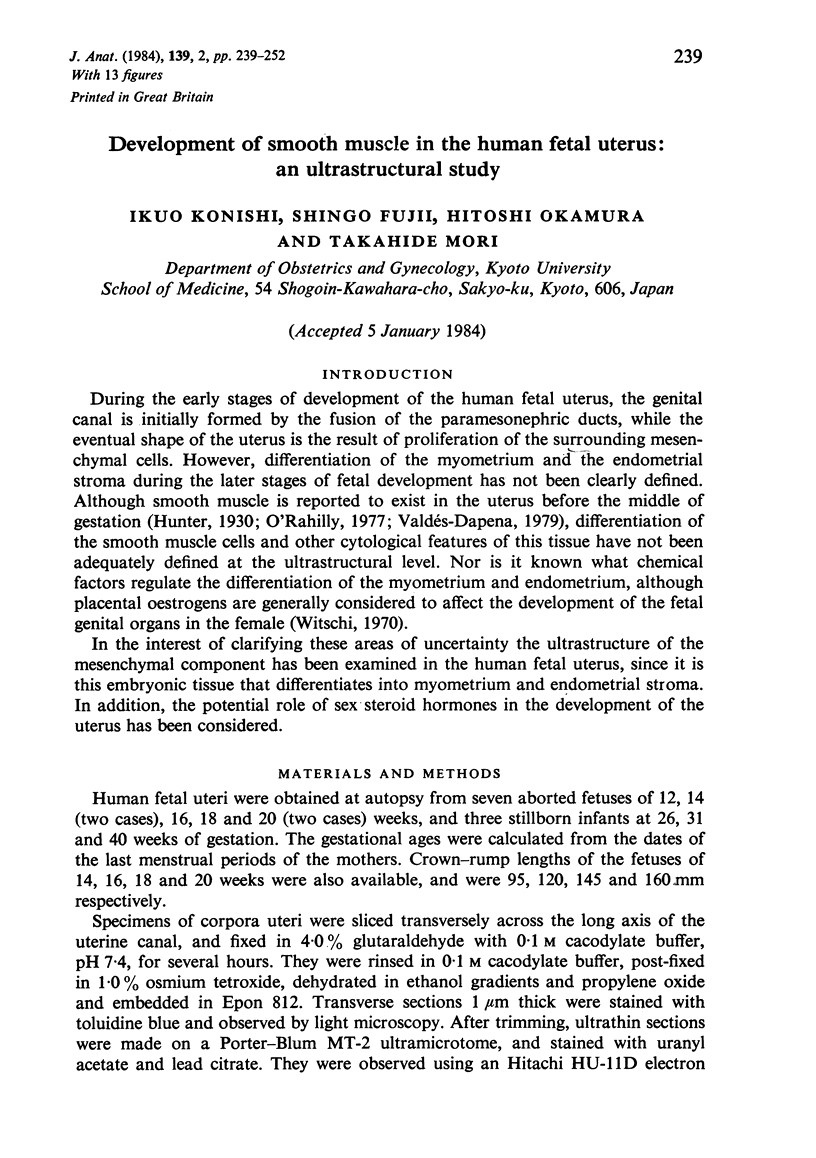
Images in this article
Selected References
These references are in PubMed. This may not be the complete list of references from this article.
- Bird C. C., Willis R. A. The production of smooth muscle by the endometrial stroma of the adult human uterus. J Pathol Bacteriol. 1965 Jul;90(1):75–81. doi: 10.1002/path.1700900108. [DOI] [PubMed] [Google Scholar]
- Fujii S., Nakashima N., Okamura H., Takenaka A., Kanzaki H., Okuda Y., Morimoto K., Nishimura T. Progesterone-induced smooth muscle-like cells in the subperitoneal nodules produced by estrogen. Experimental approach to leiomyomatosis peritonealis disseminata. Am J Obstet Gynecol. 1981 Jan 15;139(2):164–172. doi: 10.1016/0002-9378(81)90440-3. [DOI] [PubMed] [Google Scholar]
- JOST A. The role of fetal hormones in prenatal development. Harvey Lect. 1961;55:201–226. [PubMed] [Google Scholar]
- Leeson T. S., Leeson C. R. The rat ureter. Fine structural changes during its development. Acta Anat (Basel) 1965;62(1):60–79. doi: 10.1159/000142744. [DOI] [PubMed] [Google Scholar]
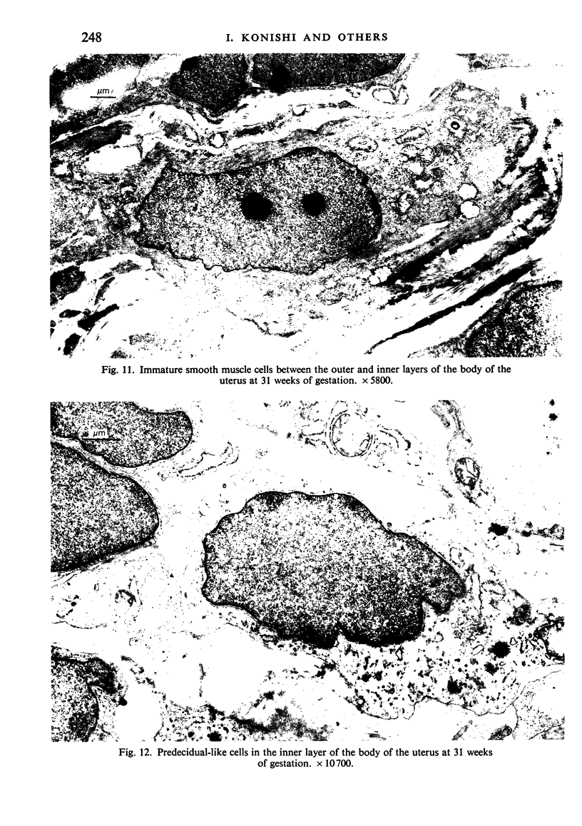
- OBER W. B., BERNSTEIN J. Observations on the endometrium and ovary in the newborn. Pediatrics. 1955 Oct;16(4):445–460. [PubMed] [Google Scholar]
- Tulchinsky D., Hobel C. J., Yeager E., Marshall J. R. Plasma estrone, estradiol, estriol, progesterone, and 17-hydroxyprogesterone in human pregnancy. I. Normal pregnancy. Am J Obstet Gynecol. 1972 Apr 15;112(8):1095–1100. doi: 10.1016/0002-9378(72)90185-8. [DOI] [PubMed] [Google Scholar]
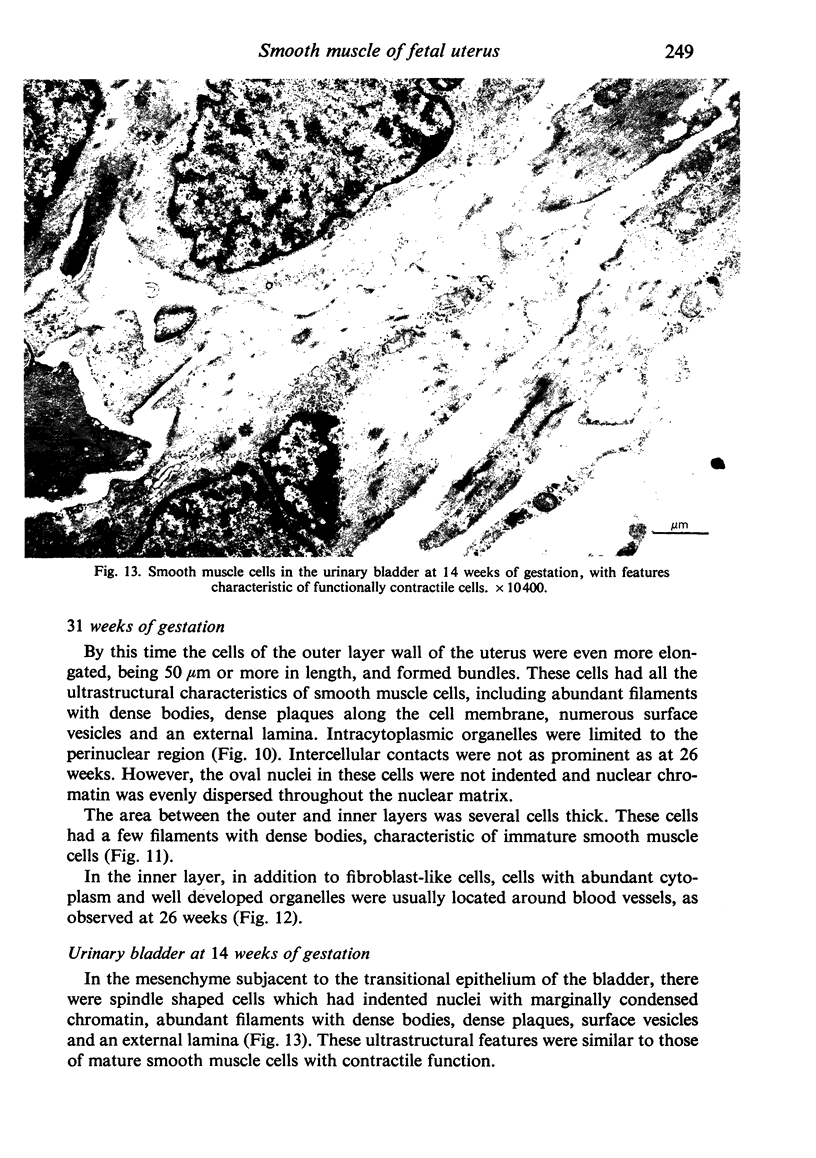
- Tulchinsky D. Placental secretion of unconjugated estrone, estradiol and estriol into the maternal and the fetal circulation. J Clin Endocrinol Metab. 1973 Jun;36(6):1079–1087. doi: 10.1210/jcem-36-6-1079. [DOI] [PubMed] [Google Scholar]
- Wienke E. C., Jr, Cavazos F., Hall D. G., Lucas F. V. Ultrastructure of the human endometrial stroma cell during the menstrual cycle. Am J Obstet Gynecol. 1968 Sep 1;102(1):65–77. doi: 10.1016/0002-9378(68)90434-1. [DOI] [PubMed] [Google Scholar]
- Wynn R. M. Ultrastructural development of the human decidua. Am J Obstet Gynecol. 1974 Mar 1;118(5):652–670. doi: 10.1016/s0002-9378(16)33740-1. [DOI] [PubMed] [Google Scholar]
- Yamauchi A., Burnstock G. Post-natal development of smooth muscle cells in the mouse vas deferens. A fine structural study. J Anat. 1969 Jan;104(Pt 1):1–15. [PMC free article] [PubMed] [Google Scholar]