Abstract
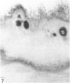
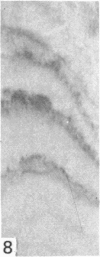
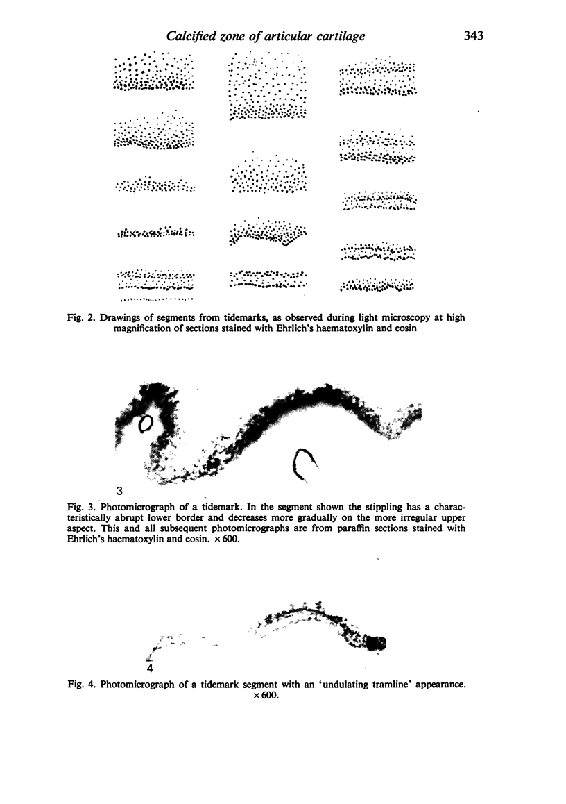
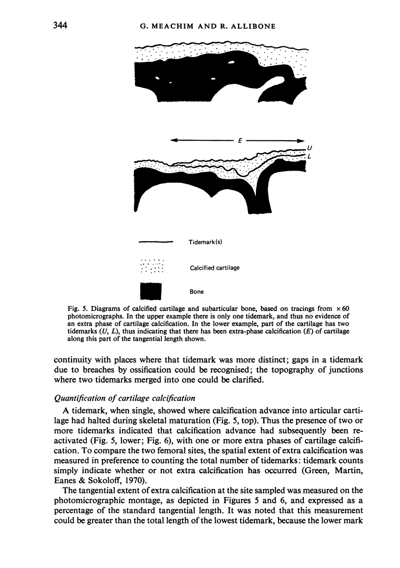
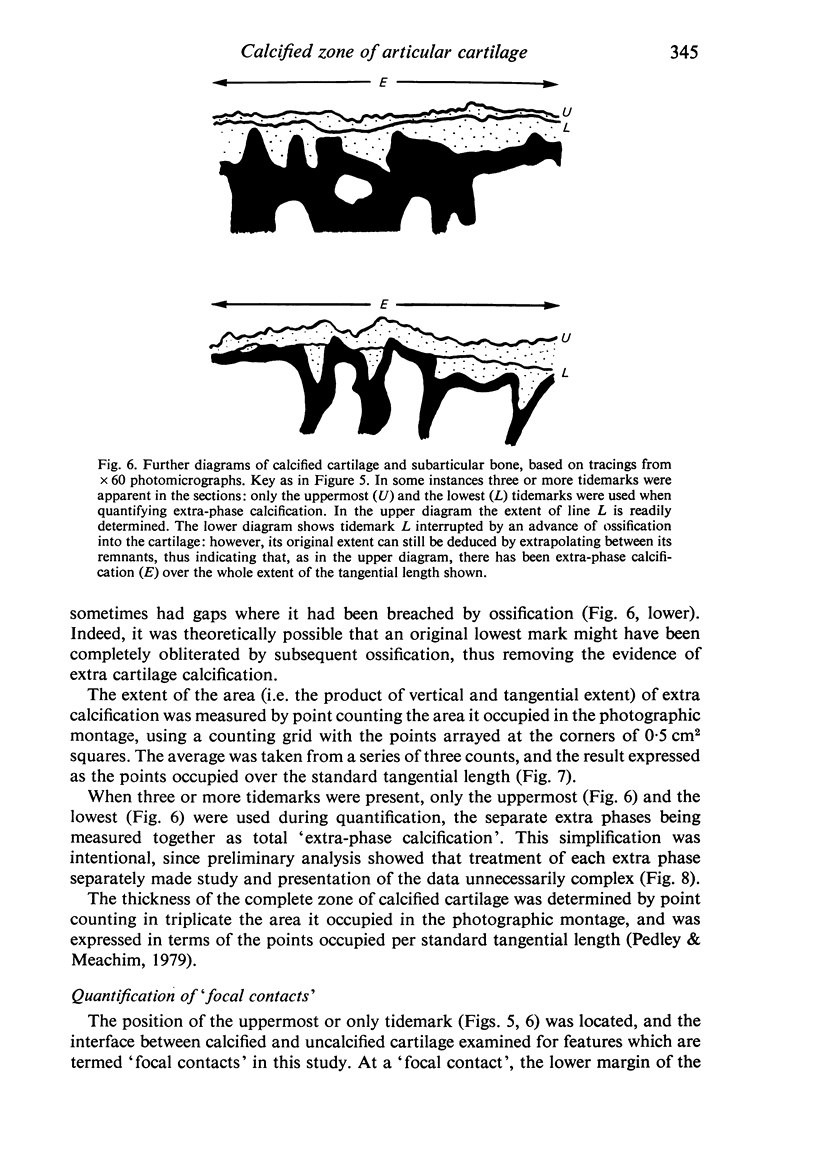
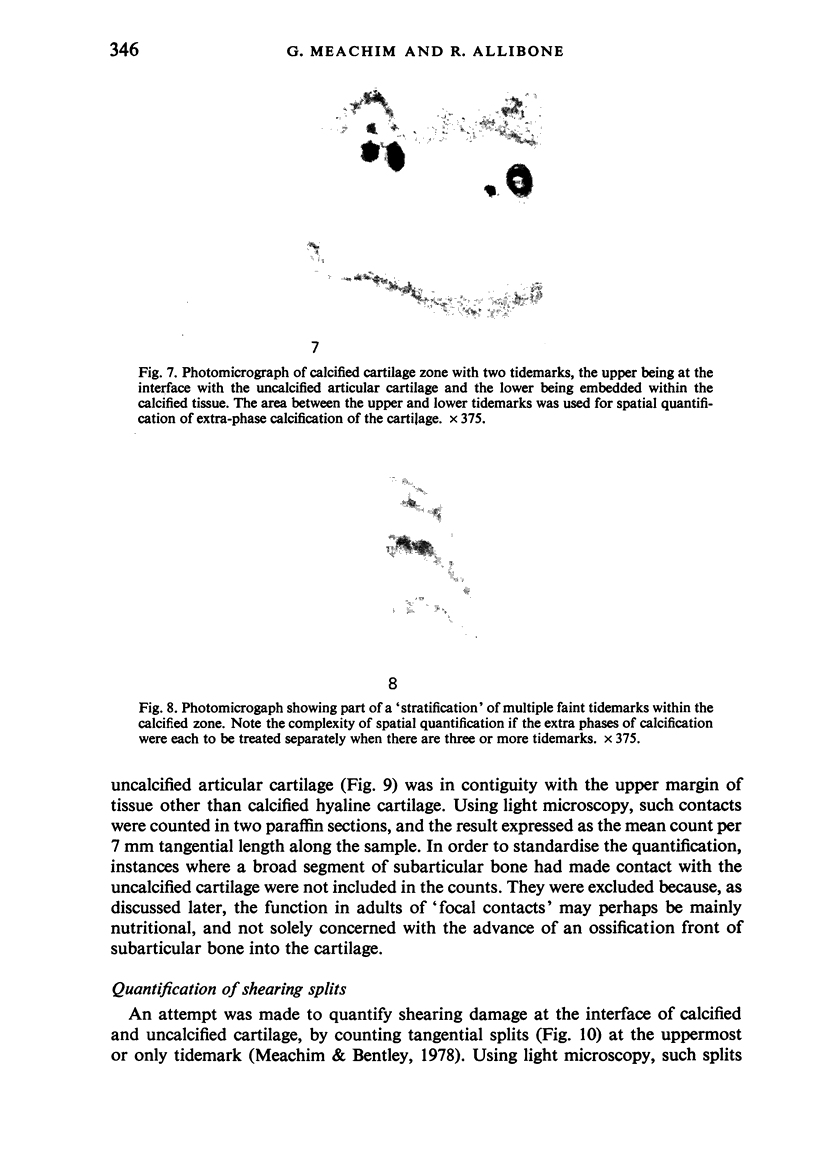
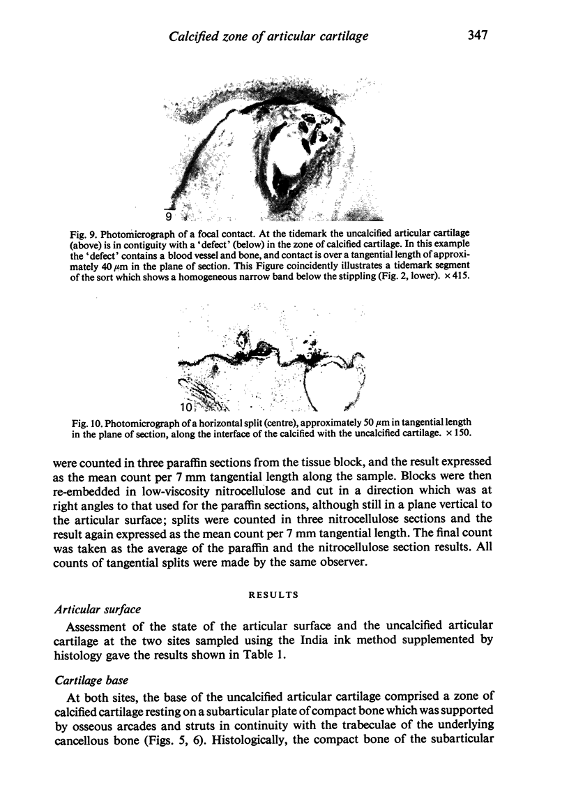
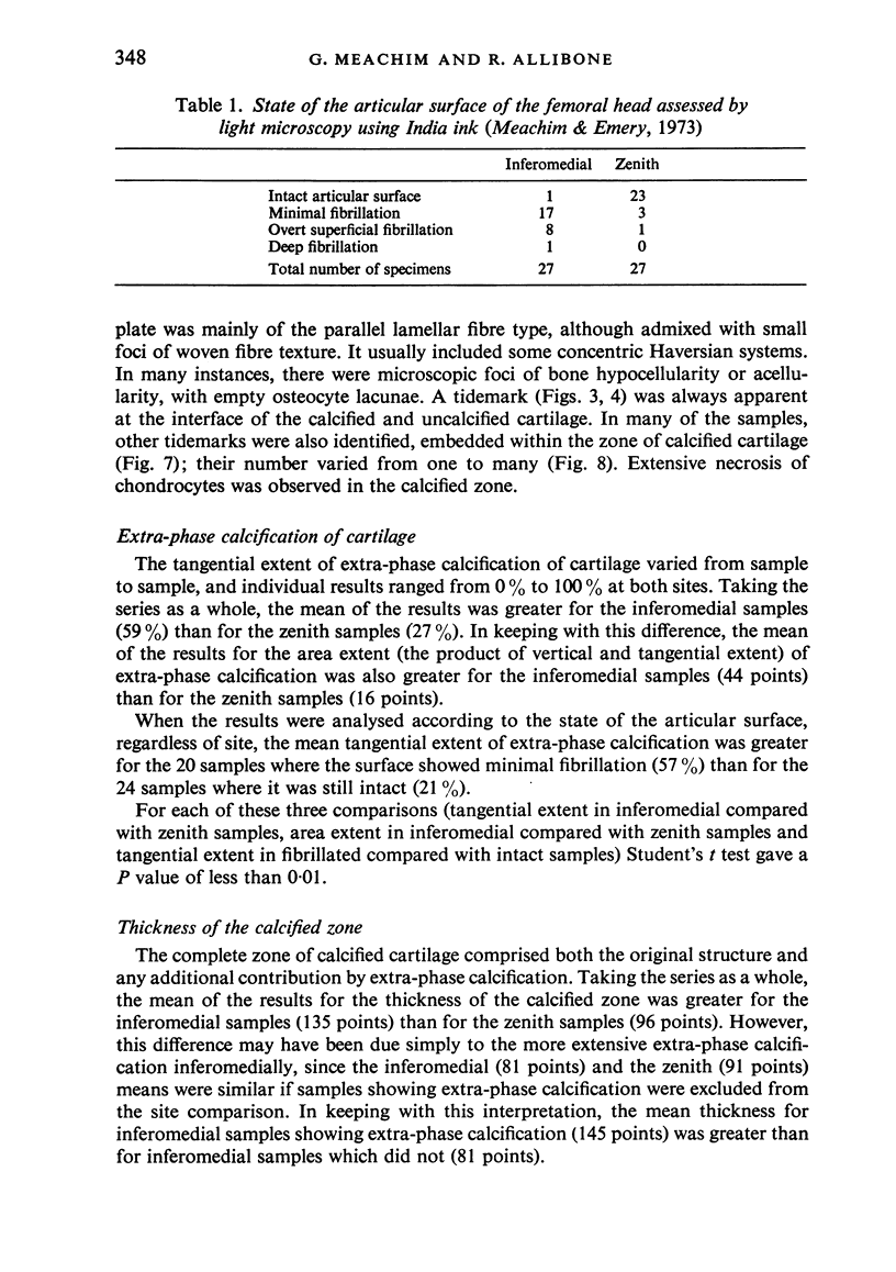
A series of 27 adult human femoral heads has been examined for topographical variation in 'remodelling' and other histological features of the calcified zone at the base of the articular cartilage. The specimens were obtained from necropsies; hip joints with osteoarthritic bone exposure were excluded. A tissue sample from the inferomedial aspect was compared with one from the femoral zenith, using a standard length along the articular contour at each site. Histological sections were cut in a plane vertical to that of the articular surface. A study was made of the various patterns seen within cartilage tidemarks when these were examined at high magnification in paraffin sections stained with Ehrlich's haematoxylin and eosin. Special attention was paid to the identification of tidemark segments which stained faintly and were not readily apparent. The tidemarks were mapped on a photomicrographic montage from each of the tissue samples. When a sample showed evidence of one or more extra phases of cartilage calcification, as indicated by the presence of more than one tidemark, the spatial extent of the extra calcification was quantified by linear measurement and by point counting on the photomicrographic montage. The mean of the results for the spatial extent of extra-phase calcification of the cartilage was greater for the inferomedial than for the zenith samples. However, it was also greater for samples where the articular surface showed minimal fibrillation than for samples where the surface was still intact, and it was noted that surface fibrillation was much more common in the inferomedial than in the zenith samples. Where there was more than one tidemark, the lowest sometimes showed gaps where it had been breached by an advance of ossification into the calcified cartilage. The mean value of the tangential extent of such gaps was similar at the two sites sampled. Focal contacts, where the uncalcified articular cartilage was in contiguity with calcified zone 'defects' containing tissue other than calcified hyaline cartilage, were more numerous at the femoral zenith than inferomedial to the fovea. Counts were also made of tangential shearing splits at the interface of the calcified and uncalcified cartilage. Subject to the reservation that genuine splitting may be difficult to distinguish from technical artifact, the mean number was closely similar at the two sites sampled. The interpretation of the findings is discussed in relation to remodelling changes in the cartilage base and to degenerative changes in the overlying articular cartilage.
Full text
PDF
Images in this article
Selected References
These references are in PubMed. This may not be the complete list of references from this article.
- Byers P. D., Contepomi C. A., Farkas T. A. A post mortem study of the hip joint. Including the prevalence of the features of the right side. Ann Rheum Dis. 1970 Jan;29(1):15–31. doi: 10.1136/ard.29.1.15. [DOI] [PMC free article] [PubMed] [Google Scholar]
- FAWNS H. T., LANDELLS J. W. Histochemical studies of rheumatic conditions. I. Observations on the fine structures of the matrix of normal bone and cartilage. Ann Rheum Dis. 1953 Jun;12(2):105–113. doi: 10.1136/ard.12.2.105. [DOI] [PMC free article] [PubMed] [Google Scholar]
- Green W. T., Jr, Martin G. N., Eanes E. D., Sokoloff L. Microradiographic study of the calcified layer of articular cartilage. Arch Pathol. 1970 Aug;90(2):151–158. [PubMed] [Google Scholar]
- JOHNSON L. C. Kinetics of osteoarthritis. Lab Invest. 1959 Nov-Dec;8:1223–1241. [PubMed] [Google Scholar]
- Lane L. B., Villacin A., Bullough P. G. The vascularity and remodelling of subchondrial bone and calcified cartilage in adult human femoral and humeral heads. An age- and stress-related phenomenon. J Bone Joint Surg Br. 1977 Aug;59(3):272–278. doi: 10.1302/0301-620X.59B3.893504. [DOI] [PubMed] [Google Scholar]
- Meachim G., Bentley G. Horizontal splitting in patellar articular cartilage. Arthritis Rheum. 1978 Jul-Aug;21(6):669–674. doi: 10.1002/art.1780210610. [DOI] [PubMed] [Google Scholar]
- Meachim G., Emery I. H. Cartilage fibrillation in shoulder and hip joints in Liverpool necropsies. J Anat. 1973 Nov;116(Pt 2):161–179. [PMC free article] [PubMed] [Google Scholar]
- Meachim G., Whitehouse G. H., Pedley R. B., Nichol F. E., Owen R. An investigation of radiological, clinical and pathological correlations in osteoarthrosis of the hip. Clin Radiol. 1980 Sep;31(5):565–574. doi: 10.1016/s0009-9260(80)80054-7. [DOI] [PubMed] [Google Scholar]
- Pedley R. B., Meachim G. Topographical variation in patellar subarticular calcified tissue density. J Anat. 1979 Jun;128(Pt 4):737–745. [PMC free article] [PubMed] [Google Scholar]
- Woods C. G., Greenwald A. S., Haynes D. W. Subchondral vascularity in the human femoral head. Ann Rheum Dis. 1970 Mar;29(2):138–142. doi: 10.1136/ard.29.2.138. [DOI] [PMC free article] [PubMed] [Google Scholar]


