Abstract
Background
Alectinib is a second generation of anaplastic lymphoma kinase (ALK) inhibitor that has been approved for the treatment of advanced non-small-cell lung cancer (NSCLC) with ALK rearrangements. Hepatotoxicity is the most common adverse drug reaction. However, there is currently no published report on the pathologic findings of alectinib-induced hyperbilirubinemia.
Case Presentation
Here, we report a case of a patient with NSCLC and chronic hepatitis B (CHB) who was treated with alectinib and developed grade 4 hyperbilirubinemia after 3 years on therapy. Alectinib was discontinued, and an artificial liver support system (ALSS) was used to decline blood bilirubin levels. The pathological manifestations from a liver biopsy showed the hepatocytes with scattered focal necrosis, bile stasis, and vesicular steatosis, bile emboli in capillaries, and star-shaped fibers proliferation in the portal area.
Conclusion
This is the first report of alectinib-induced hyperbilirubinemia which was confirmed by liver histopathology and successfully relieved by ALSS treatment and drug discontinuation.
Keywords: alectinib, ALK, hyperbilirubinemia, NSCLC, ALSS
Introduction
Alectinib is a second-generation and highly selective anaplastic lymphoma kinase (ALK) tyrosine kinase inhibitor (TKI) that has acquired accelerated approval by the US Food and Drug Administration in 2015 for the treatment of patients with advanced ALK-positive non–small cell lung cancer (NSCLC).1 Thus, alectinib has become the first-line treatment strategy for newly diagnosed advanced ALK-positive NSCLC, especially in resisting brain metastases due to it can penetrate the central nervous system. In general, Alectinib is well tolerated, but some common laboratory adverse events (AEs) observed including anemia, elevated aspartate aminotransferase (AST), elevated alkaline phosphatase (ALT), and hyperbilirubinemia.2 The occurrence of alectinib-induced hyperbilirubinemia was up to 36.2% at all grades in a clinical trial,3 and the incidence of grade 3 (5–10 times the upper limit of normal) or higher elevation is about 2%.4
Considering the long-term duration of alectinib use in patients with ALK-positive NSCLC, clinical management of adverse drug reactions is crucial to maximize the benefits. However, there are currently no findings reported on the hepatic pathology of alectinib-induced hyperbilirubinemia. Here, we report a case of alectinib-induced grade 4 hyperbilirubinemia in a Chinese male diagnosed with advanced ALK-positive NSCLC. Histological findings from a liver biopsy showed severe bile emboli in capillaries and feather-like hepatocytes with bile stasis in this patient, whose blood bilirubin was successfully decreased with ALSS treatment and drug interruption.
Case Presentation
A 48-year-old Chinese male never-smoker was diagnosed with stage IV NSCLC in March 2021 at a domestic tertiary specialized hospital. His chest CT scan revealed diffuse nodular lesions in both lungs, accompanied by multiple lymph node enlargement including mediastinal, hilar, and supraclavicular. The cytology of pulmonary alveolar lavage fluid and pathology of supraclavicular lymph node both confirmed as adenocarcinoma, and the gene testing revealed ALK rearrangements. Subsequently, Alectinib was given orally, at a dose of 600 mg twice daily from March 2021, which resulted in constant response and the patient was continuously treated for over 3 years. It is particularly pointed out that the patient with a history of chronic hepatitis B infection for 20 years and without antiviral treatment or regular follow-up.
On April 26th, 2024, the patient initially presented with jaundice, abdominal distension, pruritus, and fatigue. The laboratory result showed total bilirubin (TBIL) 263.5μmol/L, direct bilirubin (DBIL) 134.6μmol/L, and indirect bilirubin (IBIL) 134.6μmol/L, amma-glutamyl transferase (GGT) 55U/L, alkaline phosphatase (ALP) 286U/L, total bile acid (TBA) 217.2μmol/L, alanine aminotransferase (ALT) 59U/L, and aspartate aminotransferase (AST) 91U/L. After a detailed medical history inquiry, the patient denied oral traditional Chinese medicine or nonsteroidal anti-inflammatory drugs that can cause liver dysfunction, not drinking or an unclean diet recently. Next, he further conducted relevant laboratory tests and imaging scans after admission, the results presented in Table 1. Of note, HBsAg, HBeAb and HBcAb was positive, HBsAb and HBeAg was negative. Hepatitis B viral load was 1820 IU/mL by quantitative polymerase chain reaction, he was diagnosed as HBeAg negative chronic hepatitis B infection; however, it cannot explain the increased bilirubin, as the transaminase is only slightly elevated.
Table 1.
Laboratory Parameters and Imaging of the Patient at Admission
| Parameters | Parameters | ||
|---|---|---|---|
| Blood routine | Hepatitis virus-related marker | ||
| White blood cells (109/L) | 7.70 | HBsAg (IU/mL) | 294.1 (+) |
| Neutrophils (109/L) | 5.55 | HBsAb (mIU/mL) | 0.78 (−) |
| Lymphocytes (109/L) | 1.17 | HBeAg (C.O.I) | 0.06 (−) |
| Monocytes (109/L) | 0.71 | HBeAb (C.O.I) | >100.00 (+) |
| Red blood cells (1012/L) | 3.62 | HBcAb (C.O.I) | 807.58 (+) |
| Hemoglobin (g/L) | 118 | HCV antibody (C.O.I) | (−) |
| Platelet (109/L) | 223 | Hepatitis A virus IgM | (−) |
| Coagulation indicators | Hepatitis E virus IgM | (−) | |
| Prothrombin time (s) | 11.4 | Hepatitis B virus DNA (IU/mL) | 1820 |
| Activated partial thromboplastin time (s) | 24.9 | Epstein-barr virus RNA | (−) |
| Fibrinogen (g/L) | 3.08 | Cytomegalovirus RNA | (−) |
| D-Dimer (mg/L) | 0.26 | Autoimmune liver diseases | |
| Fibrinogen degradation products (μg/mL) | 0.99 | Hepatopathy-related antibody | (−) |
| Liver function | Antinuclear antibodies spectrum | (−) | |
| Total bilirubin (μmol/L) | 248.1 | Immunoglobulin | Normal |
| Direct bilirubin (μmol/L) | 118.7 | Tumor marker | |
| Indirect bilirubin (μmol/L) | 129.4 | Alpha-fetoprotein (IU/mL) | 3.10 |
| Albumin (g/L) | 36 | Carcinoembryonic antigen (ng/mL) | 3.30 |
| Alanine aminotransferase (U/L) | 52 | Cancer antigen 199 (IU/mL) | 13.6 |
| Aspartate aminotransferase (U/L) | 80 | Abdominal contrast-enhanced MRI | |
| Gamma-glutamyl transpeptidase (U/L) | 45 | Multiple hemangioma of liver, nonmetastatic lesion | |
| Alkaline phosphatase (U/L) | 269 | MRCP | |
| Total bile acid (μmol/L) | 217.2 | Non-dilated bile duct of liver inside and outside |
Abbreviations: MRI, Magnetic resonance imaging; MRCP, Magnetic resonance cholangiopancreatography.
There was no evidence to prove liver metastases or infection with other hepatitis viruses or autoimmune liver diseases. All those workup failed to identify other etiologies contributing to his acute hyperbilirubinemia; therefore, a diagnosis of alectinib-induced grade 4 hyperbilirubinemia (>10.0⨰ULN if baseline was normal, Common Terminology Criteria for Adverse Events, v.5.0) was considered, and alectinib was discontinued.
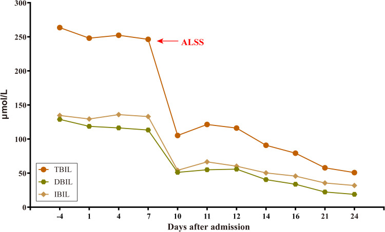
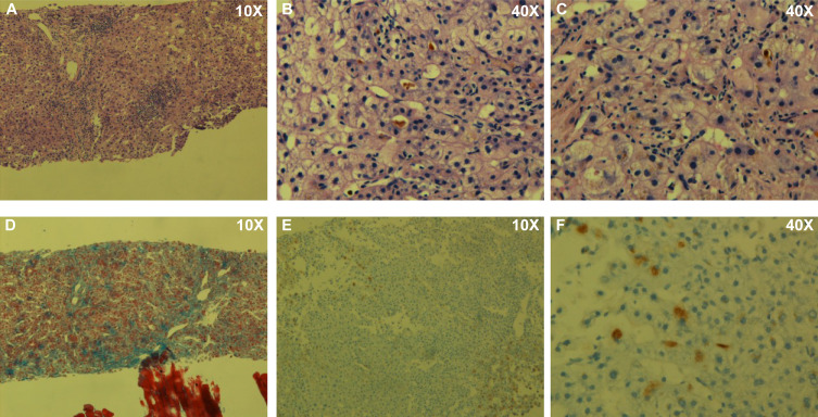
After continuous hepatoprotective treatment for 1 week comprising ursodeoxycholic acid, glycyrrhizin and polyene phosphatidylcholine, the bilirubin levels were still unable to descend, while AST and ALT levels remained similar. Considering the tendency towards liver failure and the potential risk of cancer progression after discontinuing anti-tumor therapy, he received ALSS using a double plasma molecular absorption system (DPMAS) on May 9th, 2024, to ameliorate liver dysfunction. The TBIL decreased after one course of ALSS (Figure 1). After that, a liver biopsy was performed to assess the etiology of hyperbilirubinemia, which revealed scattered focal necrosis of hepatocytes and inflammatory infiltrate in the portal area, feather-like hepatocytes with bile stasis, bile emboli in capillaries, and vesicular steatosis in hepatocytes (Figure 2A–C). The trichrome stain revealed the star-shaped fibers proliferation in the portal area (Figure 2D), and there were observed hepatic cells expressing HBsAg (Figure 2E and F). The final pathology report was HBeAg (-) CHB-G3S2 and acute cholestasis hepatitis was highly likely caused by drug-induced liver injury (DILI).
Figure 1.
Plot of total bilirubin (TBIL), direct bilirubin (DBIL), and indirect bilirubin (IBIL) against the days after admission. The time of receiving an artificial liver support system (ALSS) is pointed by the red arrow.
Figure 2.
Features of liver histopathology. (A) Scattered focal necrosis of hepatocytes and inflammatory infiltrate in the portal area at 10X magnification. (B and C) At 40X magnification, it shows feather-like hepatocytes with bile stasis, bile emboli in capillaries, and vesicular steatosis in hepatocytes. (D) The trichrome stain highlights the star shaped fibers proliferation in the portal area at 10X magnification. (E and F) HBsAg was expressed in hepatic cells.
Discussion
Since the overall incidence rate of grade 3–4 DILI of ALK inhibitors is lower than 30%, the actual progression to severe DILI is relatively uncommon.4 Alectinib, as the second-generation ALK inhibitor, was approved for the first-line treatment based on its efficacy in ALK+ NSCLC. In global5 and Asian6 Phase 3 studies of alectinib in untreated ALK-positive advanced NSCLC patients, the incidence of grade 3 or higher blood bilirubin elevations was relatively low (only 2%). In addition, hepatotoxicity occurred in our case after 3 years of treatment, which is longer than the average of patients who experienced hepatotoxicity within the first 2 months of therapy with ALK inhibitors. It is worth noting that although hepatotoxicity caused by TKI is usually not fatal, it may lead to long-term consequences such as cirrhosis.7 Therefore, patients should be carefully monitored for alectinib-induced hepatotoxicity to individually dynamic tailored appraisal of the risk/benefit. However, the etiology, management, or sequelae of alectinib-induced hepatotoxicity have yet to be elucidated. To our knowledge, no one has reported histological features of patients with grade 4 hyperbilirubinemia caused by alectinib.
In this case, we first reported the histological findings of grade 4 blood bilirubin elevations in a patient with 3-year treatment with alectinib. The liver biopsy showed hepatocytes with scattered focal necrosis, bile stasis, and vesicular steatosis, bile emboli in capillaries, and star-shaped fibers proliferation in the portal area. Based on his history of medication, considering DILI on CHB. Due to the mild increase in ALT and AST, the etiology of elevated bilirubin is highly likely caused by alectinib, not chronic hepatitis B. The specific mechanism is currently unclear and may be related to the metabolic pathway of the drug. It is pointed out that mitochondrial toxicity, glycolysis disorder and ROS-dependent DNA damage of hepatocyte cell lines were observed in other TKI drugs.8,9 In addition, as previously mentioned, long term TKI treatment may lead to liver cirrhosis. Therefore, it is difficult to assess whether the etiology of liver fibrosis in our case was caused by HBV or alectinib-induced, or both.
Hepatotoxicity induced by ALK-TKIs was the common adverse reaction that may necessitate dosage adjustment or even treatment cessation in trails.4 In our case, jaundice did not resolve after discontinuation of alectinib for 1 week, ALSS was used to alleviate jaundice to improve hepatic function. Owing to the presence of CHB and drug-induced acute grade 4 hyperbilirubinemia, we strongly advised that the patient permanently discontinuation of alectinib meanwhile undergoing nucleotide analogue antiviral therapy to avoid death of DILI or needing a transplant to survive.
Conclusion
This case highlights the importance of closely monitoring hepatotoxicity of patients who are on alectinib regardless of the duration of treatment. For alectinib-induced serious liver injury, we should take an individual analysis of the risk and benefit of discontinuation of the drug.
Highlights
Alectinib may induce serious hyperbilirubinemia.
The pathological manifestations of the liver presented as hepatocytes with scattered focal necrosis, bile stasis, and vesicular steatosis, bile emboli in capillaries in a liver biopsy of a Chinese ALK-positive NSCLC patient treated with long-term oral alectinib.
It is vital to closely monitor the hepatotoxicity alectinib-induced to avoid adverse consequences. Proper management of the ALK-TKI-induced hepatotoxicity is a new challenge for oncologists and hepatologists.
Ethical Approval Statement
The written informed consent was provided by the patient to have the case details and any accompanying images published. The publishing of the case details was also approved by the Research and Ethics Committee of the Shanghai Ninth People’s Hospital, Shanghai, China.
Disclosure
The authors declare that the research was conducted in the absence of any commercial or financial relationships that could be construed as a potential conflict of interest.
References
- 1.Larkins E, Blumenthal GM, Chen H, et al. FDA approval: alectinib for the treatment of metastatic, ALK-positive non-small cell lung cancer following crizotinib. Clin Cancer Res. 2016;22(21):5171–5176. doi: 10.1158/1078-0432.CCR-16-1293 [DOI] [PubMed] [Google Scholar]
- 2.Breadner D, Blanchette P, Shanmuganathan S, et al. Efficacy and safety of ALK inhibitors in ALK-rearranged non-small cell lung cancer: a systematic review and meta-analysis. Lung Cancer. 2020;144:57–63. doi: 10.1016/j.lungcan.2020.04.011 [DOI] [PubMed] [Google Scholar]
- 3.Tamura T, Kiura K, Seto T, et al. Three-year follow-up of an alectinib phase I/II study in ALK-positive non-small-cell lung cancer: AF-001JP. J Clin Oncol. 2017;35(14):1515–1521. doi: 10.1200/JCO.2016.70.5749 [DOI] [PMC free article] [PubMed] [Google Scholar]
- 4.Zhou F, Yang Y, Zhang L, et al. Expert consensus of management of adverse drug reactions with anaplastic lymphoma kinase tyrosine kinase inhibitors. ESMO Open. 2023;8(3):101560. doi: 10.1016/j.esmoop.2023.101560 [DOI] [PMC free article] [PubMed] [Google Scholar]
- 5.Peters S, Camidge DR, Shaw AT, et al. Alectinib versus crizotinib in untreated ALK-positive non-small-cell lung cancer. New Engl J Med. 2017;377(9):829–838. doi: 10.1056/NEJMoa1704795 [DOI] [PubMed] [Google Scholar]
- 6.Zhou C, Kim SW, Reungwetwattana T, et al. Alectinib versus crizotinib in untreated Asian patients with anaplastic lymphoma kinase-positive non-small-cell lung cancer (ALESIA): a randomised phase 3 study. Lancet Respir Med. 2019;7(5):437–446. doi: 10.1016/S2213-2600(19)30053-0 [DOI] [PubMed] [Google Scholar]
- 7.Shah RR, Morganroth J, Shah DR. Hepatotoxicity of tyrosine kinase inhibitors: clinical and regulatory perspectives. Drug Safety. 2013;36(7):491–503. doi: 10.1007/s40264-013-0048-4 [DOI] [PubMed] [Google Scholar]
- 8.Mingard C, Paech F, Bouitbir J, et al. Mechanisms of toxicity associated with six tyrosine kinase inhibitors in human hepatocyte cell lines. J appl toxicol. 2018;38(3):418–431. doi: 10.1002/jat.3551 [DOI] [PubMed] [Google Scholar]
- 9.Yan H, Du J, Chen X, et al. ROS-dependent DNA damage contributes to crizotinib-induced hepatotoxicity via the apoptotic pathway. Toxicol Appl Pharmacol. 2019;383:114768. doi: 10.1016/j.taap.2019.114768 [DOI] [PubMed] [Google Scholar]


