Abstract
Metabolic reprogramming is a prominent characteristic of tumor cells, evidenced by heightened secretion of lactate, which is linked to tumor progression. Furthermore, the accumulation of lactate in the tumor microenvironment (TME) influences immune cell activity, including the activity of macrophages, dendritic cells and T cells, fostering an immunosuppressive milieu. Anti-programmed cell death protein 1 (PD-1)/programmed death-ligand 1 (PD-L1) therapy is associated with a prolonged survival time of patients with non-small cell lung cancer. However, some patients still develop resistance to anti-PD-1/PD-L1 therapy. Lactate is associated with resistance to anti-PD-1/PD-L1 therapy. The present review summarizes what is known about lactate metabolism in tumor cells and how it affects immune cell function. In addition, the present review emphasizes the relationship between lactate secretion and immunotherapy resistance. The present review also explores the potential for targeting lactate within the TME to enhance the efficacy of immunotherapy.
Keywords: cancer immunity, immunotherapy, lactate, programmed cell death protein 1, programmed death-ligand 1, tumor environment
1. Introduction
Immune checkpoint inhibitors (ICIs) targeting programmed cell death protein 1 (PD-1) or programmed death ligand 1 (PD-L1) are used in the treatment of a wide range of tumors, such as lung cancer, cervical cancer and renal cell carcinomas (1–4). These agents are associated with a prolonged survival time (1–4). Studies such as KEYNOTE-024 and KEYNOTE-042 have been pivotal in establishing the role of pembrolizumab, a PD-1 inhibitor, in improving overall survival in patients with advanced non-small cell lung cancer (1,2). The results of the ENGOT-cx11/GOG-3047/KEYNOTE-A18 study demonstrated that the combination of pembrolizumab with chemoradiotherapy improved overall survival in patients with locally advanced cervical cancer compared with a placebo (3). Additionally, adjuvant pembrolizumab was observed to enhance overall survival in patients with renal cell carcinoma compared with a placebo (4). However, primary or acquired resistance to ICIs is commonly observed (5). Therefore, research investigating the mechanisms of PD-1/PD-L1 ICI resistance is vital to improve clinical outcomes. There have been a number of studies examining the mechanisms of resistance to PD-1/PD-L1 ICIs, which include loss of tumor antigens and antigen presentation (6), T-cell exhaustion (7), lack of interferon signaling (8), and lack of PD-L1 expression (9). Furthermore, additional pathways involved in the inhibition of immune cells within the tumor microenvironment (TME) can lead to anti-PD-1/PD-L1 resistance (10,11). For example, TYRO3 can increase the responsiveness to anti-PD-1 therapy by altering the macrophage profile towards a more M2-like state, which is facilitated by an increase in VEGF expression (10). Phospholipase C γ2 (PLCG2) serves a role in modulating the TME by reducing the infiltration of CD8+ T cells and increasing the infiltration of regulatory T cells (Treg cells), which can suppress the immune response (11). Additionally, PLCG2 contributes to the upregulation of PD-1 and PD-L1 expression. This dual action of PLCG2 facilitates immune escape and is associated with resistance to anti-PD-1 therapy (11). The immunosuppressive TME resulting from the metabolic reprogramming of tumor cells represents a barrier to the effectiveness of immunotherapy (12).
Tumor cells maintain their proliferation and cellular function through specific metabolic patterns, a process known as metabolic reprogramming (13). Under aerobic conditions, respiration in eukaryotic cells is mainly aerobic, providing energy through oxidative phosphorylation. By contrast, cancer cells prefer to generate energy through aerobic glycolysis, known as the ‘Warburg effect’, consuming large amounts of glucose and increasing the production of lactate (14). Lactate is subsequently released extracellularly, which results in an acidic TME, which can facilitate tumor growth, angiogenesis and immune evasion (14,15). Since lactate acts as a bridge linking metabolic reprogramming to immunosuppression (16), a growing number of studies have noted the impact of lactate on the response to anti-PD-1/PD-L1 therapy (17–19). The current review presents the mechanisms of resistance to PD-1/PD-L1 ICIs and takes a detailed look at the potential role of lactate in these mechanisms.
2. Mechanisms of resistance to anti-PD-1/PD-L1 therapy
The presence of PD-L1 on cancer cells facilitates immune evasion through its interaction with PD-1 on activated T cells (20). This interaction results in the phosphorylation of the immune receptor tyrosine-based switch motif and subsequent binding to the Src homology 2 (SH2) domains of SH2-containing phosphatase 2 (SHP2) (21). Once activated, SHP2 dephosphorylates proximal T-cell receptor (TCR) signaling molecules, such as ζ-associated protein of 70 kD, which is a key component of the TCR signaling cascade. This dephosphorylation event dampens the TCR signaling, leading to the suppression of T cell activation (22). In addition, PD-1 is expressed on the surface of tumor-associated macrophages, and a study has indicated that blocking the PD-1/PD-L1 axis can increase the activity of tumor-associated macrophages (20). PD-1/PD-L1 ICIs work by targeting the PD-1/PD-L1 axis, which has been shown to be a successful treatment strategy in multiple cancer types such as melanoma, renal cell carcinoma and cervical cancer (3,4,6).
There has been much discussion about the mechanisms of resistance to anti-PD-1/PD-L1 therapy. Firstly, as aforementioned, anti-PD-1/PD-L1 therapy works by targeting the PD-1/PD-L1 axis; therefore, PD-L1 expression is critical for a response to immunotherapy (6). Secondly, effective immune responses cannot be achieved without antigen expression and antigen presentation (23,24). Accordingly, a study has demonstrated that tumors with sparse immune infiltration exhibit diminished neoantigen editing function (25). Furthermore, it has been demonstrated that activation of β-catenin can suppress antitumor immune responses by impeding the recruitment of dendritic cells (DCs) (23). Antigen presentation leads to T-cell activation, which is a crucial method for the immune system to attack and eliminate cancer cells (17). Furthermore, evidence shows that an abundance of CD8+ T cells is essential for antitumor immunity (7,26). A clinical trial of pembrolizumab in microsatellite instability-high gastric cancer has demonstrated that abundant tumor-infiltrating lymphocytes were associated with a clinical benefit (27). Furthermore, preventing the activation of T cells or other mechanisms that affect the functioning of T cells can lead to low response rates to anti-PD-1/PD-L1 therapy (10). It has been shown that TYRO3 protein tyrosine kinase inhibited tumor cell ferroptosis, suppressing T-cell attack and reducing responsiveness to anti-PD-1/PD-L1 therapy (10). In addition to the aforementioned mechanisms, there are numerous studies on other aspects of resistance such as genetic mutations (28), gut microbiota (29) and metabolism (17). Although progress has been achieved in elucidating the mechanism of resistance to anti-PD-1/PD-L1 therapy, the intricate nature of the TME remains a research limitation. This complexity arises from the interactions among various cell types and molecular pathways, which collectively impact the progression of resistance to anti-PD-1/PD-L1 therapy (30). Consequently, further research is essential to address these challenges such as tumor heterogeneity and the interactions among various cell types, as well as to enhance the understanding of resistance to anti-PD-1/PD-L1 therapy.
Resistance to cancer immunotherapy is often linked to an immunosuppressive TME in which key nutrients serve a crucial role (17–19). Tumor cells and immune cells engage in competition for essential nutrients, leading to reprogramming of metabolic pathways in immune cells, which in turn suppresses antitumor immune responses (31,32). In recent years, a study has investigated the relationship between tumor metabolism, immune evasion mechanisms and resistance to immunotherapy (32). Notably, metabolic byproducts produced by tumor cells, particularly lactic acid, are known to contribute to the immunosuppressive nature of the TME (33–35). A growing body of research suggests that lactic acid in the TME is associated with resistance to immunotherapy (33–35).
3. Lactate metabolism
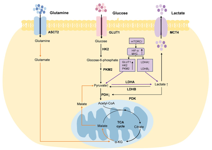
Otto Warburg noticed that cancer cells preferentially generate energy through aerobic glycolysis, which is a hallmark of tumor cell metabolism (36). Under aerobic conditions, normal cells transform glucose into pyruvate via glycolysis. This pyruvate is then transferred to the mitochondria and oxidized through the tricarboxylic acid (TCA) cycle to generate carbon dioxide, oxygen and adenosine triphosphate (37). In cancer cells, a marked proportion of the pyruvate generated through glycolysis does not enter the mitochondria but is converted into lactate (37). Cancer cells regulate lactate production and secretion in the TME through several key enzymes such as glucose transporter 1 (GLUT1), hexokinase 2 (HK2) and pyruvate kinase 2 (PKM2) (16,38) (Fig. 1).
Figure 1.
Lactate metabolism in the tumor microenvironment. In cancer cells, lactate is mainly produced through glycolysis and glutaminolysis. Glucose is converted to pyruvate in the cytoplasm, after which most pyruvate is metabolized to lactate by LDH. In addition, glutamine can be converted to glutamate, which is then transformed into α-KG. α-KG is transformed into malate via the TCA cycle, which is then translocated to the cytoplasm to provide pyruvate for lactate production. GLUT1, glucose transporter 1; HK2, hexokinase 2; PKM2, pyruvate kinase 2; PDK, pyruvate dehydrogenase kinase; PDH, pyruvate dehydrogenase; LDH, lactate dehydrogenase; MCT4, monocarboxylate transporter 4; ASCT2, alanine-serine-cysteine transporter type-2; TCA, tricarboxylic acid; α-KG, α-ketoglutarate; HIF-1α, hypoxia-inducible factor-1α.
Hypoxia-inducible factor-1α (HIF-1α) and c-Myc serve crucial roles in regulating lactate metabolism in cancer cells, and can be regulated by mTOR (39). HIF-1α and c-Myc can increase pyruvate production by promoting the activity of GLUT1, HK2 and PKM2 (16,38). GLUT1 is responsible for transporting glucose from the extracellular space to the intracellular space, and HK2 converts glucose to glucose-6-phosphate (16). PKM2 is the key enzyme in the final step of glucose conversion to pyruvate, whereas HIF-1α and c-Myc affect lactate dehydrogenase (LDH) expression (16). LDH is divided into LDHA and LDHB, which serve opposite roles; LDHA is responsible for catalyzing the transformation of pyruvate into lactate (40). HIF-1α and c-Myc can increase lactate production by upregulating LDHA expression and inhibiting LDHB expression (16). The secreted lactate can subsequently promote the phosphorylation of pyruvate dehydrogenase (PDH) by PDH kinase (PDK), thereby resulting in a greater conversion of pyruvate to lactate (41). PDK can phosphorylate PDH and inhibit its activity (42).
In addition to glycolysis, cancer cells produce lactate through glutaminolysis. Cancer cells uptake glutamine and convert it to glutamate via glutaminase, which is regulated by c-Myc. Glutamate is then converted to α-ketoglutarate, which in turn is transformed into malate via the TCA cycle. Malate is then translocated to the cytoplasm, where it is converted into pyruvate by the action of malic enzyme, ultimately facilitating lactate production (43).
Some lactate can enter the TME through the monocarboxylate transporter (MCT) at concentrations up to 40 mM (44). Lactate is a critical metabolite of glycolysis and serves a crucial role in tumorigenesis and progression of tumors (16). In particular, lactate is not only a TCA cycle carbon source for tumor cells (45–47), but also increases the uptake and metabolism of glutamine by promoting the expression of glutamine transporter and glutaminase 1 (48). In addition, lactate is an important signaling molecule, which can influence the functions of immune cells, and impact tumorigenesis (49) and/or tumor metastasis (50). For example, Xie et al (51) found that lactate could inhibit the mTOR signaling pathway and the nuclear translocation of promyelocytic leukemia zinc-finger by decreasing the extracellular pH, ultimately resulting in dysfunction of natural killer (NK) T cells, particularly characterized by a reduction in the production of IFN-γ and IL-4.
4. Impact of lactate on resistance to anti-PD-1/PD-L1 therapy
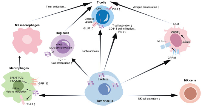
By analyzing large-scale pan-cancer data, a study has revealed that lactate metabolism-related features were negatively associated with antitumor immunity and positively associated with immunotherapy resistance (52). The positive response to ICIs in patients is linked to the presence of pre-existing intratumoral T-cell infiltration and an immunologically favorable TME characterized as ‘hot’ or T-cell-inflamed (53). Lactate can promote an immunosuppressive TME via its effects on immune cells (Fig. 2), which is associated with immune cell infiltration and response to ICIs (19).
Figure 2.
Lactate mediates the generation of an immunosuppressive TME. Accumulation of lactate induces differentiation and activation of M2-macrophages and Treg cells, inhibits the antigen-presenting function of DCs, activation of T cells and NK cells, and promotes immune escape of tumor cells. As a result, an immunosuppressive TME is formed, which affects the efficacy of anti-PD-1/PD-L1 therapy. PD-1, programmed cell death protein 1; PD-L1, programmed death-ligand 1; DCs, dendritic cells; NK cells, natural killer cells; Treg cells, regulatory T cells; MHC-II, major histocompatibility complex class II; HIF-1α, hypoxia-inducible factor-1α; GPR, Gi-protein-coupled receptor; TME, tumor microenvironment; PKA, protein kinase A; CREB, cAMP response element binding protein; NFAT, nuclear factor of activated T-cells; CMC1, C-X9-C motif containing 1; GLUT10, glucose transporter 10; CHOP, C/EBP homologous protein.
Macrophages
Macrophages are professional phagocytic cells that are capable of activating T helper cells by presenting peptide antigens through major histocompatibility complex class II (MHC-II) (54). Macrophages can be categorized into two distinct phenotypes, namely the classically activated (M1) or the alternatively activated (M2) macrophages. M1-like macrophages secrete pro-inflammatory cytokines and have the capacity to induce tumor cell death, whereas M2-like macrophages are known for their secretion of anti-inflammatory cytokines and their role in facilitating tumor progression (55). M2-like macrophages can promote malignant tumor initiation and progression (56). G protein-coupled receptor 132 (GPR132), expressed by macrophages, can sense the lactate signal in the TME (57). Upon sensing lactate, GPR132 activates G proteins coupled to it. This subsequently activates protein kinase A (PKA). The activated PKA phosphorylates cAMP response element binding protein (CREB), which then enters the nucleus (58). CREB binds to the promoter regions of M2 macrophage biomarkers, including CD206, arginase 1 (ARG1) and IL10, and promotes their expression (58). In addition, lactate can stabilize HIF-1α protein, which induces the expression of VEGF and ARG1, thereby leading to M2 macrophage polarization (59). Data have also shown that M2 macrophage polarization can be induced by lactate through the activation of the ERK/STAT3 signaling pathway, which promotes the expression of CD206 and ARG1 (55). Recent research has revealed that lactate generated by tumor cells accumulated in macrophages and induced histone H3 lysine 18 lactylation, which enhanced the expression of M2 macrophage biomarkers such as CD206, ARG1, IL10 and adrenomedullin (60). M2 macrophages can inhibit the activity of CD8+ T cells by secreting immunosuppressive factors such as IL10 and transforming growth factor β1, thereby reducing the killing effect of CD8+ T cells on tumor cells (60). The addition of lactate can increase VEGF production by macrophages, further stimulating angiogenesis (61). Furthermore, a recent study found that exogenous lactate could increase PD-L1 expression in macrophages via the activation of NF-κB (62). PD-L1 expressed on macrophages negatively regulates T-cell function and serves a crucial role in response to ICI therapy (63). Thus, lactate may influence the efficacy of immunotherapy by modulating macrophage function.
DCs
DCs are pivotal antigen-presenting cells within the TME, and are responsible for processing and presenting antigens to naïve T lymphocytes, thereby initiating an antigen-specific immune response (64). DCs have been identified as crucial players in the response to ICIs and are promising candidates for cancer immunotherapy (64). DCs are commonly categorized into plasmacytoid DCs (pDCs), monocyte-derived DCs and conventional DCs (cDCs), which encompass cDC1s and cDC2s (65). pDCs are a subset of bone marrow-derived DCs and are responsible for producing IFN-I (53). Monocyte-derived DCs are differentiated from monocytes in response to inflammation and are present under steady state conditions in specific tissues such as the gastrointestinal tract and respiratory tract (66). cDCs are derived from precursor cells originating in the bone marrow and serve a crucial role in inducing T-cell-dependent adaptive immunity (53).
Within the TME, DC immunoreactivity is typically suppressed (67). One study found that lactic acidosis impaired the function of pDCs in patients with melanoma (68). Lactate functions as a ligand for Gi-protein-coupled receptor 81 (GPR81), binding to and subsequently activating it (69). The activation of GPR81 results in downregulation of MHC-II expression on the surface of DCs, which reduces the antigen-presenting capability of DCs to T cells, thus inhibiting T cell activation and proliferation (69). Recent research has revealed that lactate could drive the formation of mature regulatory DCs (mregDCs) through activation of sterol response element binding protein 2 (70). mregDCs further inhibit antigenic cross-presentation by DCs through the secretion of soluble mediators, such as preprotein convertase kukurenine/kexin type 9, and promote Treg cell differentiation, while inhibiting activation of CD8+ T cells, thus leading to an immunosuppressive TME (70). Furthermore, reducing lactate production in DCs can increase C/EBP homologous protein expression in DCs and subsequently induce DC maturation, which promotes T-cell activation and improves the tumor response to anti-PD-1/PD-L1 therapy (71).
T cells and Treg cells
Recent research has found that elevated concentrations of lactic acid diminished the glucose uptake and antitumor efficacy of CD8+ T cells by directly interacting with the intracellular motif of GLUT10 (72). Furthermore, lactate has been shown to enhance C-X9-C motif containing 1 (CMC1) protein expression through the induction of ubiquitin specific peptidase 7, a deubiquitinating enzyme that facilitates the stabilization and deubiquitination of CMC1 protein (73). The upregulation of CMC1 expression is associated with increased levels of T-cell surface inhibitory receptors, including PD-1 and T-cell immunoglobulin and mucin-domain containing-3, indicating that CMC1 may serve a role in promoting T-cell depletion (73). Lactate is associated with impairment of T-cell cytotoxicity and IFN-γ secretion in liver kinase B1-deficient tumors, which affects the anti-PD-1/PD-L1 response in vivo (19). Additionally, lactate can inhibit CD8+ T-cell migration into tumor tissue (74). In pancreatic cancer [specifically pancreatic ductal adenocarcinoma (PDAC)], targeting of solute carrier family 4 member 4 can increase CD8+ T-cell infiltration and IFN-γ production due to the reduction of lactate production and the higher extracellular pH, which can sensitize PDAC to anti-PD-1/PD-L1 therapy (75). In hepatocellular carcinoma, inhibition of MCT4 reduces lactate output and alleviates TME acidification, which suppresses tumor growth by enhancing the infiltration and cytotoxic activity of CD8+ T cells, and has also been found to enhance the effectiveness of anti-PD-1 therapy (76). Lactate-mediated inactivation of NF-κB sensitizes cytotoxic T cells to activation-induced cell death, thereby reducing cytotoxic CD8+ T-cell infiltration and impairing the efficacy of anti-PD-1/PD-L1 therapy (77). However, it has also been proposed that lactate is an important physiological carbon source for promoting T-cell activity and that the intact function of LDH is critical for its cytotoxic function (78,79). Whether lactate itself or the resulting acidic environment mediates these different outcomes remains to be further explored.
Treg cells are central in the mediation of immune tolerance (49). Under a low-glucose and high-lactic acid environment, lactic acid can enhance the expression of PD-1 by Treg cells and inhibit PD-1 expression in effector T cells, resulting in anti-PD-1/PD-L1 therapy failure (35). Mechanistically, lactic acid enters Treg cells via MCT1 and promotes the expression of intranuclear nuclear factor of activated T-cells (NFAT)1, which positively regulates PD-1 expression (35). Recent research has shown that MOESIN lactylation levels were lower in individuals responding to anti-PD-1 therapy than in nonresponding individuals (49). Lactate can regulate the generation of Treg cells by modifying Lys72 in MOESIN, which enhances its interaction with transforming growth factor β receptor I and SMAD3 signaling (49).
NK cells
NK cells mediate immunity to pathogens independently of antigen-presenting cells (80). Lactate accumulation within the TME results in TME acidification, leading to intracellular acidification and impaired energy metabolism in NK cells upon uptake of lactate (81). Data show that the SIX homeobox 1/LDHA axis can promote the accumulation of tumor lactate in pancreatic cancer, thus inhibiting the function of NK cells (82). In breast cancer, LDHB-associated lactic acid clearance has been found to enhance NK cell activity (83). In addition, lactate and low pH reduce the cytotoxic activity of NK cells. Mechanistically, lactic acid and its dissociated hydrogen ions (H+) can lead to intracellular acidification. The activity of calcium-modulated phosphatase, which is sensitive to pH variations, may be inhibited in an acidic environment, consequently affecting the dephosphorylation and nuclear translocation of NFAT (84). By impeding NFAT activity, lactic acid diminishes the transcription and production of IFN-γ, thereby impairing the effector functions of NK cells (84). Furthermore, lactic acid indirectly hinders NK cell function by promoting the expansion of myeloid-derived suppressor cells (81). Combination strategies encompassing anti-NK cell and anti-PD-1/PD-L1 therapies show greater efficacy than anti-NK cell therapies in gastric cancer (85).
5. Clinical significance of lactate in anti-PD-1/PD-L1 therapy
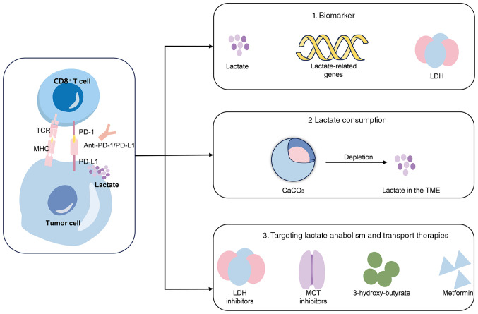
Within the TME, TCRs identify tumor antigens presented by MHC molecules, facilitating the activation of T cells to execute cytotoxic functions and eliminate cancer cells (6). Nevertheless, tumors can progressively evolve immune evasion strategies, including the upregulation of PD-L1, which impairs T-cell activity through its interaction with PD-1 receptors on T-cell surfaces (6). Anti-PD therapies employ monoclonal antibodies to inhibit the PD-1/PD-L1 signaling pathway (6). Lactate metabolism in tumor cells has been shown to be associated with immunotherapy resistance (18). Thus, further investigation is warranted to explore the potential of utilizing lactate as a predictive marker for immunotherapy efficacy, as well as the potential of targeting lactate metabolism to enhance the effectiveness of immunotherapy (Fig. 3).
Figure 3.
Clinical applications of lactate. Lactate and other key enzymes may serve a role in predicting the effectiveness of immunotherapy and combination therapy. Lactate-related genes, lactate and LDH levels have the potential to function as biomarkers for the therapeutic response. The abundance of lactate within the TME can be diminished either through direct depletion using CaCO3 or by targeting key enzymes involved in lactate metabolism, such as LDH or MCT. Additionally, the combination of 3-hydroxy-butyrate and metformin has been demonstrated to synergistically reduce serum lactate concentrations. TCR, T-cell receptor; MHC, major histocompatibility complex; LDH, lactate dehydrogenase; MCT, monocarboxylate transporter; PD-1, programmed cell death protein 1; PD-L1, programmed death-ligand 1; TME, tumor microenvironment.
A lactate metabolism-related signature associated with the prediction of responses to immunotherapy and related prognosis has been identified and validated using information from public databases; however, validation in a larger number of patients is required (52). For example, a prognostic signature was constructed for patients with renal clear cell carcinoma using three lactate-associated genes, and this could serve as a reliable predictor of prognosis and response to immunotherapy (86). Furthermore, a study has demonstrated that patients treated with pembrolizumab who exhibited elevated baseline LDH levels had a reduced overall survival compared with those with normal LDH levels, suggesting that LDH could function as a biomarker for predicting the efficacy of immunotherapy (87).
Targeting metabolism combined with immunotherapy can help to increase the effectiveness of immunotherapy (88). Lactate abundance in the TME can be reduced by affecting key enzymes in lactate metabolism such as LDH (88) or by directly depleting lactate (89). Accordingly, nanovaccines are already available that deliver CaCO3 to tumor tissue to deplete lactate (89). However, inhibition of lactic acid production in tumor cells is also required. Evidence has shown that lactate/GPR81 blockade (3-hydroxy-butyrate) combined with metformin synergistically inhibited cancer cell proliferation in vitro. Additionally, this combination has been shown to suppress glycolysis and oxidative phosphorylation metabolism, as well as impede tumor growth and reduce serum lactate levels in tumor-bearing mice. Furthermore, this treatment regimen enhances the infiltration of CD8+ T cells in tumors and augments IFN-γ secretion in lymph nodes (90). Taken together, these findings suggest a promising strategy to enhance patient responsiveness to PD-1/PD-L1 inhibition (90).
A multifunctional nanoplatform has shown effective consumption of glucose and lactate within the TME. The nanoplatform combined three components: Glucose oxidase, laccase and CpG. These were integrated into a zeolitic imidazolate framework-8 structure and then coated with a red blood cell membrane. Additionally, in conjunction with anti-PD-1/PD-L1 therapy, the nanoplatform elicited robust systemic immunity, resulting in successful eradication of tumors (91).
Targeting lactate output is also a promising therapeutic strategy (76). Diclofenac has been shown to be a powerful inhibitor of MCT1 and MCT4, which reduced lactate secretion from tumor cells, and enhanced T-cell killing induced by anti-PD-1 ICIs and the efficacy of ICI therapy (92).
Targeting lactate metabolism and its associated pathways offers novel strategies for cancer treatment, potentially providing innovative therapeutic approaches to address immunotherapy resistance. However, metabolic therapies targeting tumors may also impact cells within the TME and compromise immune cell function (93). Therefore, the synergistic interaction between metabolic therapies and antitumor immunity requires careful consideration. Given the metabolic heterogeneity of tumors, precision and personalization may represent the future direction for metabolic therapies in oncology.
6. Discussion
Immunotherapy has been linked to enhanced survival of patients with cancer (3); however, screening for immune-resistant and immune-beneficial patient populations remains a major challenge. To address this challenge, the link between metabolic reprogramming and immunotherapy has become a hot research topic. Metabolic reprogramming involves modifications in various metabolic pathways such as glycolysis, amino acid metabolism and lipid metabolism (93). Glycolysis produces lactate as a byproduct, which serves as a crucial metabolite for cellular functions (94). Amino acid and lipid metabolism are also involved in the regulation of lactate metabolism and influence tumor immunity (43). For instance, amino acids such as glutamine can serve as precursors for lactate production and are converted to lactate in tumor cells (43). Intermediates produced during lipid metabolism can also influence the glycolytic process. Valerate and butyrate enhance mTOR activity, while mTORC1-mediated glutamine uptake suppresses the expression of glycolytic genes such as GLUT1 and HK2 (95). In tumor cells, increased production of acetyl-CoA via fatty acid oxidation may inhibit the activity of PDH, reducing the conversion of pyruvate to acetyl-CoA, and consequently increasing lactate production (96,97). The present review has improved the understanding of the effects of lactate on tumor cell proliferation and function; however, specific regulatory mechanisms remain to be explored. For instance, the regulatory effects of lactate on immune cells have the potential to suppress antitumor immunity and contribute to resistance against immunotherapy. Understanding the mechanisms underlying metabolic reprogramming in tumors, as well as the interactions facilitating their immune evasion, is pivotal for devising strategies to enhance the efficacy of immunotherapy.
Briefly, the mechanisms through which lactate influences immune cell function within the TME are as follows (33,93,98): i) The induction of an acidic environment that impairs the activity of immune cells; ii) the modulation of immune cell signaling pathways, such as NF-κB and HIF-1α; iii) the utilization of lactate as a substrate for lactylation, which modifies proteins, including histones, thereby impacting immune cell gene expression and function; iv) the promotion of recruitment and stimulation of immunosuppressive cells such as Treg cells; and v) the regulation of the metabolic state of immune cells by either providing energy as a metabolic substrate or affecting metabolic pathways such as glycolysis and oxidative phosphorylation. Overall, the role of lactate within the TME is multifaceted and diverse. Current research mainly emphasizes the contributions of lactate to tumor progression and immune evasion (16,50). However, under certain conditions, lactate can also serve as an energy source and provide survival support to immune cells. It is expected that future studies will reveal more specific mechanisms of the role of lactate in tumor progression and metastasis, providing a theoretical basis for the development of novel therapeutic strategies.
In summary, most current research indicates that lactate within the TME may impact the efficacy of anti-PD-1/PD-L1 therapies through its role in mediating immunosuppression (71–75). This finding implies that biomarkers associated with lactate metabolism could serve as predictive indicators of the response to anti-PD-1/PD-L1 treatment (99). A study has demonstrated that immunotherapy efficacy can be altered by modulating lactate metabolism (19). Modifying lactate metabolism might enhance the responsiveness of patients to anti-PD-1/PD-L1 therapies. Lactate, a byproduct of tumor cell metabolism, is also involved in the metabolic processes of immune cells. Consequently, elucidating the metabolic crosstalk between tumor cells and immune cells is crucial for generating novel insights and therapeutic targets to enhance the efficacy of anti-PD-1/PD-L1 therapies. However, research on the impact of targeting metabolic pathways in tumor cells on immune cell metabolism within the TME remains limited. Further studies are needed to identify novel targeted agents capable of more effectively and selectively modulating immune responses within the TME.
Acknowledgements
Not applicable.
Funding Statement
The present study was supported by the National Natural Science Foundation of China (grant nos. 82103004 and 82273323).
Availability of data and materials
Not applicable.
Authors' contributions
YZ and YH contributed to conceptualization and writing of the manuscript. QT, LP, JW and FT contributed to the design of figures and revising the manuscript. XD conceptualized the article and reviewed the manuscript. Data authentication is not applicable. All authors have read and approved the final manuscript.
Ethics approval and consent to participate
Not applicable.
Patient consent for publication
Not applicable.
Competing interests
The authors declare that they have no competing interests.
References
- 1.Reck M, Rodríguez-Abreu D, Robinson AG, Hui R, Csőszi T, Fülöp A, Gottfried M, Peled N, Tafreshi A, Cuffe S, et al. Pembrolizumab versus chemotherapy for PD-L1-positive non-small-cell lung cancer. N Engl J Med. 2016;375:1823–1833. doi: 10.1056/NEJMoa1606774. [DOI] [PubMed] [Google Scholar]
- 2.Mok TSK, Wu YL, Kudaba I, Kowalski DM, Cho BC, Turna HZ, Castro G, Jr, Srimuninnimit V, Laktionov KK, Bondarenko I, et al. Pembrolizumab versus chemotherapy for previously untreated, PD-L1-expressing, locally advanced or metastatic non-small-cell lung cancer (KEYNOTE-042): A randomised, open-label, controlled, phase 3 trial. Lancet. 2019;393:1819–1830. doi: 10.1016/S0140-6736(18)32409-7. [DOI] [PubMed] [Google Scholar]
- 3.Lorusso D, Xiang Y, Hasegawa K, Scambia G, Leiva M, Ramos-Elias P, Acevedo A, Sukhin V, Cloven N, Pereira de Santana Gomes AJ, et al. Pembrolizumab or placebo with chemoradiotherapy followed by pembrolizumab or placebo for newly diagnosed, high-risk, locally advanced cervical cancer (ENGOT-cx11/GOG-3047/KEYNOTE-A18): Overall survival results from a randomised, double-blind, placebo-controlled, phase 3 trial. Lancet. 2024;404:1321–1332. doi: 10.1016/S0140-6736(24)01808-7. [DOI] [PubMed] [Google Scholar]
- 4.Choueiri TK, Tomczak P, Park SH, Venugopal B, Ferguson T, Symeonides SN, Hajek J, Chang YH, Lee JL, Sarwar N, et al. Overall survival with adjuvant pembrolizumab in renal-cell carcinoma. N Engl J Med. 2024;390:1359–1371. doi: 10.1056/NEJMoa2312695. [DOI] [PubMed] [Google Scholar]
- 5.Yi M, Zheng X, Niu M, Zhu S, Ge H, Wu K. Combination strategies with PD-1/PD-L1 blockade: Current advances and future directions. Mol Cancer. 2022;21:28. doi: 10.1186/s12943-021-01489-2. [DOI] [PMC free article] [PubMed] [Google Scholar]
- 6.Vesely MD, Zhang T, Chen L. Resistance mechanisms to anti-PD cancer immunotherapy. Annu Rev Immunol. 2022;40:45–74. doi: 10.1146/annurev-immunol-070621-030155. [DOI] [PubMed] [Google Scholar]
- 7.Peng DH, Rodriguez BL, Diao L, Chen L, Wang J, Byers LA, Wei Y, Chapman HA, Yamauchi M, Behrens C, et al. Collagen promotes anti-PD-1/PD-L1 resistance in cancer through LAIR1-dependent CD8+ T cell exhaustion. Nature Commun. 2020;11:4520. doi: 10.1038/s41467-020-18298-8. [DOI] [PMC free article] [PubMed] [Google Scholar]
- 8.Yu M, Peng Z, Qin M, Liu Y, Wang J, Zhang C, Lin J, Dong T, Wang L, Li S, et al. Interferon-γ induces tumor resistance to anti-PD-1 immunotherapy by promoting YAP phase separation. Mol Cell. 2021;81:1216–1230.e9. doi: 10.1016/j.molcel.2021.01.010. [DOI] [PubMed] [Google Scholar]
- 9.Zhou X, Zou L, Liao H, Luo J, Yang T, Wu J, Chen W, Wu K, Cen S, Lv D, et al. Abrogation of HnRNP L enhances anti-PD-1 therapy efficacy via diminishing PD-L1 and promoting CD8+ T cell-mediated ferroptosis in castration-resistant prostate cancer. Acta Pharm Sin B. 2022;12:692–707. doi: 10.1016/j.apsb.2021.07.016. [DOI] [PMC free article] [PubMed] [Google Scholar]
- 10.Jiang Z, Lim SO, Yan M, Hsu JL, Yao J, Wei Y, Chang SS, Yamaguchi H, Lee HH, Ke B, et al. TYRO3 induces anti-PD-1/PD-L1 therapy resistance by limiting innate immunity and tumoral ferroptosis. J Clin Invest. 2021;131:e139434. doi: 10.1172/JCI139434. [DOI] [PMC free article] [PubMed] [Google Scholar]
- 11.Zhou X, Lin J, Shao Y, Zheng H, Yang Y, Li S, Fan X, Hong H, Mao Z, Xue P, et al. Targeting PLCG2 suppresses tumor progression, orchestrates the tumor immune microenvironment and potentiates immune checkpoint blockade therapy for colorectal cancer. Int J Biol Sci. 2024;20:5548–5575. doi: 10.7150/ijbs.98200. [DOI] [PMC free article] [PubMed] [Google Scholar]
- 12.Dai Y, Guo Z, Leng D, Jiao G, Chen K, Fu M, Liu Y, Shen Q, Wang Q, Zhu L, Zhao Q. Metal-coordinated NIR-II nanoadjuvants with nanobody conjugation for potentiating immunotherapy by tumor metabolism reprogramming. Adv Sci (Weinh) 2024;11:e2404886. doi: 10.1002/advs.202404886. [DOI] [PMC free article] [PubMed] [Google Scholar]
- 13.Pavlova Natalya N, Thompson Craig B. The emerging hallmarks of cancer metabolism. Cell Metab. 2016;23:27–47. doi: 10.1016/j.cmet.2015.12.006. [DOI] [PMC free article] [PubMed] [Google Scholar]
- 14.Nisar H, Sanchidrián González PM, Brauny M, Labonté FM, Schmitz C, Roggan MD, Konda B, Hellweg CE. Hypoxia changes energy metabolism and growth rate in non-small cell lung cancer cells. Cancers (Basel) 2023;15:2472. doi: 10.3390/cancers15092472. [DOI] [PMC free article] [PubMed] [Google Scholar]
- 15.Li X, Wenes M, Romero P, Huang SCC, Fendt SM, Ho PC. Navigating metabolic pathways to enhance antitumour immunity and immunotherapy. Nat Rev Clin Oncol. 2019;16:425–441. doi: 10.1038/s41571-019-0203-7. [DOI] [PubMed] [Google Scholar]
- 16.Zhang Y, Zhai Z, Duan J, Wang X, Zhong J, Wu L, Li A, Cao M, Wu Y, Shi H, et al. Lactate: The mediator of metabolism and immunosuppression. Front Endocrinol (Lausanne) 2022;13:901495. doi: 10.3389/fendo.2022.901495. [DOI] [PMC free article] [PubMed] [Google Scholar]
- 17.Shergold AL, Millar R, Nibbs RJB. Understanding and overcoming the resistance of cancer to PD-1/PD-L1 blockade. Pharmacol Res. 2019;145:104258. doi: 10.1016/j.phrs.2019.104258. [DOI] [PubMed] [Google Scholar]
- 18.Cao Z, Xu D, Harding J, Chen W, Liu X, Wang Z, Wang L, Qi T, Chen S, Guo X, et al. Lactate oxidase nanocapsules boost T cell immunity and efficacy of cancer immunotherapy. Sci Transl Med. 2023;15:eadd2712. doi: 10.1126/scitranslmed.add2712. [DOI] [PMC free article] [PubMed] [Google Scholar]
- 19.Qian Y, Galan-Cobo A, Guijarro I, Dang M, Molkentine D, Poteete A, Zhang F, Wang Q, Wang J, Parra E, et al. MCT4-dependent lactate secretion suppresses antitumor immunity in LKB1-deficient lung adenocarcinoma. Cancer Cell. 2023;41:1363–1380.e7. doi: 10.1016/j.ccell.2023.05.015. [DOI] [PMC free article] [PubMed] [Google Scholar]
- 20.Gordon SR, Maute RL, Dulken BW, Hutter G, George BM, McCracken MN, Gupta R, Tsai JM, Sinha R, Corey D, et al. PD-1 expression by tumour-associated macrophages inhibits phagocytosis and tumour immunity. Nature. 2017;545:495–499. doi: 10.1038/nature22396. [DOI] [PMC free article] [PubMed] [Google Scholar]
- 21.Marasco M, Berteotti A, Weyershaeuser J, Thorausch N, Sikorska J, Krausze J, Brandt HJ, Kirkpatrick J, Rios P, Schamel WW, et al. Molecular mechanism of SHP2 activation by PD-1 stimulation. Sci Adv. 2020;6:eaay4458. doi: 10.1126/sciadv.aay4458. [DOI] [PMC free article] [PubMed] [Google Scholar]
- 22.Yokosuka T, Takamatsu M, Kobayashi-Imanishi W, Hashimoto-Tane A, Azuma M, Saito T. Programmed cell death 1 forms negative costimulatory microclusters that directly inhibit T cell receptor signaling by recruiting phosphatase SHP2. J Exp Med. 2012;209:1201–1217. doi: 10.1084/jem.20112741. [DOI] [PMC free article] [PubMed] [Google Scholar]
- 23.Ruiz de Galarreta M, Bresnahan E, Molina-Sánchez P, Lindblad KE, Maier B, Sia D, Puigvehi M, Miguela V, Casanova-Acebes M, Dhainaut M, et al. β-Catenin activation promotes immune escape and resistance to anti-PD-1 therapy in hepatocellular carcinoma. Cancer Discov. 2019;9:1124–1141. doi: 10.1158/2159-8290.CD-19-0074. [DOI] [PMC free article] [PubMed] [Google Scholar]
- 24.Zhou L, Mudianto T, Ma X, Riley R, Uppaluri R. Targeting EZH2 enhances antigen presentation, antitumor Immunity, and circumvents anti-PD-1 resistance in head and neck cancer. Clin Cancer Res. 2020;26:290–300. doi: 10.1158/1078-0432.CCR-19-1351. [DOI] [PMC free article] [PubMed] [Google Scholar]
- 25.Rosenthal R, Cadieux EL, Salgado R, Bakir MA, Moore DA, Hiley CT, Lund T, Tanić M, Reading JL, Joshi K, et al. Neoantigen-directed immune escape in lung cancer evolution. Nature. 2019;567:479–485. doi: 10.1038/s41586-019-1032-7. [DOI] [PMC free article] [PubMed] [Google Scholar]
- 26.Tumeh PC, Harview CL, Yearley JH, Shintaku IP, Taylor EJM, Robert L, Chmielowski B, Spasic M, Henry G, Ciobanu V, et al. PD-1 blockade induces responses by inhibiting adaptive immune resistance. Nature. 2014;515:568–571. doi: 10.1038/nature13954. [DOI] [PMC free article] [PubMed] [Google Scholar]
- 27.Kwon M, An M, Klempner SJ, Lee H, Kim KM, Sa JK, Cho HJ, Hong JY, Lee T, Min YW, et al. Determinants of response and intrinsic resistance to PD-1 blockade in microsatellite instability-high gastric cancer. Cancer Discov. 2021;11:2168–2185. doi: 10.1158/2159-8290.CD-21-0219. [DOI] [PubMed] [Google Scholar]
- 28.Skoulidis F, Goldberg ME, Greenawalt DM, Hellmann MD, Awad MM, Gainor JF, Schrock AB, Hartmaier RJ, Trabucco SE, Gay L, et al. STK11/LKB1 mutations and PD-1 inhibitor resistance in KRAS-mutant lung adenocarcinoma. Cancer Discov. 2018;8:822–835. doi: 10.1158/2159-8290.CD-18-0099. [DOI] [PMC free article] [PubMed] [Google Scholar]
- 29.Messaoudene M, Pidgeon R, Richard C, Ponce M, Diop K, Benlaifaoui M, Nolin-Lapalme A, Cauchois F, Malo J, Belkaid W, et al. A natural polyphenol exerts antitumor activity and circumvents anti-PD-1 resistance through effects on the gut microbiota. Cancer Discov. 2022;12:1070–1087. doi: 10.1158/2159-8290.CD-21-0808. [DOI] [PMC free article] [PubMed] [Google Scholar]
- 30.Lei Q, Wang D, Sun K, Wang L, Zhang Y. Resistance mechanisms of anti-PD1/PDL1 therapy in solid tumors. Front Cell Dev Biol. 2020;8:672. doi: 10.3389/fcell.2020.00672. [DOI] [PMC free article] [PubMed] [Google Scholar]
- 31.Murciano-Goroff YR, Warner AB, Wolchok JD. The future of cancer immunotherapy: Microenvironment-targeting combinations. Cell Res. 2020;30:507–519. doi: 10.1038/s41422-020-0337-2. [DOI] [PMC free article] [PubMed] [Google Scholar]
- 32.Zou W, Green DR. Beggars banquet: Metabolism in the tumor immune microenvironment and cancer therapy. Cell Metab. 2023;35:1101–1113. doi: 10.1016/j.cmet.2023.06.003. [DOI] [PMC free article] [PubMed] [Google Scholar]
- 33.Wang ZH, Peng WB, Zhang P, Yang XP, Zhou Q. Lactate in the tumour microenvironment: From immune modulation to therapy. EBioMedicine. 2021;73:103627. doi: 10.1016/j.ebiom.2021.103627. [DOI] [PMC free article] [PubMed] [Google Scholar]
- 34.Jedlička M, Feglarová T, Janstová L, Hortová-Kohoutková M, Frič J. Lactate from the tumor microenvironment-A key obstacle in NK cell-based immunotherapies. Front Immunol. 2022;13:932055. doi: 10.3389/fimmu.2022.932055. [DOI] [PMC free article] [PubMed] [Google Scholar]
- 35.Kumagai S, Koyama S, Itahashi K, Tanegashima T, Lin YT, Togashi Y, Kamada T, Irie T, Okumura G, Kono H, et al. Lactic acid promotes PD-1 expression in regulatory T cells in highly glycolytic tumor microenvironments. Cancer Cell. 2022;40:201–218.e9. doi: 10.1016/j.ccell.2022.01.001. [DOI] [PubMed] [Google Scholar]
- 36.Warburg O, Wind F, Negelein E. Über den stoffwechsel von tumoren im körper. Klin Wochenschr. 1926;5:829–832. doi: 10.1007/BF01726240. [DOI] [Google Scholar]
- 37.Paul S, Ghosh S, Kumar S. Tumor glycolysis, an essential sweet tooth of tumor cells. Semin Cancer Biol. 2022;86:1216–1230. doi: 10.1016/j.semcancer.2022.09.007. [DOI] [PubMed] [Google Scholar]
- 38.Luo B, Song L, Chen L, Cai Y, Zhang M, Wang S. Ganoderic acid D attenuates gemcitabine resistance of triple-negative breast cancer cells by inhibiting glycolysis via HIF-1alpha destabilization. Phytomedicine. 2024;129:155675. doi: 10.1016/j.phymed.2024.155675. [DOI] [PubMed] [Google Scholar]
- 39.Mossmann D, Park S, Hall MN. mTOR signalling and cellular metabolism are mutual determinants in cancer. Nat Rev Cancer. 2018;18:744–757. doi: 10.1038/s41568-018-0074-8. [DOI] [PubMed] [Google Scholar]
- 40.Zhang P, Wan Y, Ma J, Gong J, Zhong Z, Cui Y, Zhang H, Da Y, Ma J, Li C, et al. Epigenetic silencing of LDHB promotes hepatocellular carcinoma by remodeling the tumor microenvironment. Cancer Immunol Immunother. 2024;73:127. doi: 10.1007/s00262-024-03717-2. [DOI] [PMC free article] [PubMed] [Google Scholar]
- 41.Hong SM, Lee YK, Park I, Kwon SM, Min S, Yoon G. Lactic acidosis caused by repressed lactate dehydrogenase subunit B expression down-regulates mitochondrial oxidative phosphorylation via the pyruvate dehydrogenase (PDH)-PDH kinase axis. J Biol Chem. 2019;294:7810–7820. doi: 10.1074/jbc.RA118.006095. [DOI] [PMC free article] [PubMed] [Google Scholar]
- 42.Yue J, Xu J, Yin Y, Shu Y, Li Y, Li T, Zou Z, Wang Z, Li F, Zhang M, et al. Targeting the PDK/PDH axis to reverse metabolic abnormalities by structure-based virtual screening with in vitro and in vivo experiments. Int J Biol Macromol. 2024;262:129970. doi: 10.1016/j.ijbiomac.2024.129970. [DOI] [PubMed] [Google Scholar]
- 43.Li X, Yang Y, Zhang B, Lin X, Fu X, An Y, Zou Y, Wang JX, Wang Z, Yu T. Lactate metabolism in human health and disease. Signal Transduct Target Ther. 2022;7:305. doi: 10.1038/s41392-022-01151-3. [DOI] [PMC free article] [PubMed] [Google Scholar]
- 44.Tian LR, Lin MZ, Zhong HH, Cai YJ, Li B, Xiao ZC, Shuai XT. Nanodrug regulates lactic acid metabolism to reprogram the immunosuppressive tumor microenvironment for enhanced cancer immunotherapy. Biomater Sci. 2022;10:3892–3900. doi: 10.1039/D2BM00650B. [DOI] [PubMed] [Google Scholar]
- 45.Tasdogan A, Faubert B, Ramesh V, Ubellacker JM, Shen B, Solmonson A, Murphy MM, Gu Z, Gu W, Martin M, et al. Metabolic heterogeneity confers differences in melanoma metastatic potential. Nature. 2020;577:115–120. doi: 10.1038/s41586-019-1847-2. [DOI] [PMC free article] [PubMed] [Google Scholar]
- 46.Hui S, Ghergurovich JM, Morscher RJ, Jang C, Teng X, Lu W, Esparza LA, Reya T, Le Zhan, Yanxiang Guo J, et al. Glucose feeds the TCA cycle via circulating lactate. Nature. 2017;551:115–118. doi: 10.1038/nature24057. [DOI] [PMC free article] [PubMed] [Google Scholar]
- 47.Faubert B, Li KY, Cai L, Hensley CT, Kim J, Zacharias LG, Yang C, Do QN, Doucette S, Burguete D, et al. Lactate metabolism in human lung tumors. Cell. 2017;171:358–371.e9. doi: 10.1016/j.cell.2017.09.019. [DOI] [PMC free article] [PubMed] [Google Scholar]
- 48.Pérez-Escuredo J, Dadhich RK, Dhup S, Cacace A, Van Hée VF, De Saedeleer CJ, Sboarina M, Rodriguez F, Fontenille MJ, Brisson L, et al. Lactate promotes glutamine uptake and metabolism in oxidative cancer cells. Cell Cycle. 2016;15:72–83. doi: 10.1080/15384101.2015.1120930. [DOI] [PMC free article] [PubMed] [Google Scholar]
- 49.Gu J, Zhou J, Chen Q, Xu X, Gao J, Li X, Shao Q, Zhou B, Zhou H, Wei S, et al. Tumor metabolite lactate promotes tumorigenesis by modulating MOESIN lactylation and enhancing TGF-β signaling in regulatory T cells. Cell Rep. 2022;39:110986. doi: 10.1016/j.celrep.2022.110986. [DOI] [PubMed] [Google Scholar]
- 50.Ippolito L, Comito G, Parri M, Iozzo M, Duatti A, Virgilio F, Lorito N, Bacci M, Pardella E, Sandrini G, et al. Lactate rewires lipid metabolism and sustains a metabolic-epigenetic axis in prostate cancer. Cancer Res. 2022;82:1267–1282. doi: 10.1158/0008-5472.CAN-21-0914. [DOI] [PMC free article] [PubMed] [Google Scholar]
- 51.Xie D, Zhu S, Bai L. Lactic acid in tumor microenvironments causes dysfunction of NKT cells by interfering with mTOR signaling. Sci China Life Sci. 2016;59:1290–1296. doi: 10.1007/s11427-016-0348-7. [DOI] [PubMed] [Google Scholar]
- 52.Chen D, Liu P, Lu X, Li J, Qi D, Zang L, Lin J, Liu Y, Zhai S, Fu D, et al. Pan-cancer analysis implicates novel insights of lactate metabolism into immunotherapy response prediction and survival prognostication. J Exp Clin Cancer Res. 2024;43:125. doi: 10.1186/s13046-024-03042-7. [DOI] [PMC free article] [PubMed] [Google Scholar]
- 53.Marciscano AE, Anandasabapathy N. The role of dendritic cells in cancer and anti-tumor immunity. Semin Immunol. 2021;52:101481. doi: 10.1016/j.smim.2021.101481. [DOI] [PMC free article] [PubMed] [Google Scholar]
- 54.Christofides A, Strauss L, Yeo A, Cao C, Charest A, Boussiotis VA. The complex role of tumor-infiltrating macrophages. Nat Immunol. 2022;23:1148–1156. doi: 10.1038/s41590-022-01267-2. [DOI] [PMC free article] [PubMed] [Google Scholar]
- 55.Mu X, Shi W, Xu Y, Xu C, Zhao T, Geng B, Yang J, Pan J, Hu S, Zhang C, et al. Tumor-derived lactate induces M2 macrophage polarization via the activation of the ERK/STAT3 signaling pathway in breast cancer. Cell Cycle. 2018;17:428–438. doi: 10.1080/15384101.2018.1444305. [DOI] [PMC free article] [PubMed] [Google Scholar]
- 56.Zhang A, Xu Y, Xu H, Ren J, Meng T, Ni Y, Zhu Q, Zhang WB, Pan YB, Jin J, et al. Lactate-induced M2 polarization of tumor-associated macrophages promotes the invasion of pituitary adenoma by secreting CCL17. Theranostics. 2021;11:3839–3852. doi: 10.7150/thno.53749. [DOI] [PMC free article] [PubMed] [Google Scholar]
- 57.Chen P, Zuo H, Xiong H, Kolar MJ, Chu Q, Saghatelian A, Siegwart DJ, Wan Y. Gpr132 sensing of lactate mediates tumor-macrophage interplay to promote breast cancer metastasis. Proc Natl Acad Sci USA. 2017;114:580–585. doi: 10.1073/pnas.1614035114. [DOI] [PMC free article] [PubMed] [Google Scholar]
- 58.Jiang H, Wei H, Wang H, Wang Z, Li J, Ou Y, Xiao X, Wang W, Chang A, Sun W, et al. Zeb1-induced metabolic reprogramming of glycolysis is essential for macrophage polarization in breast cancer. Cell Death Dis. 2022;13:206. doi: 10.1038/s41419-022-04632-z. [DOI] [PMC free article] [PubMed] [Google Scholar]
- 59.Colegio OR, Chu NQ, Szabo AL, Chu T, Rhebergen AM, Jairam V, Cyrus N, Brokowski CE, Eisenbarth SC, Phillips GM, et al. Functional polarization of tumour-associated macrophages by tumour-derived lactic acid. Nature. 2014;513:559–563. doi: 10.1038/nature13490. [DOI] [PMC free article] [PubMed] [Google Scholar]
- 60.Cai J, Song L, Zhang F, Wu S, Zhu G, Zhang P, Chen S, Du J, Wang B, Cai Y, et al. Targeting SRSF10 might inhibit M2 macrophage polarization and potentiate anti-PD-1 therapy in hepatocellular carcinoma. Cancer Commun (Lond) 2024;44:1231–1260. doi: 10.1002/cac2.12607. [DOI] [PMC free article] [PubMed] [Google Scholar]
- 61.Zhang J, Muri J, Fitzgerald G, Gorski T, Gianni-Barrera R, Masschelein E, D'Hulst G, Gilardoni P, Turiel G, Fan Z, et al. Endothelial lactate controls muscle regeneration from ischemia by inducing M2-like macrophage polarization. Cell Metab. 2020;31:1136–1153.e7. doi: 10.1016/j.cmet.2020.05.004. [DOI] [PMC free article] [PubMed] [Google Scholar]
- 62.Morrissey SM, Zhang F, Ding C, Montoya-Durango DE, Hu X, Yang C, Wang Z, Yuan F, Fox M, Zhang HG, et al. Tumor-derived exosomes drive immunosuppressive macrophages in a pre-metastatic niche through glycolytic dominant metabolic reprogramming. Cell Metab. 2021;33:2040–2058.e10. doi: 10.1016/j.cmet.2021.09.002. [DOI] [PMC free article] [PubMed] [Google Scholar]
- 63.Tang H, Liang Y, Anders RA, Taube JM, Qiu X, Mulgaonkar A, Liu X, Harrington SM, Guo J, Xin Y, et al. PD-L1 on host cells is essential for PD-L1 blockade-mediated tumor regression. J Clin Invest. 2018;128:580–588. doi: 10.1172/JCI96061. [DOI] [PMC free article] [PubMed] [Google Scholar]
- 64.Del Prete A, Salvi V, Soriani A, Laffranchi M, Sozio F, Bosisio D, Sozzani S. Dendritic cell subsets in cancer immunity and tumor antigen sensing. Cell Mol Immunol. 2023;20:432–447. doi: 10.1038/s41423-023-00990-6. [DOI] [PMC free article] [PubMed] [Google Scholar]
- 65.See P, Dutertre CA, Chen J, Gunther P, McGovern N, Irac SE, Gunawan M, Beyer M, Händler K, Duan K, et al. Mapping the human DC lineage through the integration of high-dimensional techniques. Science. 2017;356:eaag3009. doi: 10.1126/science.aag3009. [DOI] [PMC free article] [PubMed] [Google Scholar]
- 66.Rigamonti A, Villar J, Segura E. Monocyte differentiation within tissues: A renewed outlook. Trends Immunol. 2023;44:999–1013. doi: 10.1016/j.it.2023.10.005. [DOI] [PubMed] [Google Scholar]
- 67.Peng X, He Y, Huang J, Tao Y, Liu S. Metabolism of dendritic cells in tumor microenvironment: for immunotherapy. Front Immunol. 2021;12:613492. doi: 10.3389/fimmu.2021.613492. [DOI] [PMC free article] [PubMed] [Google Scholar]
- 68.Monti M, Vescovi R, Consoli F, Farina D, Moratto D, Berruti A, Specchia C, Vermi W. Plasmacytoid dendritic cell impairment in metastatic melanoma by lactic acidosis. Cancers (Basel) 2020;12:2085. doi: 10.3390/cancers12082085. [DOI] [PMC free article] [PubMed] [Google Scholar]
- 69.Brown TP, Bhattacharjee P, Ramachandran S, Sivaprakasam S, Ristic B, Sikder MOF, Ganapathy V. The lactate receptor GPR81 promotes breast cancer growth via a paracrine mechanism involving antigen-presenting cells in the tumor microenvironment. Oncogene. 2020;39:3292–3304. doi: 10.1038/s41388-020-1216-5. [DOI] [PubMed] [Google Scholar]
- 70.Plebanek MP, Xue Y, Nguyen YV, DeVito NC, Wang X, Holtzhausen A, Beasley GM, Theivanthiran B, Hanks BA. A lactate-SREBP2 signaling axis drives tolerogenic dendritic cell maturation and promotes cancer progression. Sci Immunol. 2024;9:eadi4191. doi: 10.1126/sciimmunol.adi4191. [DOI] [PubMed] [Google Scholar]
- 71.Wang Z, Xu F, Hu J, Zhang H, Cui L, Lu W, He W, Wang X, Li M, Zhang H, et al. Modulation of lactate-lysosome axis in dendritic cells by clotrimazole potentiates antitumor immunity. J Immunother Cancer. 2021;9:e002155. doi: 10.1136/jitc-2020-002155. [DOI] [PMC free article] [PubMed] [Google Scholar]
- 72.Liu Y, Wang F, Peng D, Zhang D, Liu L, Wei J, Yuan J, Zhao L, Jiang H, Zhang T, et al. Activation and antitumor immunity of CD8+ T cells are supported by the glucose transporter GLUT10 and disrupted by lactic acid. Sci Transl Med. 2024;16:eadk7399. doi: 10.1126/scitranslmed.adk7399. [DOI] [PubMed] [Google Scholar]
- 73.Chen Y, Gao J, Ma M, Wang K, Liu F, Yang F, Yang F, Zou X, Cheng Z, Wu D. The potential role of CMC1 as an immunometabolic checkpoint in T cell immunity. Oncoimmunology. 2024;13:2344905. doi: 10.1080/2162402X.2024.2344905. [DOI] [PMC free article] [PubMed] [Google Scholar]
- 74.Sasaki K, Nishina S, Yamauchi A, Fukuda K, Hara Y, Yamamura M, Egashira K, Hino K. Nanoparticle-mediated delivery of 2-deoxy-D-glucose induces antitumor immunity and cytotoxicity in liver tumors in mice. Cell Mol Gastroenterol Hepatol. 2021;11:739–762. doi: 10.1016/j.jcmgh.2020.10.010. [DOI] [PMC free article] [PubMed] [Google Scholar]
- 75.Cappellesso F, Orban MP, Shirgaonkar N, Berardi E, Serneels J, Neveu MA, Di Molfetta D, Piccapane F, Caroppo R, Debellis L, et al. Targeting the bicarbonate transporter SLC4A4 overcomes immunosuppression and immunotherapy resistance in pancreatic cancer. Nat Cancer. 2022;3:1464–1483. doi: 10.1038/s43018-022-00470-2. [DOI] [PMC free article] [PubMed] [Google Scholar]
- 76.Fang Y, Liu W, Tang Z, Ji X, Zhou Y, Song S, Tian M, Tao C, Huang R, Zhu G, et al. Monocarboxylate transporter 4 inhibition potentiates hepatocellular carcinoma immunotherapy through enhancing T cell infiltration and immune attack. Hepatology. 2023;77:109–123. doi: 10.1002/hep.32348. [DOI] [PubMed] [Google Scholar]
- 77.Liu H, Liang Z, Cheng S, Huang L, Li W, Zhou C, Zheng X, Li S, Zeng Z, Kang L. Mutant KRAS drives immune evasion by sensitizing cytotoxic T-cells to activation-induced cell death in colorectal cancer. Adv Sci (Weinh) 2023;10:e2203757. doi: 10.1002/advs.202203757. [DOI] [PMC free article] [PubMed] [Google Scholar]
- 78.Kaymak I, Luda KM, Duimstra LR, Ma EH, Longo J, Dahabieh MS, Faubert B, Oswald BM, Watson MJ, Kitchen-Goosen SM, et al. Carbon source availability drives nutrient utilization in CD8(+) T cells. Cell Metab. 2022;34:1298–1311.e6. doi: 10.1016/j.cmet.2022.07.012. [DOI] [PMC free article] [PubMed] [Google Scholar]
- 79.Notarangelo G, Spinelli JB, Perez EM, Baker GJ, Kurmi K, Elia I, Stopka SA, Baquer G, Lin JR, Golby AJ, et al. Oncometabolite d-2HG alters T cell metabolism to impair CD8(+) T cell function. Science. 2022;377:1519–1529. doi: 10.1126/science.abj5104. [DOI] [PMC free article] [PubMed] [Google Scholar]
- 80.Wang H, Grzywacz B, Sukovich D, McCullar V, Cao Q, Lee AB, Blazar BR, Cornfield DN, Miller JS, Verneris MR. The unexpected effect of cyclosporin A on CD56+CD16- and CD56+CD16+ natural killer cell subpopulations. Blood. 2007;110:1530–1539. doi: 10.1182/blood-2006-10-048173. [DOI] [PMC free article] [PubMed] [Google Scholar]
- 81.Miao L, Lu C, Zhang B, Li H, Zhao X, Chen H, Liu Y, Cui X. Advances in metabolic reprogramming of NK cells in the tumor microenvironment on the impact of NK therapy. J Transl Med. 2024;22:229. doi: 10.1186/s12967-024-05033-w. [DOI] [PMC free article] [PubMed] [Google Scholar]
- 82.Ge W, Meng L, Cao S, Hou C, Zhu X, Huang D, Li Q, Peng Y, Jiang K. The SIX1/LDHA axis promotes lactate accumulation and leads to NK cell dysfunction in pancreatic cancer. J Immunol Res. 2023;2023:6891636. doi: 10.1155/2023/6891636. [DOI] [PMC free article] [PubMed] [Google Scholar]
- 83.Luo Z, Huang X, Xu X, Wei K, Zheng Y, Gong K, Li W. Decreased LDHB expression in breast tumor cells causes NK cell activation and promotes tumor progression. Cancer Biol Med. 2024;21:513–540. doi: 10.20892/j.issn.2095-3941.2023.0382. [DOI] [PMC free article] [PubMed] [Google Scholar]
- 84.Brand A, Singer K, Koehl GE, Kolitzus M, Schoenhammer G, Thiel A, Matos C, Bruss C, Klobuch S, Peter K, et al. LDHA-associated lactic acid production blunts tumor immunosurveillance by T and NK cells. Cell Metab. 2016;24:657–671. doi: 10.1016/j.cmet.2016.08.011. [DOI] [PubMed] [Google Scholar]
- 85.Abdolahi S, Ghazvinian Z, Muhammadnejad S, Ahmadvand M, Aghdaei HA, Ebrahimi-Barough S, Ai J, Zali MR, Verdi J, Baghaei K. Adaptive NK cell therapy modulated by anti-PD-1 antibody in gastric cancer model. Front Pharmacol. 2021;12:733075. doi: 10.3389/fphar.2021.733075. [DOI] [PMC free article] [PubMed] [Google Scholar]
- 86.Sun Z, Tao W, Guo X, Jing C, Zhang M, Wang Z, Kong F, Suo N, Jiang S, Wang H. Construction of a lactate-related prognostic signature for predicting prognosis, tumor microenvironment, and immune response in kidney renal clear cell carcinoma. Front Immunol. 2022;13:818984. doi: 10.3389/fimmu.2022.818984. [DOI] [PMC free article] [PubMed] [Google Scholar]
- 87.Wagner NB, Forschner A, Leiter U, Garbe C, Eigentler TK. S100B and LDH as early prognostic markers for response and overall survival in melanoma patients treated with anti-PD-1 or combined anti-PD-1 plus anti-CTLA-4 antibodies. Br J Cancer. 2018;119:339–346. doi: 10.1038/s41416-018-0167-x. [DOI] [PMC free article] [PubMed] [Google Scholar]
- 88.Heuser C, Renner K, Kreutz M, Gattinoni L. Targeting lactate metabolism for cancer immunotherapy-a matter of precision. Semin Cancer Biol. 2023;88:32–45. doi: 10.1016/j.semcancer.2022.12.001. [DOI] [PubMed] [Google Scholar]
- 89.Ding Y, Yang J, Wei H, Wang J, Huang S, Yang S, Guo Y, Li B, Shuai X. Construction of pH-sensitive nanovaccines encapsulating tumor cell lysates and immune adjuvants for breast cancer therapy. Small. 2023;19:e2301420. doi: 10.1002/smll.202301420. [DOI] [PubMed] [Google Scholar]
- 90.Chen S, Zhou X, Yang X, Li W, Li S, Hu Z, Ling C, Shi R, Liu J, Chen G, et al. Dual blockade of lactate/GPR81 and PD-1/PD-L1 pathways enhances the anti-tumor effects of metformin. Biomolecules. 2021;11:1373. doi: 10.3390/biom11091373. [DOI] [PMC free article] [PubMed] [Google Scholar]
- 91.Ji P, Jin XK, Deng XC, Zhang SM, Liang JL, Li QR, Chen WH, Zhang XZ. Metabolic regulation-mediated reversion of the tumor immunosuppressive microenvironment for potentiating cooperative metabolic therapy and immunotherapy. Nano Lett. 2024;24:4691–701. doi: 10.1021/acs.nanolett.4c01307. [DOI] [PubMed] [Google Scholar]
- 92.Renner K, Bruss C, Schnell A, Koehl G, Becker HM, Fante M, Menevse AN, Kauer N, Blazquez R, Hacker L, et al. Restricting glycolysis preserves T Cell effector functions and augments checkpoint therapy. Cell Rep. 2019;29:135–150.e9. doi: 10.1016/j.celrep.2019.08.068. [DOI] [PubMed] [Google Scholar]
- 93.Zheng Y, Xu R, Chen X, Lu Y, Zheng J, Lin Y, Zheng J, Lin Y, Lin P, Zhao X, Cui L. Metabolic gatekeepers: Harnessing tumor-derived metabolites to optimize T cell-based immunotherapy efficacy in the tumor microenvironment. Cell Death Dis. 2024;15:775. doi: 10.1038/s41419-024-07122-6. [DOI] [PMC free article] [PubMed] [Google Scholar]
- 94.Liu Y, Zhao Y, Song H, Li Y, Liu Z, Ye Z, Zhao J, Wu Y, Tang J, Yao M. Metabolic reprogramming in tumor immune microenvironment: Impact on immune cell function and therapeutic implications. Cancer Lett. 2024;597:217076. doi: 10.1016/j.canlet.2024.217076. [DOI] [PubMed] [Google Scholar]
- 95.Li J, Zhao J, Tian C, Dong L, Kang Z, Wang J, Zhao S, Li M, Tong X. Mechanisms of regulation of glycolipid metabolism by natural compounds in plants: Effects on short-chain fatty acids. Nutr Metab (Lond) 2024;21:49. doi: 10.1186/s12986-024-00829-5. [DOI] [PMC free article] [PubMed] [Google Scholar]
- 96.Wang J, Yang Y, Shao F, Meng Y, Guo D, He J, Lu Z. Acetate reprogrammes tumour metabolism and promotes PD-L1 expression and immune evasion by upregulating c-Myc. Nat Metab. 2024;6:914–932. doi: 10.1038/s42255-024-01037-4. [DOI] [PubMed] [Google Scholar]
- 97.Bose S, Ramesh V, Locasale JW. Acetate metabolism in physiology, cancer, and beyond. Trends Cell Biol. 2019;29:695–703. doi: 10.1016/j.tcb.2019.05.005. [DOI] [PMC free article] [PubMed] [Google Scholar]
- 98.Burgdorf S, Porubsky S, Marx A, Popovic ZV. Cancer acidity and hypertonicity contribute to dysfunction of tumor-associated dendritic cells: Potential impact on antigen cross-presentation machinery. Cancers (Basel) 2020;12:2403. doi: 10.3390/cancers12092403. [DOI] [PMC free article] [PubMed] [Google Scholar]
- 99.Shang S, Wang MZ, Xing Z, He N, Li S. Lactate regulators contribute to tumor microenvironment and predict prognosis in lung adenocarcinoma. Front Immunol. 2022;13:1024925. doi: 10.3389/fimmu.2022.1024925. [DOI] [PMC free article] [PubMed] [Google Scholar]
Associated Data
This section collects any data citations, data availability statements, or supplementary materials included in this article.
Data Availability Statement
Not applicable.


