Abstract
Although trastuzumab deruxtecan improves the prognosis of patients with HER2-low breast cancer, the characteristics and prognostic value of low HER2 status remains to be elucidated. A prospective database of patients with clinical stage I to III breast cancer who underwent surgery between September 2012 and October 2022 at Teikyo University Hospital (Tokyo, Japan) were analyzed. HER2 was evaluated using fluorescence in situ hybridization assay, and HER2-low and HER2-negative was defined as HER2/CEP17 ratio ≥1.0, and <1.0, respectively. The median age and Ki67 score of the 1,024 patients were 56.0 years (range, 23.0–93.0 years) and 15.0% (range, 0.5–99.0%), respectively. Overall, 908 (88.7%) patients were hormone receptor positive. Among all patients, 902 (88.1%) had HER2-low tumors and 122 (11.9%) had HER2-negative tumors. Positive rates for estrogen receptor (ER) and progesterone receptor (PgR) were significantly higher in HER2-low compared with HER2-negative patients [ER: 804 (89.1%) patients vs. 99 (81.1%) patients, P=0.021; PgR: 723 (80.1%) patients vs. 86 (70.5%) patients, P=0.023]. The median Ki67 score was significantly lower in HER2-low compared with HER2-negative patients (14.5 vs. 18.5%; P=0.013). With a median follow-up time of 46.2 months, the overall survival (OS) was significantly improved in HER2-low compared with HER2-negative patients (97.4 vs. 96.7%; P=0.029). Multivariate logistic regression analyses revealed that HER2-low status was not an independent factor for OS. The findings of the present study suggest that HER2-low status may not have a significant association with prognosis, despite a significant association between Ki67 and hormone receptor expression.
Keywords: breast cancer, HER2, HER2-low status, HER2 negative, fluorescence in situ hybridization
Introduction
The HER2 gene, located on the long arm of chromosome 17, encodes a transmembrane tyrosine kinase receptor protein (1,2). Although normal breast epithelial cells have approximately 20,000 receptors per cell, the number of receptors is estimated to be about 2 million in HER2 overexpressing breast cancer (1). The HER2 receptor has a tyrosine kinase within its intracellular domain. Upon dimerization, HER2 stimulates tyrosine kinase, resulting in the activation of signaling cascades. This cascade is involved in the proliferation, invasion, and metastasis of HER2 positive breast cancer (3). HER2 overexpression was found in 15% of breast cancers and correlated with poor prognosis (4). Identification of the mechanism and aggressive feature of HER2 positive breast cancer has led to the development of trastuzumab, which has improved the prognosis of HER2 positive breast cancer.
NSABP-B31 showed an improvement in disease-free survival (DFS) and overall survival (OS) with trastuzumab in combination with adjuvant chemotherapy after surgery for HER2 positive early stage breast cancer (5). A reanalysis of HER2 expression in patients in this trial showed that 174 of 1787 patients were HER2 negative, and further subgroup analysis showed an additive effect of trastuzumab in HER2 negative patients [relative risk for DFS, 0.34; 95% confidence interval (CI), 0.14 to 0.80; P=0.014] (6). NSABP B-47, a randomized trial of adjuvant chemotherapy in combination with or without trastuzumab for HER2-negative patients showed no additive effect of trastuzumab [hazard ratio (HR), 0.98; 95% CI, 0.76 to 1.25; P=0.85] (7).
HER2 negative breast cancer account for approximately 80% of all breast cancers. Moreover, HER2 expression is observed in HER2 negative breast cancers, albeit at low levels (HER2-low breast cancer) (8), accounting for approximately half of all breast cancer cases (9). Recently, trastuzumab deruxtecan was shown to improve the progression-free survival (PFS) and OS of patients with HER2-low breast cancer in DESTINY Breast04 trial. This new classification is very interesting and noteworthy. In addition, the DESTINY Breast06 trial showed that trastuzumab deruxtecan improve PFS in patients with HER2-low breast cancer as well as HER2-ultralow breast cancer. Nevertheless, the clinicopathological features and prognosis of HER2-low breast cancer remain poorly defined. Many other studies (3,7,10–12) regarding the clinical characteristics of HER2-low breast cancer have used immunohistochemistry (IHC) to determine HER2 status.
In this study, we retrospectively analyzed the clinicopathologic characteristics and prognosis of HER2-low breast cancer using fluorescence in situ hybridization (FISH). This is the first study to evaluate the HER2-low status by FISH assay.
Materials and methods
Study design and population
A prospective database of patients with clinical stage I to III breast cancer who underwent surgery between September 2012 and October 2022 at Teikyo University Hospital was analyzed. Patients with pathologically diagnosed invasive carcinoma without HER2 amplification on presurgical core needle biopsy or surgical specimens were included. HER2 gene amplification was evaluated by SRL Inc., Japan using the PathVysion HER-2 DNA probe kit (Abbott Molecular Inc., IL). The PathVysion HER-2 DNA probe kit consists of Locus Specific Identifier (LSI) HER-2/neu spectrum orange DNA probe and Chromosome Enumeration Probe (CEP) 17 spectrum green DNA probe. The LSI HER-2/neu DNA probe is a 226 Kb SpectrumOrange directly-labeled, fluorescent DNA probe specific for the HER-2/neu gene locus (17q11.2-q12). The CEP17 DNA probe is a 5.4 Kb SpectrumGreen directly-labeled, fluorescent DNA probe specific for the alpha satellite DNA sequence at the centromeric region of chromosome 17 (17p11.1-q11.1). Tissues were fixed in 10% neutral buffered formalin solution for 24–48 h, de-alcoholized, and then paraffin-embedded. Section slides were prepared with a thickness of 4 to 6 µm. Next, the samples were immersed in protease solution (37±1°C) for 10–60 min for enzymatic treatment, and then immersed in 10% neutral buffered formalin solution for 10 min at room temperature for fixation. After that, hybridization was performed. 10 µl of DNA probe was added to the sample and incubated at 37±1°C for 14–18 h. The samples were washed by immersion in posthybridization wash buffer at 72±1°C for 2 min, and 10 µl of 4, 6-diamidino-2-phenylindole counterstain was added for measurement. The signal was measured using a fluorescent microscope. The number of LSI HER-2/neu and CEP17 signals in 20 nuclei were counted using a 63× or 100× objective lens. HER2-low and HER2-negative was defined as HER2/CEP17 ratio ≧1.0, and <1.0, respectively. Patients were considered hormone receptor (HR)-positive if more than 1% of the infiltrating tumor cells showed immunostaining for estrogen receptor (ER) or progesterone receptor (PgR). Lymph node metastasis was assessed in presurgical core needle biopsy or surgical specimens. Pathologic complete response (pCR) was defined as ypT0/isN0.
Statistical analysis
To compare the patients' clinicopathological characteristics, continuous variables are expressed as medians and categorical variables are expressed as numbers and percentages. Pearson χ2 test were used to compared categorical variables. Two-sides P-values of <0.05 were considered statistically significant. OS was defined as time from the date of surgery to time of death or last follow-up. DFS was defined as time from the date of surgery to the date of disease recurrence or death or last follow-up. Prognostic analysis was performed using the Kaplan-Meier curves and the log-rank test. Multivariate logistic regression analyses were used to identify independent prognostic factors. All statistical analyses were performed using the SPSS Statistics 28.0.0.0 (IBM, Armonk, NY, USA).
Results
Clinicopathologic characteristics
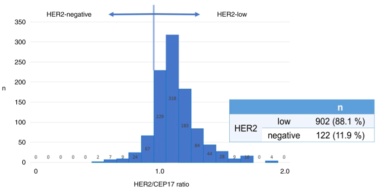
The median age, tumor size, and ki67 of the entire 1024 patients was 56.0 years (range=20.0–93.0), 2.0 cm (range=0.3–15.0 cm), and 15.0% (range=0.5–99.0%), respectively. 155 (15.1%) patients had lymph node metastasis. Additionally, 908 (88.7%) patients revealed HR positive (ER positive; 903 patients, PgR positive; 810 patients). The number of HER2-low and HER2-negative patients was 902 (88.1%) and 122 (11.9%), respectively (Fig. 1).
Figure 1.
Distribution of HER2/CEP ratio in all patients.
No significant differences were observed between HER2-low and HER2-negative patients in factors of age, tumor size, clinical T stage, lymph node metastasis, nuclear grade, rate of neoadjuvant chemotherapy (NAC), and adjuvant therapy (Table I). Positive rates for ER and PgR were significantly higher in HER2-low patients compared to HER2-negative patients [ER: 804 (89.0%) patients vs. 99 (82.0%) patients; P=0.021, PgR: 723 (80.2%) patients vs. 86 (71.3%) patients; P=0.023]. The median ki67 was significantly lower for HER2-low compared to HER2-negative (14.5 vs. 18.5%, P=0.013) patients.
Table I.
Patient characteristics.
| Characteristic | Group | HER2-low (n=902) | HER2-negative (n=122) | P-value |
|---|---|---|---|---|
| Median age, years (range) | 56.0 (20–93) | 54.0 (23–90) | 0.286 | |
| Median tumor size, cm | 2.0 | 1.9 | 0.546 | |
| cT stage, n (%) | 1 | 482 (53.4) | 66 (54.1) | 0.955 |
| 2 | 357 (39.6) | 46 (37.7) | ||
| 3 | 20 (2.2) | 5 (4.1) | ||
| 4 | 43 (4.8) | 5 (4.1) | ||
| Lymph node metastasis, n (%) | Yes | 134 (14.9) | 21 (17.2) | 0.468 |
| No | 768 (85.1) | 101 (82.8) | ||
| Nuclear grade, n (%) | 1 | 571 (63.3) | 71 (58.2) | 0.097 |
| 2 | 141 (15.6) | 20 (16.4) | ||
| 3 | 126 (14.0) | 25 (20.5) | ||
| Unknown | 64 (7.1) | 6 (4.9) | ||
| Estrogen receptor, n (%) | Positive | 804 (89.0) | 99 (82.0) | 0.021 |
| Progesterone receptor, n (%) | Positive | 723 (80.2) | 86 (71.3) | 0.023 |
| Median Ki67 (%) | 14.5 | 18.5 | 0.013 | |
| Neoadjuvant chemotherapy, n (%) | Yes | 168 (18.6) | 29 (23.8) | 0.160 |
| No | 735 (81.5) | 92 (75.4) | ||
| Adjuvant therapy, n (%) | Chemotherapy | 141 (15.6) | 19 (15.6) | 0.894 |
| Endocrine | 815 (90.4) | 102 (83.6) | 0.056 | |
| Radiation | 681 (75.5) | 100 (82.0) | 0.088 |
NAC was performed on 197 patients, including 168 HER2-low and 29 HER2-negative patients (Table II). Although the median ki67 was significantly higher for HER2-negative compared to HER2-low in patients treated with NAC (35.0 vs. 30.0%, P=0.036), the pCR rates were not significantly different between the two groups [16.1% (27/168 patients) vs. 17.2% (5/29 patients), P=0.528].
Table II.
Patient characteristics of patients treated with neoadjuvant chemotherapy.
| Characteristics | Groups | HER2-low (n=168) | HER2-negative (n=29) | P-value |
|---|---|---|---|---|
| Median age, years | 55.5 | 53.0 | 0.474 | |
| Median tumor size, cm | 3.2 | 3.2 | 0.824 | |
| Lymph node metastasis, n (%) | Yes | 103 (61.3) | 19 (65.5) | 0.667 |
| No | 65 (38.7) | 10 (34.5) | ||
| HR, n (%) | Positive | 105 (62.5) | 14 (48.3) | 0.148 |
| Negative | 63 (37.5) | 15 (51.7) | ||
| Nuclear grade, n (%) | 1.2 | 89 (53.0) | 14 (48.3) | 0.444 |
| 3 | 70 (41.7) | 15 (51.7) | ||
| Unknown | 9 (5.4) | 0 (0) | ||
| Median Ki67 (%) | 30.0 | 35.0 | 0.036 | |
| pCR rate (%) | 16.1 | 17.2 | 0.528 |
HR, hormone receptor; pCR, pathologic complete response.
Prognostic analysis
At a median follow-up time of 46.2 months, the OS was significantly better in HER2-low compared to HER2-negative (97.4 vs. 96.7%, P=0.029, Fig. 2A) patient. DFS was not significantly different between the two groups (92.1 vs. 89.5%, P=0.294, Fig. 2B). In univariate analysis, lymph node metastasis, HR expression, HER2 status, nuclear grade, and ki67 were significantly associated with OS. In the multivariate logistic analysis, lymph node metastasis and HR expression were significantly associated with OS (Table III).
Figure 2.
Kaplan-Meier survival analysis for (A) OS and (B) DFS according to HER2 status. P-values were obtained from the stratified log rank test. OS, overall survival; DFS, disease-free survival.
Table III.
Analysis of prognostic factor for OS.
| Factor | Group | OS events, no. of events/entire patients (%) | Univariate analysis P-value | Multivariate logistic regression analyses P-value |
|---|---|---|---|---|
| Age | ≥56.0 years | 21/529 (4.0) | 0.125 | |
| <56.0 years | 12/496 (2.4) | |||
| Lymph node metastasis | Yes | 16/155 (10.3) | <0.001 | <0.001 |
| No | 17/869 (2.0) | |||
| HR | Positive | 20/908 (2.2) | <0.001 | 0.005 |
| Negative | 13/116 (11.2) | |||
| HER2 | Low | 26/903 (2.9) | 0.029 | 0.340 |
| Negative | 7/121 (5.8) | |||
| NG | 1.2 | 17/803 (2.1) | <0.001 | 0.231 |
| 3 | 13/151 (8.6) | |||
| Ki67 | ≥15% | 21/463 (4.5) | 0.014 | 0.256 |
| <15% | 8/458 (1.7) |
HR, hormone receptor; NG, nuclear grade.
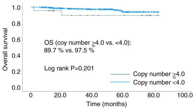
Among the HER2-low patients, 23 cases had a HER2 copy number of ≥4.0, and 662 cases had <4.0. Meanwhile, among the HER2-negative patients, one case had a HER2 copy number of ≥4.0, and 99 cases had <4.0. No significant difference was observed in the OS between the high copy number (≥4.0) and the low copy number (<4.0) (89.7 vs. 97.5% P=0.201, Fig. 3) group among the HER2-low patients.
Figure 3.

Kaplan-Meier survival analysis for OS according to HER2 copy number. Comparison of high (≥4.0) and low (<4.0) copy number in HER2-low patients. OS, overall survival.
Among HR-positive patients, HER2-low and HER2-negative patients were 808 (89.0%) and 100 (11.0%), respectively. Among HR-negative patients, HER2-low and HER2-negative patients were 94 (81.0%) and 22 (19.0%), respectively. There was no significant difference in the OS between HER2-low patients and HER2-negative patients in both HR positive and HR negative cases (HR positive: 98.2 vs. 95.0%, P=0.332; HR negative: 92.4 vs. 83.3%, P=0.311, Fig. 4).
Figure 4.
Kaplan-Meier survival analysis for overall survival of (A) HR-positive and (B) HR-negative patients according to HER2 status. HR, hormone receptor.
Discussion
In this study, the positive rates of ER and PgR were significantly higher, and the median ki67 was significantly lower in HER2-low compared to HER2-negative patients. With a median follow-up time of 46.2 months, the OS was significantly better in HER2-low patients compared to HER2-negative patients, and HER2-low expression was not independently related factors for OS.
Schettini et al reported that HER2-low tumors were more frequently found within HR-positive disease compared to triple-negative breast cancer (65.4 vs. 36.5%, P<0.001) (13). Tarantino et al reported that HER2-low breast cancer patients had a significantly higher HR-positive rate than HER2-negative (90.6 vs. 81.8%, P<0.001) which is consistent with our study (89.6 vs. 82.0%, P=0.011) (10). Moreover, they analyzed the rate of HER2-low breast cancer by ER expression levels (negative, 0%; low, 1–9%; moderate, 10–49%; high, 50–95%; very high, >95%) and found a positive correlation between ER expression and the rate of HER2-low breast cancer (negative, 40.1%; low, 46.3%; moderate, 55.2%; high, 57.8%; very high, 62.1%; P<0.001) (10). A study analyzing clinicopathologic characteristics of inflammatory breast cancer with HER2-low also showed ER expressing tumors were more common in patients with HER2-low vs. HER2-negative (65 vs. 38%, P<0.01) (14). In addition, considering that the median ki67 was significantly lower in HER2-low than in HER2-negative (14.5 vs. 18.5%, P=0.013), HER2-low was considered luminal-like and HER2-negative was considered triple negative breast cancer-like. The PAM50 gene expression profile showed a lower percentage of basal-like and higher percentage of luminal A in HER2-low compared to HER2-negative breast cancer patients (luminal A, 50.8 vs. 28.7%; basal-like, 13.3 vs. 43.7%, P<0.001) (13). In other words, most of these studies showed that HER2-low breast cancer is luminal-like and HER2-negative breast cancer is triple-negative-like. This finding is consistent with the significantly higher positive rate of ER and PgR in HER2-low patients compared to HER2-negative patients in our study. Therefore, luminal-like feature might be one of the clinicopathologic characteristics of HER2-low breast cancer.
There are various views on the prognostic impact of HER2-low. Retrospective study of 3169 Japanese breast cancer patients without HER2 overexpression found no statistically significant differences between the prognosis of HER2-low and HER2-0 patients, regardless of HR status, although patients in the HER2-low group tended to have better prognosis than those in the HER2-0 group (5 year OS: HR-positive, 96.7 vs. 94.9%, P=0.215; HR-negative, 86.5 vs. 79.3%, P=0.152) (15). Another retrospective study of 804 primary breast cancer patients also confirmed no significant differences in progression-free interval (PFI) between HER2-low and HER2-subtype (5 year PFI rate: HER2-low/HR+ vs. HER2-/HR+, 82.6 vs. 78.0%, P=0.65; HER2-low/HR-vs. HER2-/HR-, 63 vs. 65%, P=0.81) (16). Gampenrieder et al analyzed the prognostic value of low HER2 expression in metastatic breast cancer and reported that low HER2 expression was not associated with OS regardless of HR expression (11). A study analyzing clinicopathologic characteristics of inflammatory breast cancer (IBC) with HER2-low reported that differences in OS were small, both among ER-positive HER2-negative vs. HER2-low IBC (48 month OS: 80 vs. 81%, HR: 0.82, 95%CI:0.39–1.73) and ER-negative HER2-negative vs. HER2-low IBC (48 month OS: 34 vs. 47%, HR: 1.34, 95%CI: 0.74–2.41) (14). Moreover, Mouabbi et al reported that progression-free survival and OS were not statistically different between the patients with HER2-low and HER2-negative treated with targeted therapy (cyclin-dependent kinase 4/6 inhibitors, everolimus, or alpelisib) plus endocrine therapy (17). Tarantino et al reported that patients with HER2-low breast cancer had a significantly better OS than patients with HER2-negative breast cancer. However, this significance disappeared when HR-positive and HR-negative were analyzed separately or when adjusted for menopausal status, HR status, tumor grade, and histology [HR-positive, 1.34 (P=0.26); HR-negative, 0.88 (P=0.65); overall, 1.14 (P=0.52)] (10). On the other hand, Park et al retrospectively analyzed 2452 non-metastatic triple negative breast cancer and revealed that OS was significantly better in patients with HER2-low compared to HER2-0 (HR: 0.698, 95%CI: 0.517–0.943, P=0.019) (18). In our study, although OS was significantly better in HER2-low, HER2-low was not significant associated factor for OS in multivariate analysis. The high rate of HR positivity in HER2-low patients and the positive correlation between HER2 amplification and ER expression suggest that HR status may be a confounding factor in prognosis.
Denkert et al performed a pooled analysis of a cohort study of patients with HER2 non-amplified primary breast cancer who underwent NAC, and found a significantly higher pCR rate for HER2-negative compared to HER2-low across 2310 patients (39.0 vs. 29.2%, P=0.0002) (19). Tarantino et al analyzed inflammatory breast cancer and reported the higher pCR rate for HER2-negative breast cancer compared to HER2-low breast cancer (11 vs. 6%, OR: 1.8, 95%CI:0.6–5.3) (14). In our study, there was no significant difference in the pCR rate between the two groups (17.2 vs. 16.1% vs., P=0.528). This could be attributed to a small number of patients in our study.
This study had several limitations, the most important limitation of this study is that the cutoff value for HER2-low was defined as a HER2/CEP17 ratio ≧1.0. The definition of HER2-low status using FISH assay requires careful consideration. In general, HER2-low is defined as an IHC score of 1+ or 2+ with negative FISH assay. Among 1024 cases in our study, HER2 status was evaluated by both IHC and FISH assay in 280 patients, and there was no significant correlation between IHC and FISH assay (HER2/CEP17 ratio; IHC 0, 0.8–1.7; 1+, 0.5–1.7; 2+, 0.0–1.7, P=0.118). In this study, HER2-low was defined as HER2/CEP17 ratio ≧1.0 since copy number of HER2 is more than the CEP. The validity of this definition using FISH requires further investigation. The other limitation is that the data for this study was collected at a single site, so the results are exploratory. Moreover, our study was underpowered to find statistical significance due to the small sample size. Additionally, because of the short observation period, caution is needed in interpreting the prognosis, especially considering that most of the patients in our study were HR positive. Therefore, this study is ongoing with additional patients and an extended observation period. In the near future, more solid data will be provided.
Many other studies (3,7,10–12) regarding the clinical characteristics of HER2-low breast cancer have used IHC to determine HER2 status. To the best of our knowledge, this study is the only study evaluating HER2 status using FISH assay, and this new definition of HER2-low is a strength of this study. In the future, this new definition may help guide treatment strategies for HER2-low breast cancer.
In this analysis, HER2-low patients had a significantly higher HR-positive rate and lower median ki67 compared to HER2-negative patients. Multivariate logistic regression analyses showed that HER2-low status was not an independent factor for OS. These data suggested that HER2-low may not be established as an independent subtype.
Acknowledgements
Not applicable.
Funding Statement
Funding: No funding was received.
Availability of data and materials
The data generated in the present study may be requested from the corresponding author.
Authors' contributions
AS and HJ contributed to the concept and design of the study and were major contributors to writing the manuscript. AM contributed to the study design. YM, AM and TI contributed to data acquisition. HJ contributed to review and editing. AS, YM, AM, TI and HJ confirm the authenticity of all the data. All authors read and approved the final version of the manuscript.
Ethics approval and consent to participate
The present study was approved by The Medical Ethics Committee of Teikyo University School of Medicine (Tokyo, Japan; approval no. 22-113). Patient informed consent was waived.
Patient consent for publication
Not applicable.
Competing interests
The authors declare that they have no competing interests.
References
- 1.Ross JS, Fletcher JA, Linette GP, Stec J, Clark E, Ayers M, Symmans WF, Pusztai L, Bloom KJ. The Her-2/neu gene and protein in breast cancer 2003: Biomarker and target of therapy. Oncologist. 2003;8:307–325. doi: 10.1634/theoncologist.8-4-307. [DOI] [PubMed] [Google Scholar]
- 2.Grant S, Qiao L, Dant P. Roles of ERBB family receptor tyrosine kinases, and downstream signaling pathways, in the control of cell growth and survival. Front Biosci. 2002;7:d376–d389. doi: 10.2741/A782. [DOI] [PubMed] [Google Scholar]
- 3.Zhang H, Katerji H, Turner BM, Hicks DG. HER2-low breast cancers: New opportunities and challenges. Am J Clin Pathol. 2022;157:328–336. doi: 10.1093/ajcp/aqab117. [DOI] [PubMed] [Google Scholar]
- 4.Slamon DJ, Clark GM, Wong SG, Levin WJ, Ullrich A, McGuire WL. Human breast cancer: Correlation of relapse and survival with amplification of the HER-2/neu oncogene. Science. 1987;235:177–182. doi: 10.1126/science.3798106. [DOI] [PubMed] [Google Scholar]
- 5.Romond EH, Perez EA, Bryant J, Suman VJ, Geyer CE, Jr, Davidson NE, Tan-Chiu E, Martino S, Paik S, Kaufman PA, et al. Trastuzumab plus adjuvant chemotherapy for operable HER2-positive breast cancer. N Engl J Med. 2005;353:1673–1684. doi: 10.1056/NEJMoa052122. [DOI] [PubMed] [Google Scholar]
- 6.Paik S, Kim C, Wolmark N. HER2 status and benefit from adjuvant trastuzumab in breast cancer. N Engl J Med. 2008;358:1409–1411. doi: 10.1056/NEJMc0801440. [DOI] [PubMed] [Google Scholar]
- 7.Fehrenbacher L, Cecchini RS, Geyer CE, Jr, Rastogi P, Costantino JP, Atkins JN, Crown JP, Polikoff J, Boileau JF, Provencher L, et al. NSABP B-47/NRG oncology phase III randomized trial comparing adjuvant chemotherapy with or without trastuzumab in high-risk invasive breast cancer negative for HER2 by FISH and with IHC 1+ or 2. J Clin Oncol. 2020;38:444–453. doi: 10.1200/JCO.19.01455. [DOI] [PMC free article] [PubMed] [Google Scholar]
- 8.Ergun Y, Ucar G, Akagunduz B. Comparison of HER2-zero and HER2-low in terms of clinicopathological factors and survival in early-stage breast cancer: A systematic review and meta-analysis. Cancer Treat Rev. 2023;115:102538. doi: 10.1016/j.ctrv.2023.102538. [DOI] [PubMed] [Google Scholar]
- 9.Tarantino P, Hamilton E, Tolaney SM, Cortes J, Morganti S, Ferraro E, Marra A, Viale G, Trapani D, Cardoso F, et al. HER2-low breast cancer: Pathological and clinical landscape. J Clin Oncol. 2020;38:1951–1962. doi: 10.1200/JCO.19.02488. [DOI] [PubMed] [Google Scholar]
- 10.Tarantino P, Jin Q, Tayob N, Jeselsohn RM, Schnitt SJ, Vincuilla J, Parker T, Tyekucheva S, Li T, Lin NU, et al. Prognostic and biologic significance of ERBB2-low expression in early-stage breast cancer. JAMA Oncol. 2022;8:1177–1183. doi: 10.1001/jamaoncol.2022.2286. [DOI] [PMC free article] [PubMed] [Google Scholar]
- 11.Gampenrieder SP, Rinnerthaler G, Tinchon C, Petzer A, Balic M, Heibl S, Schmitt C, Zabernigg AF, Egle D, Sandholzer M, et al. Landscape of HER2-low metastatic breast cancer (MBC): Results from the Austrian AGMT_MBC-registry. Breast Cancer Res. 2021;23:112. doi: 10.1186/s13058-021-01492-x. [DOI] [PMC free article] [PubMed] [Google Scholar]
- 12.Miglietta F, Griguolo G, Bottosso M, Giarratano T, Lo Mele M, Fassan M, Cacciatore M, Genovesi E, De Bartolo D, Vernaci G, et al. HER2-low-positive breast cancer: Evolution from primary tumor to residual disease after neoadjuvant treatment. NPJ Breast Cancer. 2022;8:66. doi: 10.1038/s41523-022-00434-w. [DOI] [PMC free article] [PubMed] [Google Scholar]
- 13.Schettini F, Chic N, Brasó-Maristany F, Paré L, Pascual T, Conte B, Martínez-Sáez O, Adamo B, Vidal M, Barnadas E, et al. Clinical, pathological, and PAM50 gene expression features of HER2-low breast cancer. NPJ Breast Cancer. 2021;7:1. doi: 10.1038/s41523-020-00208-2. [DOI] [PMC free article] [PubMed] [Google Scholar]
- 14.Tarantino P, Niman S, Erick T, Priedigkeit N, Harrison BT, Giordano A, Nakhlis F, Bellon JR, Parker T, Strauss S, et al. HER2-low inflammatory breast cancer: Clinicopathologic features and prognostic implications. Eur J Cancer. 2022;174:277–286. doi: 10.1016/j.ejca.2022.07.001. [DOI] [PubMed] [Google Scholar]
- 15.Horisawa N, Adachi Y, Takatsuka D, Nozawa K, Endo Y, Ozaki Y, Sugino K, Kataoka A, Kotani H, Yoshimura A, et al. The frequency of low HER2 expression in breast cancer and a comparison of prognosis between patients with HER2-low and HER2-negative breast cancer by HR status. Breast Cancer. 2022;29:234–241. doi: 10.1007/s12282-021-01303-3. [DOI] [PubMed] [Google Scholar]
- 16.Agostinetto E, Rediti M, Fimereli D, Debien V, Piccart M, Aftimos P, Sotiriou C, de Azambuja E. HER2-low breast cancer: Molecular characteristics and prognosis. Cancers (Basel) 2021;13:2824. doi: 10.3390/cancers13112824. [DOI] [PMC free article] [PubMed] [Google Scholar]
- 17.Mouabbi J, Singareeka Raghavendra A, Bassett R, Jr, Hassan A, Tripathy D, Layman RM. Survival outcomes in patients with hormone receptor-positive metastatic breast cancer with low or no ERBB2 expression treated with targeted therapies plus endocrine therapy. JAMA Netw Open. 2023;6:e2313017. doi: 10.1001/jamanetworkopen.2023.13017. [DOI] [PMC free article] [PubMed] [Google Scholar]
- 18.Park WK, Nam SJ, Kim SW, Lee JE, Yu J, Lee SK, Ryu JM, Chae BJ. The prognostic impact of HER2-low and menopausal status in triple-negative breast cancer. Cancers (Basel) 2024;16:2566. doi: 10.3390/cancers16142566. [DOI] [PMC free article] [PubMed] [Google Scholar]
- 19.Denkert C, Seither F, Schneeweiss A, Link T, Blohmer JU, Just M, Wimberger P, Forberger A, Tesch H, Jackisch C, et al. Clinical and molecular characteristics of HER2-low-positive breast cancer: Pooled analysis of individual patient data from four prospective, neoadjuvant clinical trials. Lancet Oncol. 2021;22:1151–1161. doi: 10.1016/S1470-2045(21)00548-9. [DOI] [PubMed] [Google Scholar]
Associated Data
This section collects any data citations, data availability statements, or supplementary materials included in this article.
Data Availability Statement
The data generated in the present study may be requested from the corresponding author.


