Abstract
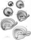
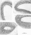
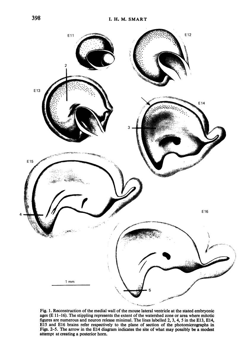
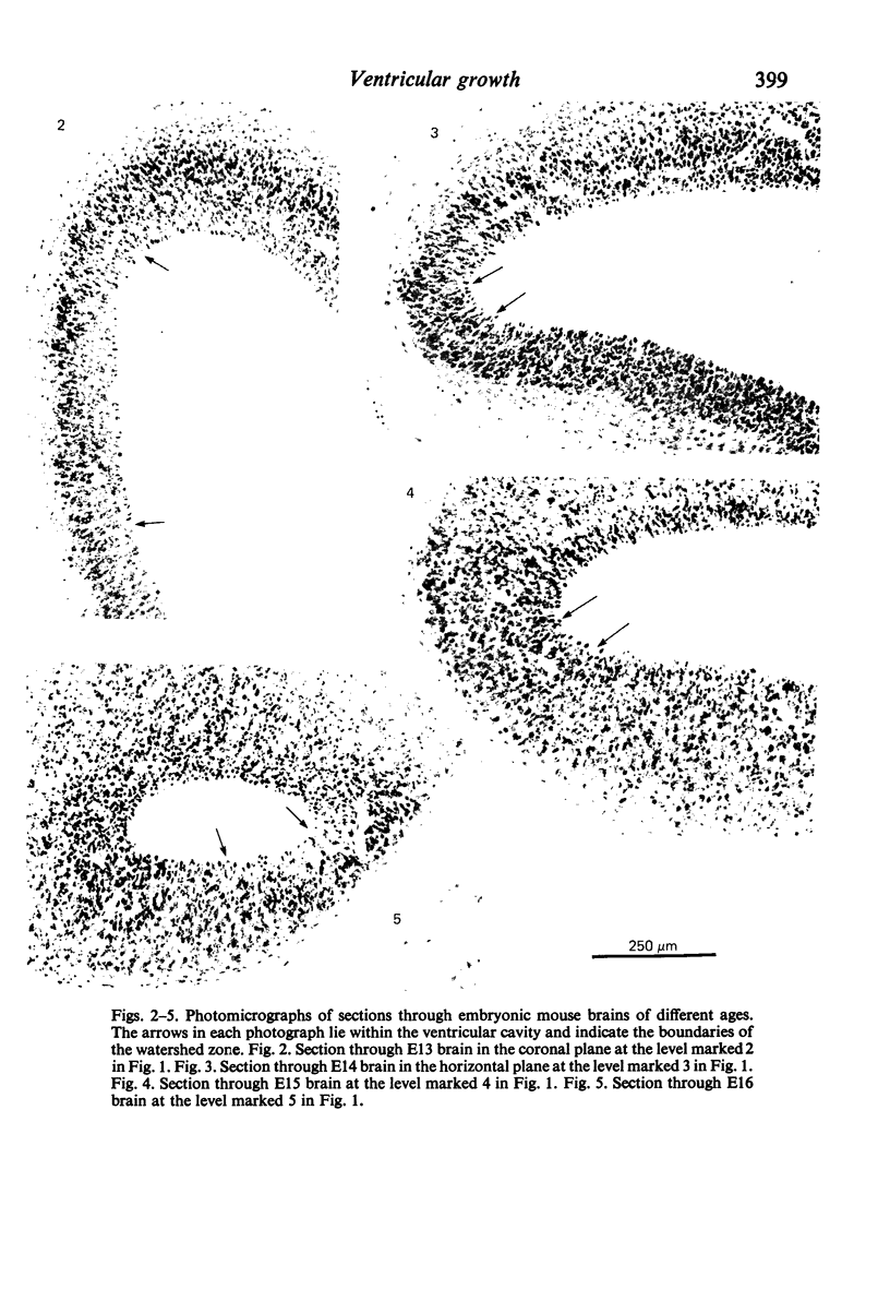
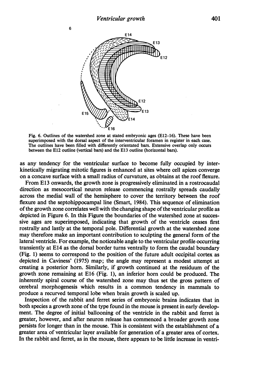
In sections of the prenatal mouse brain, sites of maximum area increase of the lateral ventricle were mapped onto reconstructions of the ventricular surface. This was done by identifying areas of ventricular layer where mitotic density was high and the adjacent intermediate layer either absent or thinly populated with neurons. It was assumed that in these areas, cell division was producing ventricular cells rather than neurons and that they were therefore gaining in area, whereas sites against which neurons were accumulating were either ceasing to increase in area or at least were increasing more slowly. Such an area occupied a zone at the junction between the medial and lateral telencephalic walls. The zone was eliminated during development in a rostrocaudal direction. It is suggested that modulation of growth along this zone may be an important factor in fashioning the form of the ventricular cavity.
Full text
PDF
Images in this article
Selected References
These references are in PubMed. This may not be the complete list of references from this article.
- Caviness V. S., Jr Architectonic map of neocortex of the normal mouse. J Comp Neurol. 1975 Nov 15;164(2):247–263. doi: 10.1002/cne.901640207. [DOI] [PubMed] [Google Scholar]
- Smart I. H. Histogenesis of the mesocortical area of the mouse telencephalon. J Anat. 1984 May;138(Pt 3):537–552. [PMC free article] [PubMed] [Google Scholar]
- Smart I. H. Proliferative characteristics of the ependymal layer during the early development of the spinal cord in the mouse. J Anat. 1972 Apr;111(Pt 3):365–380. [PMC free article] [PubMed] [Google Scholar]
- Smart I. H. Three dimensional growth of the mouse isocortex. J Anat. 1983 Dec;137(Pt 4):683–694. [PMC free article] [PubMed] [Google Scholar]


